NRF1 / Nuclear respiratory factor 1 · IHC design guide

Design Immunohistochemistry for NRF1

Plan paraffin-section NRF1 IHC around the nuclear staining reported in most tissues (HPA tissue IHC). This guide pairs catalog antibody conditions with tissue controls and flags the medium consistency between staining and RNA expression (datasheet A01129-2; HPA tissue IHC).

Evidence assembled Oct 2026 · For research use; verify linked source records and product datasheet before use
Immunohistochemistry protocol sheet for NRF1 (IHC for NRF1): expected localisation Nuclear staining in most tissues (HPA tissue IHC), antibody A01129-2, validated IHC image, and IHC protocol steps
Printable NRF1 IHC protocol sheet — expected localisation Nuclear staining in most tissues (HPA tissue IHC), antibody A01129-2, controls and protocol steps. Open the full NRF1 IHC guide →

NRF1 Immunohistochemistry Experimental Design Guide

Expected localisation, validated protocols, controls and antibodies — the at-a-glance facts below, then the full design guide.

Must know before staining
Expected localisation Nuclear staining in most tissues (HPA tissue IHC)
Staining pattern Nuclear signal in cells across most tissues (HPA tissue IHC)
Antigen retrieval Citrate pH 6 HIER, heat-mediated (datasheet A01129-2)
Positive control ⓘ Adrenal gland+4 more · see all
Negative control ⓘ Adipose tissue+4 more · see all
Important caveats
Reasons your staining may differ from the expected pattern.
Fixation Keep fixation consistent across sections (standard IHC practice; not target-specific); assess its effect empirically.
Caveat Skeletal muscle is strongest in UniProt, yet myocytes are undetected by IHC (UniProt; HPA tissue IHC)
Regulation Broad expression, strongest in skeletal muscle (UniProt)
Isoform / epitope 4 isoforms; antibody epitope coverage is unspecified (UniProt)
Section 1

Recommended NRF1 IHC & IF Protocols

The catalog antibody’s IHC-P protocol is accompanied by a published nuclear respiratory factor 1 protocol using breast cancer tissue microarrays (PMC5774493).

Recommended immunohistochemistry (IHC-P) protocol parameters
SampleParaffin-embedded human ovary cancer tissue; fixative not specified (datasheet A01129-2)
FixationImage fixative and duration unreported (datasheet A01129-2); verify before use.
Sectioning4–5 µm sections on charged slides (standard)
DeparaffinisationXylene, graded ethanol series to water (standard)
Antigen retrievalHeat retrieval: Citrate pH 6, 20 min (datasheet A01129-2)
Peroxidase block3% H2O2, 10 min, room temperature (standard)
Blocking10% goat serum (datasheet A01129-2)
Primary antibodyRabbit anti-NRF1, 0.5-1μg/ml (datasheet A01129-2)
Primary incubationOvernight at 4 °C (datasheet A01129-2)
DetectionStreptavidin-biotin complex (SABC), DAB chromogen (datasheet A01129-2)
CounterstainHematoxylin, blue, dehydrate and mount (standard)
Expected resultNRF1-positive staining in glandular cells of adrenal gland (HPA tissue IHC: Medium). HPA tissue profile: Nuclear expression in most tissues. No signal in the no-primary control.
💡Decision noteStart with heat-mediated citrate retrieval at pH 6 (datasheet A01129-2); the published protocol also used citrate at pH 6.0 (PMC5774493).
Section 2

What Is the Expected NRF1 Staining Pattern?

NRF1 is a nuclear transcription factor with no transmembrane segment (UniProt Q16656: subcellular location, topology). In paraffin sections, expect nuclear staining in many cell types, including glandular cells in colon and breast and glial cells in cerebral cortex (HPA tissue IHC: Medium). HPA describes nuclear expression in most tissues, but rates its tissue IHC evidence Approved, with medium agreement between staining and RNA data and external verification pending (HPA tissue IHC).

What am I looking at on my slide?
Nuclear staining in colon glandular cells, with little signal outside nuclei.This matches the expected compartment and an observed positive cell population (UniProt Q16656: Nucleus; HPA tissue IHC: colon glandular cells, Medium). Judge the nuclear pattern within the tissue architecture, rather than treating every cell on the section as an equivalent positive control.
Strong, predominantly membranous staining, with nuclei largely unstained.A membrane dominant pattern conflicts with NRF1's annotated nuclear location and absence of a transmembrane segment (UniProt Q16656: location, topology). Treat it as suspect until the nuclear pattern is reproduced with appropriate controls; the pattern alone cannot identify the source of artefact.
Staining is confined to skeletal myocytes, while an included known positive cell population is unstained.Do not infer a convincing positive result from the myocyte signal alone. HPA reports skeletal myocytes as Not detected, despite UniProt describing strongest expression in skeletal muscle (HPA tissue IHC; UniProt Q16656: tissue specificity). The sources disagree on this tissue, so it is a poor sole positive control.
Brown color appears broadly across nuclei, cytoplasm, and tissue spaces.A diffuse deposit obscures the expected nuclear distribution (UniProt Q16656: Nucleus). Assess background with a no primary control and inspect tissue morphology (general IHC practice). Cytoplasmic color by itself is inconclusive because HPA notes an additional, uncertain cytosolic location in ICC-IF (HPA subcellular).
No nuclear signal in colon glandular cells on an otherwise interpretable section.This is unexpected for a population reported at Medium staining, though HPA's Approved tissue pattern remains pending external verification (HPA tissue IHC: colon; reliability). Check the assay and specimen before calling NRF1 absent; one negative section cannot resolve technical failure versus biological variation.
💡Expected NRF1 appearanceCall a result convincing when nuclei of an HPA positive population, such as colon glandular cells, show clear Medium staining with limited background (HPA tissue IHC: colon); predominantly membranous color or color in tissue spaces is suspect against the nuclear annotation (UniProt Q16656: location, topology).
How each factor affects the staining
Which compartment should lead interpretation?Nuclear staining should dominate in tissue IHC (HPA tissue IHC: nuclear expression in most tissues; UniProt Q16656: Nucleus). A weak cytoplasmic component should be interpreted cautiously: ICC-IF lists cytosol as an additional location with uncertain support (HPA subcellular).
Which tissue can serve as a positive comparator?Colon and breast glandular cells and cerebral cortex glial cells have reported Medium staining (HPA tissue IHC). Choose a comparator whose relevant cells are identifiable on the section; HPA's tissue assessment is Approved with external verification pending (HPA tissue IHC: reliability).
Which apparent negative needs special care?Skeletal myocytes are Not detected by HPA tissue IHC, while UniProt calls skeletal muscle the site of strongest expression (HPA tissue IHC: skeletal muscle; UniProt Q16656: tissue specificity). Record this discrepancy rather than using skeletal muscle alone to accept or reject an assay.
How strong is the pattern evidence?The reported tissue profile is Approved, with medium agreement between antibody staining and RNA expression and external verification pending (HPA tissue IHC: reliability). The listed antibody, HPA029329, is Approved for IHC (HPA antibodies); that status supports use of its pattern as a guide, not proof that every stained structure contains NRF1.
What should IF/ICC show?Expect mainly nucleoplasmic signal; additional cytosolic localisation is uncertain (HPA subcellular: ICC-IF). This is a localisation cross-check only: the primary application here is chromogenic IHC on paraffin sections.
Why is my staining missing, weak or wrong?
SituationLikely causeNext action
No nuclear staining in a known positive comparator.The readout may have failed, or the selected section may differ from the reported HPA pattern; a single absence does not distinguish those possibilities (HPA tissue IHC: Approved, external verification pending).Confirm that the comparator contains the expected cell population, inspect morphology, and check the IHC run controls and general retrieval and detection steps (general IHC practice). Repeat before interpreting the specimen as NRF1 negative.
Membrane dominant staining replaces the expected nuclear pattern.The location conflicts with nuclear NRF1 and its lack of a transmembrane segment (UniProt Q16656: location, topology); the image alone cannot assign a specific artefact.Compare a known positive tissue and a no primary control on the same run, then review primary antibody and detection settings (general IHC practice). Require a reproducible nuclear pattern before scoring positivity.
An HPA Not detected cell population stains strongly.The staining may reflect cross-reactivity or detection background, although HPA's tissue assessment has medium consistency and awaits external verification (HPA tissue IHC: negative populations, reliability).Check the precise cell identity and compartment, then compare a reported positive population and no primary control (general IHC practice). Do not label the unexpected cell population NRF1 positive from color alone.
Brown deposits spread through tissue spaces or obscure cell borders.Diffuse chromogen can make compartment scoring unreliable (general IHC practice). If the detection system uses peroxidase, endogenous enzyme activity is one possible contributor (general IHC practice).Inspect a no primary control; if it also develops color, review blocking and detection controls for the system used (general IHC practice). Reassess nuclear staining only after background permits cell level interpretation.
Only skeletal muscle is available as a putative positive control.The sources conflict: HPA reports myocytes Not detected, while UniProt reports strongest expression in skeletal muscle (HPA tissue IHC; UniProt Q16656: tissue specificity).Use an HPA reported positive cell population, such as colon glandular cells, for the assay comparator when available (HPA tissue IHC: colon, Medium). Document the skeletal muscle result separately rather than using it as the sole pass or fail criterion.
Weak cytoplasmic color accompanies otherwise clear nuclear staining.HPA reports cytosol as an additional but uncertain ICC-IF location, while its tissue IHC profile is nuclear (HPA subcellular; HPA tissue IHC: profile).Score the interpretable nuclear component and document the cytoplasmic component separately (general IHC practice). Compare background controls before assigning biological meaning to the weaker extra compartment.

Sample controls for NRF1 IHC & IF

🧪Run adrenal gland first and assess nuclear staining in its glandular cells (HPA: Medium in adrenal gland glandular cells; UniProt Q16656: Nucleus). Use adipose tissue adipocytes as the biological negative (HPA: Not detected in adipocytes). On the adrenal slide, cells selected as internal negatives should show counterstain without nuclear chromogen; verify that interpretation against the no-primary slide (standard IHC control).
Positive control tissue: Adrenal gland (Glandular cells, HPA Medium)
Negative control tissue: Adipose tissue (HPA Not detected)
ICC-IF cell lines (HPA subcellular resource): HPA ICC-IF images show NRF1 in A-431, U-251MG, with annotated localisation: Nucleoplasm (supported) (HPA subcellular).
Technical controls: Include a no-primary, secondary-only control, a host-matched rabbit IgG isotype control, and NRF1-knockout material where available; peptide competition is an alternative if the immunizing peptide is available (selected A01129-2 caption: rabbit primary; standard IHC controls). For the adrenal section, block endogenous peroxidase and check for endogenous biotin background when using the caption’s biotin–streptavidin/DAB detection system (selected A01129-2 caption: biotinylated secondary, SABC and DAB; standard IHC practice).
⚠️Feasibility: A target-specific fixation window is unreported, and the selected A01129-2 paraffin-section caption does not state a fixative (selected A01129-2 caption). Start retrieval optimization with citrate buffer at pH 6 for 20 minutes, as reported for the catalog antibody; its effect in adrenal gland remains to be checked (selected A01129-2 caption: ovary cancer section; HPA: adrenal gland glandular cells, Medium). The supplied evidence does not establish that frozen sections or IF are easier, or document an adrenal-specific artefact; use nuclear localization when judging signal (HPA: nucleoplasm supported; selected A01129-2 caption: paraffin-section IHC).

HPA tissue IHC evidence for NRF1

Comprehensive Human Protein Atlas IHC scoring per tissue (reliability: Approved — Medium consistency between antibody staining and RNA expression data. Pending external verification.). Rows are taken directly from the HPA tissue chart — click any row's HPA link to view the source.

Positive expression · recommended positive controls

TissueCell typeLevelEvidenceSource
Adrenal gland Glandular cells Medium Protein (IHC) HPA →
Appendix Glandular cells Medium Protein (IHC) HPA →
Breast Glandular cells Medium Protein (IHC) HPA →
Caudate Glial cells Medium Protein (IHC) HPA →
Cerebellum Cells in granular layer Medium Protein (IHC) HPA →

Undetected expression · recommended negative controls

TissueCell typeLevelEvidenceSource
Adipose tissue Adipocytes Not detected Protein (IHC) HPA →
Bone marrow Hematopoietic cells Not detected Protein (IHC) HPA →
Esophagus Squamous epithelial cells Not detected Protein (IHC) HPA →
Liver Cholangiocytes Not detected Protein (IHC) HPA →
Salivary gland Glandular cells Not detected Protein (IHC) HPA →
Section 3

Advanced NRF1 IHC Tips

Troubleshoot chromogenic NRF1 IHC in paraffin sections by checking retrieval, nuclear localisation, controls and cell-specific scoring (datasheet A01129-2; UniProt Q16656; HPA tissue IHC).

How should I retrieve NRF1 when nuclear staining is weak?
Start with heat-mediated citrate buffer at pH 6 for 20 minutes in paraffin sections (datasheet A01129-2). Run a known staining control alongside the test section so a weak result can be distinguished from a retrieval failure; the product image demonstrates staining in human ovary cancer tissue under those retrieval conditions (datasheet A01129-2). If staining remains weak, adjust heating and cooling consistently across sections, then compare nuclear signal with tissue damage and background before trying another retrieval condition (standard IHC practice; UniProt Q16656: nucleus). Keep antibody concentration and detection constant during that comparison: the pictured workflow used 1 μg/ml primary antibody and DAB detection (datasheet A01129-2).
Could fixation explain weak or uneven NRF1 staining?
The selected paraffin-section caption does not state a fixative, so NRF1-specific fixation sensitivity is unknown for this antibody (datasheet A01129-2). Record the actual fixative and fixation interval for every specimen, and compare sections processed together before attributing differences to biology (standard IHC practice). Check whether weak nuclear staining follows thick tissue regions, poorly preserved areas or section edges; those patterns can indicate processing variation rather than a change in NRF1 abundance (standard IHC practice; UniProt Q16656: nucleus). Retest with matched controls and the documented citrate pH 6 retrieval for 20 minutes, changing one processing variable at a time (datasheet A01129-2; standard IHC practice).
What should I make of predominantly cytoplasmic DAB staining?
Score nuclear staining first: NRF1 is annotated as nuclear, and tissue IHC shows nuclear expression in most tissues (UniProt Q16656: nucleus; HPA tissue IHC). A cytoplasmic pattern alone needs scrutiny because HPA supports nucleoplasmic localisation while listing cytosolic localisation as uncertain in its subcellular data (HPA subcellular). Inspect the hematoxylin counterstain to confirm that DAB lies within intact nuclei, and compare the same cell type across sections processed in one run (standard IHC practice). If cytoplasmic staining dominates, check the no-primary control, retrieval damage and DAB background before reporting a localisation shift; no transmembrane segment is annotated for NRF1 (standard IHC practice; UniProt Q16656 topology).
Can a negative section reflect isoform or epitope differences?
NRF1 has 4 annotated isoforms—Long, Short, 3 and 4—so antibody recognition across isoforms depends on the mapped epitope (UniProt Q16656: isoforms). The supplied product caption does not identify that epitope; do not treat a negative cell population as proof that it lacks every NRF1 isoform (datasheet A01129-2; UniProt Q16656: isoforms). Check available epitope documentation and compare staining after the stated citrate pH 6 retrieval, holding detection conditions fixed (datasheet A01129-2; standard IHC practice). Five annotated modified residues include CK2-associated phosphoserines at positions 39, 44, 46, 47 and 52; whether these affect this antibody's binding is unestablished (UniProt Q16656: modified residues; datasheet A01129-2).
How can IF help assess a disputed NRF1 IHC pattern?
Use IF as a separate corroboration experiment, pairing NRF1 with a marker that identifies the expected cell population; HPA reports medium IHC staining in colon glandular cells (HPA tissue IHC). Choose spectrally separated fluorophores and assess tissue autofluorescence in an unstained control before assigning weak signal to NRF1 (standard IF practice). For an antibody targeting an intracellular epitope, use permeabilisation appropriate for nuclear access; NRF1 is nuclear and has no annotated transmembrane segment, but this antibody's epitope location is unspecified (UniProt Q16656: localisation and topology; datasheet A01129-2). Compare nuclear colocalisation with a DNA counterstain, then validate the IF result independently before using it to explain chromogenic IHC (HPA subcellular; standard IF practice).
How do I reduce diffuse brown staining without losing nuclei?
Start by comparing a no-primary section with the stained section, then identify whether brown deposit tracks tissue edges, damaged regions or the expected nuclei (standard IHC practice; UniProt Q16656: nucleus). The pictured workflow used 10% goat serum block, 1 μg/ml primary antibody overnight at 4°C, and biotinylated secondary detection with DAB (datasheet A01129-2). If background persists, optimise primary concentration and washing while retaining a positive control, and include a peroxidase block before chromogen development as a general chromogenic IHC step (standard IHC practice). With biotin-based detection, assess endogenous biotin as a possible source of signal; the caption establishes that detection chemistry, not NRF1-specific biotin interference (datasheet A01129-2; standard IHC practice).
How should NRF1 staining be scored across samples? ⚠ ANSWER MARKED FOR VERIFICATION
Define the scored compartment as intact nuclei, consistent with NRF1's nuclear annotation and the predominantly nuclear tissue pattern (UniProt Q16656: nucleus; HPA tissue IHC). For each prespecified cell population, report the percentage of positive nuclei and an intensity-weighted H-score, recording the scoring threshold before examining group differences (standard IHC practice). Normalise positive counts to the number of evaluable nuclei in that same cell population, or report positive nuclear density per mm² of viable tissue when area is the denominator (standard IHC practice). Use matched staining runs and exclude necrotic or damaged regions; HPA rates its tissue staining Approved with medium RNA consistency pending external verification (standard IHC practice; HPA tissue IHC).
When is an NRF1-positive result convincing rather than artefactual?
A convincing result places DAB within preserved nuclei in an identifiable cell population, matching NRF1's nuclear annotation and HPA's mainly nuclear tissue profile (UniProt Q16656: nucleus; HPA tissue IHC). Compare the population with its own controls: HPA reports medium staining in colon glandular cells but no detected staining in adipocytes, illustrating why cell identity matters (HPA tissue IHC). Distrust signal confined to section edges, necrotic areas or the no-primary control, and investigate endogenous peroxidase or biotin contributions to this DAB, biotin-based workflow (standard IHC practice; datasheet A01129-2). Treat unexpected cytoplasmic staining cautiously because HPA marks cytosolic localisation uncertain and its tissue IHC reliability awaits external verification (HPA subcellular; HPA tissue IHC).
Boster reagents

Best NRF1 / Nuclear respiratory factor 1 IHC Antibodies

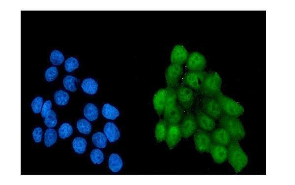
The catalog shows NRF1 IHC in human, mouse and rat tissues (catalog IHC captions), IF in MCF7 cells (M01129-1 IF caption), and ICC in A549 cells (PA1948 ICC caption).

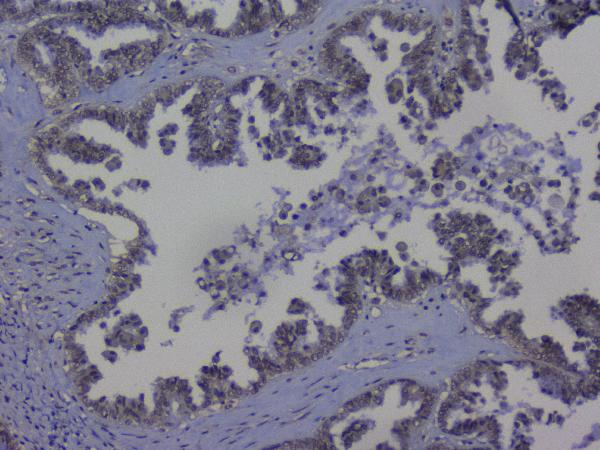
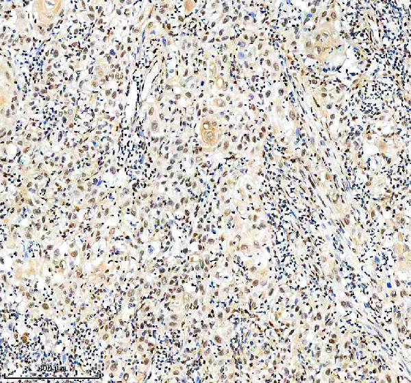
Real IHC data IHC analysis of NRF1 using anti-NRF1 antibody (A01129-2). NRF1 was detected in paraffin-embedded section of human ovary cancer tissue. Heat mediated antigen retrieval was performed in citrate buffer (pH6, epitope retrieval solution) for 20 mins. The tissue section was blocked with 10% goat serum. The tissue section was then incubated with 1μg/ml rabbit anti-NRF1 Antibody (A01129-2) overnight at 4°C. Biotinylated goat anti-rabbit IgG was used as secondary antibody and incubated for 30 minutes at 37°C. The tissue section was developed using Strepavidin-Biotin-Complex (SABC)(Catalog # SA1022) with DAB as the chromogen.
Anti-NRF1 Antibody ®
Cat # A01129-2
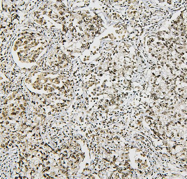
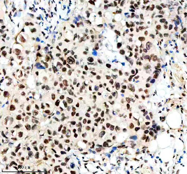
Real IHC data IHC analysis of NRF1 using anti-NRF1 antibody (M01129-1). NRF1 was detected in paraffin-embedded section of human rectal cancer tissues. Heat mediated antigen retrieval was performed in citrate buffer (pH6, epitope retrieval solution) for 20 mins. The tissue section was blocked with 10% goat serum. The tissue section was then incubated with 1μg/ml mouse anti-NRF1 Antibody (M01129-1) overnight at 4°C. Biotinylated goat anti-mouse IgG was used as secondary antibody and incubated for 30 minutes at 37°C. The tissue section was developed using Strepavidin-Biotin-Complex (SABC)(Catalog # SA1021) with DAB as the chromogen.
Anti-NRF1 Antibody ® (monoclonal, 2G4)
Cat # M01129-1
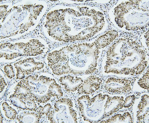
Real IHC data IHC analysis of NRF1 using anti-NRF1 antibody (M01129). NRF1 was detected in a paraffin-embedded section of human bladder cancer tissue. Heat mediated antigen retrieval was performed in EDTA buffer (pH 8.0, epitope retrieval solution). The tissue section was blocked with 10% goat serum. The tissue section was then incubated with 1:50 rabbit anti-NRF1 Antibody (M01129) overnight at 4°C. Peroxidase Conjugated Goat Anti-rabbit IgG was used as secondary antibody and incubated for 30 minutes at 37°C. The tissue section was developed using HRP Conjugated Rabbit IgG Super Vision Assay Kit (Catalog # SV0002) with DAB as the chromogen.
Anti-NRF1 Rabbit Monoclonal Antibody
Cat # M01129
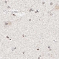
Real IHC data Anti-NRF1 antibody, PA1948, IHC(P) IHC(P): Human Mammary Cancer Tissue
Anti-Nuclear respiratory factor 1 NRF1 Antibody ®
Cat # PA1948

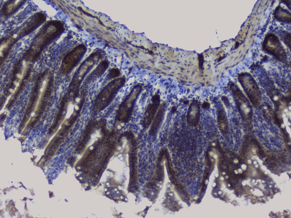
A01129-2 has paraffin-section IHC images from human ovary cancer, mouse intestine and spleen, and rat heart (A01129-2 IHC captions); M01129-1 has human cancer tissue IHC images and an MCF7 IF image (M01129-1 image captions). M01129 has paraffin-section IHC images from human cancers (M01129 IHC captions); PA1948 has IHC images from human mammary and intestinal cancers and rat intestine, plus an A549 ICC image (PA1948 image captions).

Which to pick: For human tissue IHC, M01129 offers a rabbit monoclonal antibody with a paraffin-section image using EDTA retrieval (M01129 catalog entry and IHC caption); PA1948 offers human and rat IHC images (PA1948 IHC captions). For IF/ICC, choose M01129-1 when an IF application and MCF7 IF image are needed (M01129-1 catalog entry and IF caption). For cross-species tissue IHC, A01129-2 has human, mouse and rat paraffin-section images (A01129-2 IHC captions); the fixative is unreported in the IHC captions for all four SKUs (A01129-2, M01129-1, M01129 and PA1948 IHC captions).

Each figure is that product's own IHC / IF validation image from its datasheet.

References

  1. UniProt Consortium. UniProt entry Q16656 (NRF1_HUMAN, Nuclear respiratory factor 1).
  2. Human Protein Atlas. NRF1 tissue IHC expression (reliability: Approved).
  3. Human Protein Atlas. NRF1 subcellular location (ICC-IF): Mainly localized to the nucleoplasm. In addition localized to the cytosol..
  4. Human Protein Atlas. NRF1 antibody validation summary (1 antibodies).
  5. NRF1 and NRF2 mRNA and Protein Expression Decrease Early during Melanoma Carcinogenesis: An Insight into Survival and MicroRNAs. Oxidative medicine and cellular longevity 2019 — PMC6794976.
  6. NRF1 is upregulated by docosahexaenoic acid to ameliorate MASH through the inhibition of ER stress. Cell death & disease 2026 — PMC12811613.
  7. Increased expression of mitochondrial transcription factor A and nuclear respiratory factor-1 predicts a poor clinical outcome of breast cancer. Oncology letters 2018 — PMC5774493.
  8. TALENs-directed knockout of the full-length transcription factor Nrf1α that represses malignant behaviour of human hepatocellular carcinoma (HepG2) cells. Scientific reports 2016 — PMC4827396.
  9. PubMed PMID:8253388 — UniProt-cited evidence.
  10. PubMed PMID:8034649 — UniProt-cited evidence.
  11. PubMed PMID:7629110 — UniProt-cited evidence.