This website uses cookies to ensure you get the best experience on our website.


- Table of Contents
Real validated NTF3 Western blot protocols, expected-band and isoform facts, troubleshooting for weak or shifted signal, and recommended anti-NTF3 WB antibodies. Everything you need to plan the experiment before you commit precious samples.
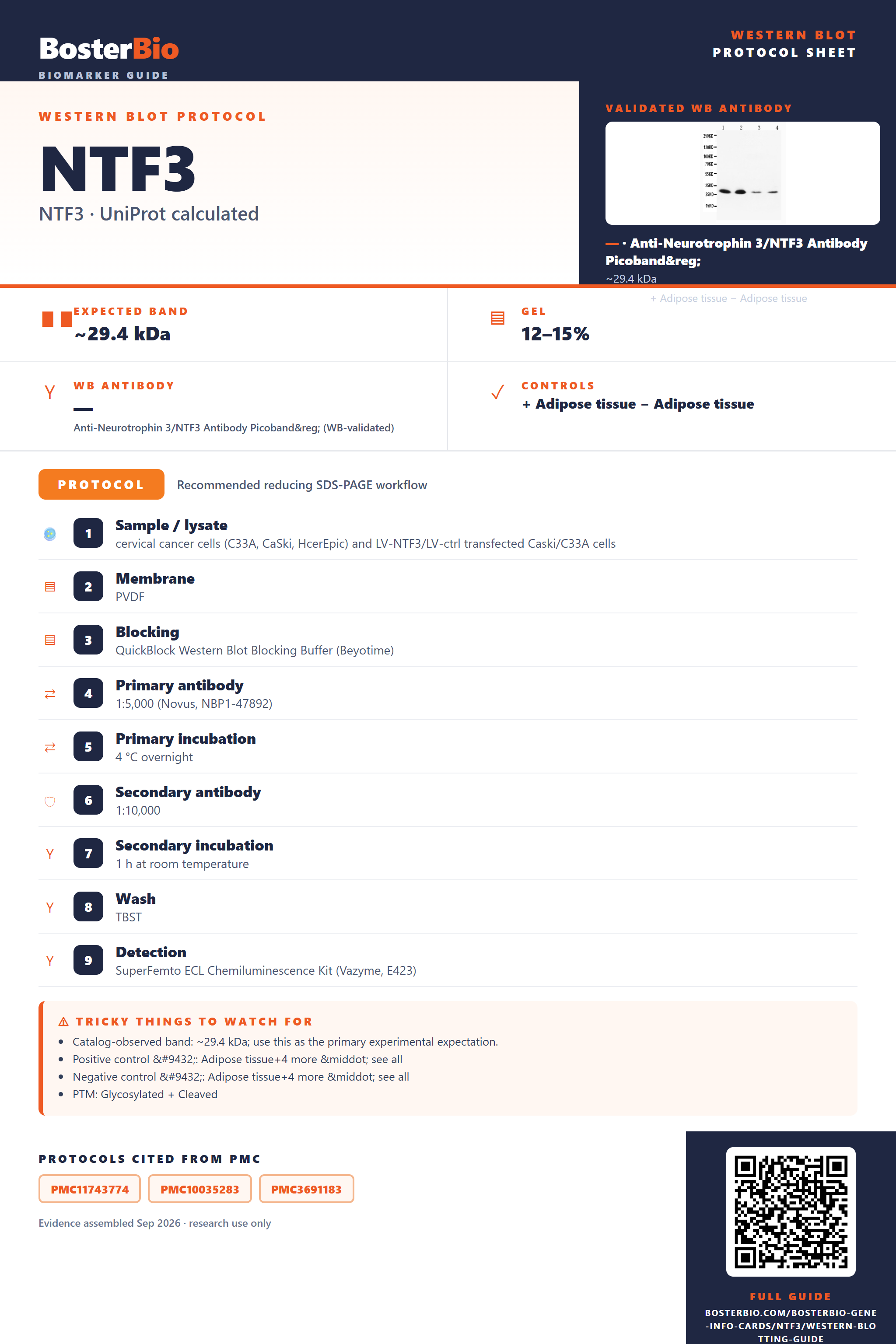
Expected bands, validated protocols, controls and antibodies — the at-a-glance facts below, then the full design guide.
| Expected band | ~29.4 kDa | |
| Observed band | ~34 kDa | |
| Gel | 12–15% | |
| Positive control | Adipose tissue+4 more · see all | |
| Negative control | Adipose tissue+4 more · see all |
| PTM | Glycosylated + Cleaved | |
| Caveat | Glycosylation adds mass | |
| Regulation | Kras signaling dn | |
| Isoform | 2 isoform(s) |
Literature-validated Western blot parameters for NTF3 — gel percentage, transfer, blocking, antibody incubation and detection, extracted from published methods.
| Sample / lysate | cervical cancer cells (C33A, CaSki, HcerEpic) and LV-NTF3/LV-ctrl transfected Caski/C33A cells |
| Membrane | PVDF |
| Blocking | QuickBlock Western Blot Blocking Buffer (Beyotime) |
| Primary antibody | 1:5,000 (Novus, NBP1-47892) |
| Primary incubation | 4 °C overnight |
| Secondary antibody | 1:10,000 |
| Secondary incubation | 1 h at room temperature |
| Wash | TBST |
| Detection | SuperFemto ECL Chemiluminescence Kit (Vazyme, E423) |
NTF3 has a 29.4 kDa predicted backbone but runs at ~34 kDa on blots due to N-glycosylation at Asn131 and cleavage of its signal peptide/propeptide during secretion.
| single band around 34 kDa | represents the mature, N-glycosylated secreted form of NTF3, running above the 29.4 kDa predicted backbone due to the N131 glycan |
| no band near the ~29 kDa full-length precursor | the 18-residue signal peptide and the propeptide (residues 19-138) are cleaved during maturation/secretion, so only the smaller mature domain is detected |
| faint or absent band in whole-cell lysate | NTF3 is a secreted protein, so most immunoreactive material is released into conditioned medium rather than retained intracellularly |
| two bands at slightly different apparent masses | corresponds to the two annotated NTF3 isoforms produced by alternative splicing |
| band mobility shifts or becomes diffuse with incomplete reduction | the three intrachain disulfide bonds stabilize the mature domain's compact fold, so under-reduced samples can migrate differently than fully reduced/denatured ones |
| Predicted mass (UniProt) | 29.4 kDa backbone for the full 257-residue precursor before any modification |
| N-glycosylation at Asn131 | adds mass and can broaden the mature band above the unmodified predicted size, consistent with the observed ~34 kDa band |
| Signal peptide and propeptide cleavage (residues 1-138) | removes the N-terminal signal sequence and propeptide during secretion, giving a smaller mature band than the uncleaved full-length precursor |
| Secreted subcellular localization | shifts most detectable protein into conditioned medium/extracellular fractions, reducing or eliminating signal in standard whole-cell lysates |
| Alternative splicing (isoforms 1 and 2) | can produce two bands of differing apparent size on the same blot |
| Intrachain disulfide bonds (152-217, 195-246, 205-248) | stabilize the mature domain's folded structure, so incomplete reduction can alter apparent mobility compared with fully reduced samples |
| Situation | Likely cause | Next action |
|---|---|---|
| No band in lysate | NTF3 is secreted and does not accumulate intracellularly in appreciable amounts | probe conditioned medium or a concentrated secreted fraction instead of whole-cell lysate |
| Band higher than expected | N-glycosylation at Asn131 adds mass above the 29.4 kDa predicted backbone | treat the sample with PNGase F and confirm the band shifts toward the predicted mass |
| Multiple bands | the two annotated splice isoforms migrate at different apparent masses | confirm isoform identity using isoform-specific antibodies or transcript context |
| Broad smear instead of sharp band | heterogeneous glycosylation at the single N-glycosylation site produces glycoform microheterogeneity | treat with PNGase F or run a higher-resolution gradient gel to sharpen the band |
| Fragments below expected size | normal proteolytic removal of the signal peptide and propeptide (residues 1-138) during maturation | check whether the antibody epitope lies within the mature domain before treating this as degradation |
| Weak or no signal | NTF3 is a low-abundance secreted growth factor diluted across a large lysate or medium volume | concentrate conditioned medium, e.g. by TCA precipitation, before loading |


Comprehensive Human Protein Atlas IHC scoring per tissue. Rows are taken directly from the HPA tissue chart — click any row's HPA link to view the source.
| Tissue | Cell type | Level | Evidence | Source |
|---|---|---|---|---|
| Adipose tissue | adipocytes | Not detected | Protein (IHC) | HPA → |
| Adrenal gland | glandular cells | Not detected | Protein (IHC) | HPA → |
| Appendix | glandular cells | Not detected | Protein (IHC) | HPA → |
| Bone marrow | hematopoietic cells | Not detected | Protein (IHC) | HPA → |
| Breast | adipocytes | Not detected | Protein (IHC) | HPA → |
| Tissue | Cell type | Level | Evidence | Source |
|---|---|---|---|---|
| Adipose tissue | adipocytes | Not detected | Protein (IHC) | HPA → |
| Adrenal gland | glandular cells | Not detected | Protein (IHC) | HPA → |
| Appendix | glandular cells | Not detected | Protein (IHC) | HPA → |
| Bone marrow | hematopoietic cells | Not detected | Protein (IHC) | HPA → |
| Breast | adipocytes | Not detected | Protein (IHC) | HPA → |
Deeper troubleshooting and optimisation questions for NTF3, answered from its protein features.
BosterBio's NTF3 antibodies are among the best-performing WB antibodies on the market — well cited, thoroughly validated, and orthogonally cross-validated against negative tissues and complementary methods.
These NTF3 antibodies are top-performing reagents, widely cited in published research, and thoroughly validated for Western blot specificity, with orthogonal cross-validation across multiple tissue and cell lysates confirming reliable, reproducible detection.
Which to pick: Two validated NTF3 antibodies are listed. PA1062 shows WB bands across rat brain tissue and MCF-7/HeLa lysates, good for broad tissue screening. A03348-2 provides a dedicated NTF3 WB image with detailed SDS-PAGE conditions for protocol replication.