This website uses cookies to ensure you get the best experience on our website.


- Table of Contents
Plan chromogenic ORAI1 IHC in paraffin sections using skin keratinocytes as a high staining reference (HPA tissue IHC). Interpret the observed cytoplasmic staining alongside ORAI1’s annotated cell membrane location (HPA tissue IHC; UniProt).

Expected localisation, validated protocols, controls and antibodies — the at-a-glance facts below, then the full design guide.
| Expected localisation | Cytoplasm observed; cell membrane annotated (HPA tissue IHC; UniProt) | |
| Staining pattern | Strong keratinocyte cytoplasm; cytoplasmic in most tissues (HPA tissue IHC) | |
| Antigen retrieval | Citrate pH 6.0 HIER, 95–98 °C, 20 min (rule: cytoplasmic / membrane antigen) | |
| Positive control | Skin+4 more · see all | |
| Negative control | Adipose tissue+4 more · see all |
| Fixation | Keep fixation consistent across sections. (standard IHC practice; not target-specific) | |
| Caveat | Cytoplasmic IHC can complicate membrane interpretation (HPA tissue IHC; UniProt) | |
| Regulation | Similar in naive and effector T helper cells (UniProt) | |
| Isoform / epitope | 2 isoforms; check whether the epitope is extracellular or cytoplasmic (UniProt) |
The catalog antibody’s IHC-P protocol is accompanied by four published ORAI1 chromogenic IHC protocols (PMC8739746; PMC3871607; PMC5190020; PMC5342032).
| Sample | Tissue sections; selected-image fixative not specified (standard IHC workflow) |
| Fixation | Image fixative and duration unreported (datasheet A00909-1); verify before use. |
| Sectioning | 4–5 µm sections on charged slides (standard) |
| Deparaffinisation | Xylene, graded ethanol series to water (standard) |
| Antigen retrieval | Heat-induced epitope retrieval in citrate buffer, pH 6.0, 20 min at 95–98 °C (standard rule: cytoplasmic / membrane antigen) |
| Peroxidase block | 3% H2O2, 10 min, room temperature (standard) |
| Blocking | 10% normal serum of the secondary host, 30 min, room temperature (standard) |
| Primary antibody | Rabbit anti-ORAI1, 10 μg/mL (datasheet A00909-1) |
| Primary incubation | Overnight at 4 °C (standard) |
| Detection | HRP-polymer secondary, DAB chromogen 5–10 min (standard) |
| Counterstain | Hematoxylin, blue, dehydrate and mount (standard) |
| Expected result | ORAI1-positive staining in keratinocytes of skin (HPA tissue IHC: High). HPA tissue profile: Cytoplasmic expression in most tissues, highly abundant in skin. No signal in the no-primary control. |
ORAI1 is a four-pass cell-membrane protein, with basolateral localisation also annotated (UniProt Q96D31 topology and subcellular location). In paraffin-section IHC, expect the clearest staining in skin keratinocytes and testicular Leydig cells (HPA: High in both). HPA also reports cytoplasmic staining in most tissues. Its tissue IHC assessment is Approved, with medium consistency against RNA data and external verification pending (HPA: tissue IHC reliability).
| Keratinocytes or Leyig cells show strong staining, with a membrane-associated component. | These are the best-supported positive cell populations (HPA: High in skin keratinocytes and testis Leydig cells). Membrane localisation agrees with ORAI1 topology (UniProt Q96D31). Assess the cells themselves; colour in adjacent spaces does not establish ORAI1 expression (standard IHC practice). |
| Positive cells show cytoplasmic staining without a sharp membrane outline. | This can agree with the reported tissue-IHC profile, which describes cytoplasmic expression in most tissues (HPA: tissue IHC profile). Record compartment and cell type separately. A membrane outline is biologically plausible, but should not be imposed as the sole acceptable paraffin-IHC pattern (UniProt Q96D31 subcellular location). |
| Signal is predominantly nuclear or appears outside cells. | Neither location matches the annotated cell-membrane protein or the reported cytoplasmic tissue-IHC profile (UniProt Q96D31 subcellular location; HPA: tissue IHC profile). Treat it as suspect until section morphology, counterstain, and a detection-only control have been checked (standard IHC practice). |
| Strong signal appears in adipocytes or glomerular cells. | Those sampled cell populations were not detected in HPA tissue IHC (HPA: adipose adipocytes; kidney glomerular cells). Consider cross-reactivity or endogenous detection activity, especially if a no-primary control also stains (standard IHC practice). A discrepant slide alone cannot identify which cause applies. |
| Colour covers broad regions or known-positive cells remain unstained. | Uniform colour obscures cell-specific interpretation (standard IHC practice). Absence of signal in skin keratinocytes or Leydig cells conflicts with HPA's High observations, but does not by itself prove that ORAI1 is absent (HPA: skin; testis). Check controls and slide quality before scoring. |
| Compartment and assay | UniProt places ORAI1 at the cell membrane, including basolateral membrane, while HPA tissue IHC describes cytoplasmic staining (UniProt Q96D31; HPA: tissue IHC profile). Interpret a paraffin section against both observations and its cell morphology; neither source establishes that every positive cell must show a crisp membrane rim. |
| Antibody epitope and topology | ORAI1 has four transmembrane segments with extracellular and cytoplasmic loops and termini (UniProt Q96D31 topology). The supplied record does not locate the IHC antibody's epitope, so it cannot predict which retrieval or permeabilisation condition exposes that epitope. Optimize those steps empirically with tissue controls (standard IHC practice). |
| Cell state and puncta | After calcium-store depletion, ORAI1 can colocalise with STIM1 in membrane puncta at ER–plasma-membrane junctions (UniProt Q96D31 subcellular location). The supplied HPA paraffin-IHC profile does not establish puncta as an expected tissue-section pattern; do not require them to call an ordinary section positive. |
| Isoforms and tissue expression | UniProt lists alpha and beta isoforms and protein expression in naive CD4 and CD8 T cells (UniProt Q96D31). HPA reports hematopoietic cells in bone marrow as Not detected in its tissue-IHC samples (HPA: bone marrow). Those observations concern different specimens and methods; the supplied antibody data do not establish isoform discrimination. |
| IF/ICC Q&A: what localisation is supported? | HPA summarises ORAI1 as membrane-localised in ICC-IF, but supplies no main-location assignment or cell-line images here (HPA: subcellular ICC-IF). Use that as a limited localisation cross-check; this section provides no IF/ICC protocol or cell-line-specific expected intensity. |
| Situation | Likely cause | Next action |
|---|---|---|
| No stain in skin keratinocytes or Leydig cells. | A failed run is possible because HPA observed High staining in these cells (HPA: skin; testis); absence alone does not isolate the failed step. | Check tissue preservation, positive-control performance, primary-antibody step, detection reagents, and counterstain; adjust retrieval or antibody concentration with controls (standard IHC practice). |
| All tissue compartments develop similar diffuse colour. | The distribution lacks the cell specificity needed to interpret HPA's keratinocyte and Leydig-cell observations (HPA: skin; testis). Excess background is possible (standard IHC practice). | Inspect the no-primary control; review blocking, washes, detection time, and primary concentration using the same tissue batch (standard IHC practice). |
| Signal is concentrated in nuclei. | Predominantly nuclear staining conflicts with UniProt's membrane localisation and HPA's cytoplasmic tissue profile (UniProt Q96D31; HPA: tissue IHC profile). | Compare with morphology and counterstain, then repeat with a detection-only control and a known-positive tissue (standard IHC practice). |
| Adipocytes or glomerular cells stain strongly. | HPA lists those cell populations as Not detected; cross-reactivity or endogenous detection activity may account for the discrepancy (HPA: adipose; kidney; standard IHC practice). | Check a no-primary control, inspect which cells carry signal, and compare with positive skin or testis on the same run (HPA: skin; testis; standard IHC practice). |
| Cytoplasmic colour is present but the membrane rim is weak. | HPA describes cytoplasmic tissue-IHC expression despite UniProt's membrane annotation (HPA: tissue IHC profile; UniProt Q96D31). The contrast is not sufficient evidence of assay failure. | Score cell type, intensity, and compartment separately; compare with HPA-positive cells and confirm that the no-primary control is clear (HPA: skin; testis; standard IHC practice). |
| Bone-marrow cells are negative despite expected T-cell expression. | UniProt reports ORAI1 protein in naive T cells, while HPA's sampled bone-marrow hematopoietic cells were Not detected by tissue IHC (UniProt Q96D31 tissue specificity; HPA: bone marrow). | Do not use bone-marrow negativity alone to reject the run. Verify staining in skin or testis and interpret any T-cell result in its specimen and assay context (HPA: skin; testis; standard IHC practice). |
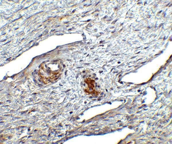
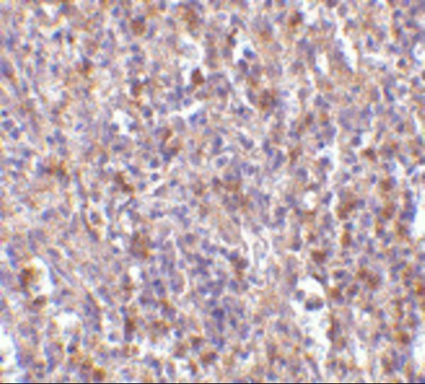
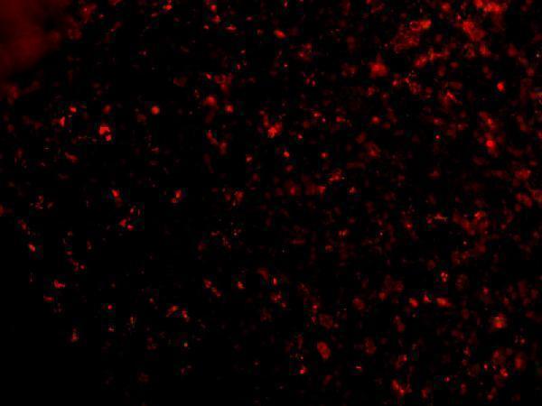
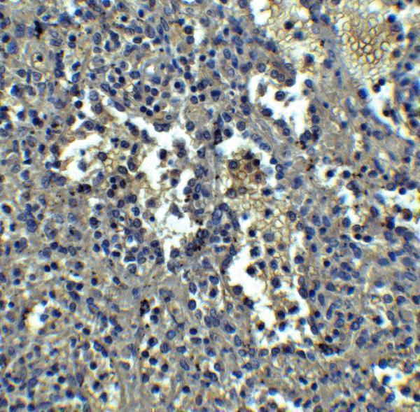
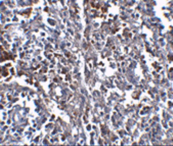
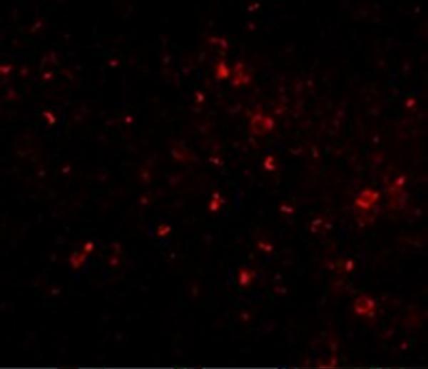
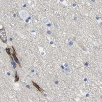


Comprehensive Human Protein Atlas IHC scoring per tissue (reliability: Approved — Medium consistency between antibody staining and RNA expression data. Pending external verification.). Rows are taken directly from the HPA tissue chart — click any row's HPA link to view the source.
| Tissue | Cell type | Level | Evidence | Source |
|---|---|---|---|---|
| Skin | Keratinocytes | High | Protein (IHC) | HPA → |
| Testis | Leydig cells | High | Protein (IHC) | HPA → |
| Cervix | Squamous epithelial cells | Medium | Protein (IHC) | HPA → |
| Duodenum | Glandular cells | Medium | Protein (IHC) | HPA → |
| Esophagus | Squamous epithelial cells | Medium | Protein (IHC) | HPA → |
| Tissue | Cell type | Level | Evidence | Source |
|---|---|---|---|---|
| Adipose tissue | Adipocytes | Not detected | Protein (IHC) | HPA → |
| Bone marrow | Hematopoietic cells | Not detected | Protein (IHC) | HPA → |
| Caudate | Glial cells | Not detected | Protein (IHC) | HPA → |
| Cerebellum | Cells in granular layer | Not detected | Protein (IHC) | HPA → |
| Endometrium | Cells in endometrial stroma | Not detected | Protein (IHC) | HPA → |
Troubleshoot ORAI1 chromogenic IHC in paraffin sections using matched controls, compartment aware scoring and the antibody’s documented tissue evidence (UniProt Q96D31; HPA; caption A00909-1).
Two anti-ORAI1 antibodies have human tissue IHC images and human IF images (catalog captions); both list Human and Mouse reactivity, while M00909 also lists Rat (catalog reactivity).
A00909-1 lists IHC-P and IF, with IHC shown in human ovary and IF shown in human spleen cells (catalog applications and image captions). M00909 lists IHC-P and IF, with both images from human spleen samples (catalog applications and image captions).
Which to pick: For tissue IHC, choose A00909-1 for the human ovary example or M00909 for the human spleen example; both list IHC-P, but their image captions do not report the fixative (catalog applications and IHC image captions). For IF, both list IF and show human spleen samples; neither payload establishes a separate ICC validation (catalog applications and IF image captions). Choose M00909 when Rat reactivity or a mouse monoclonal is needed; A00909-1 is a rabbit antibody listing Human and Mouse reactivity (catalog reactivity, host and clone).