This website uses cookies to ensure you get the best experience on our website.
- Table of Contents
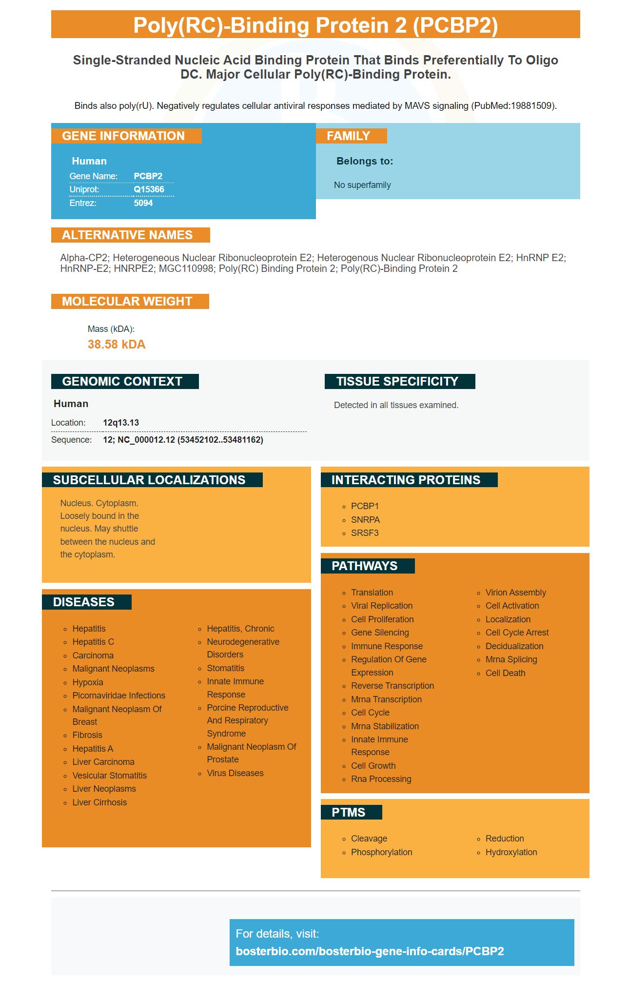
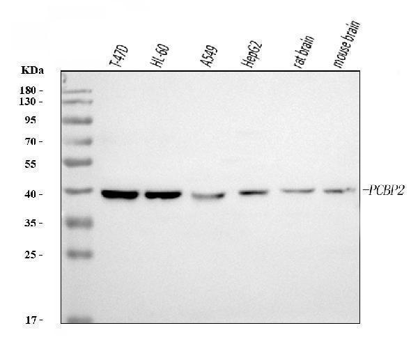
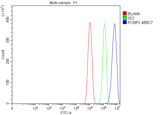
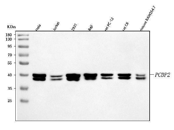
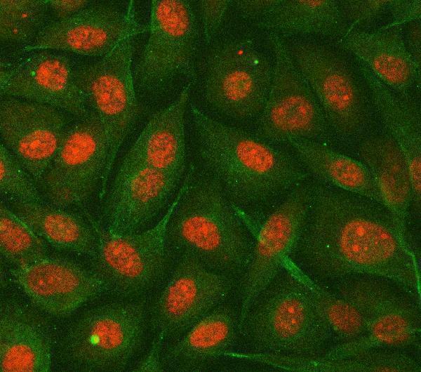
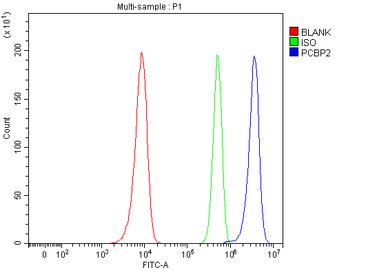
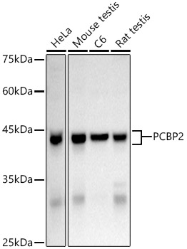
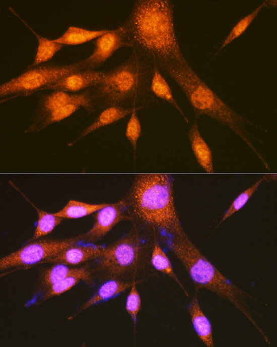
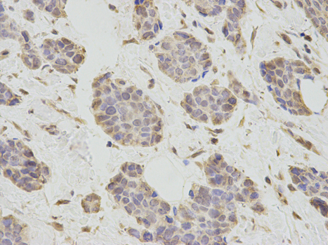
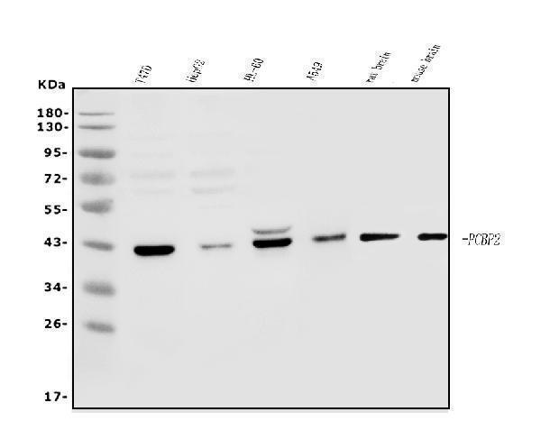
Facts about Poly(rC)-binding protein 2.
Binds also poly(rU). Negatively regulates cellular antiviral responses mediated by MAVS signaling (PubMed:19881509).
Human | |
---|---|
Gene Name: | PCBP2 |
Uniprot: | Q15366 |
Entrez: | 5094 |
Belongs to: |
---|
No superfamily |
alpha-CP2; Heterogeneous nuclear ribonucleoprotein E2; heterogenous nuclear ribonucleoprotein E2; hnRNP E2; hnRNP-E2; HNRPE2; MGC110998; poly(rC) binding protein 2; poly(rC)-binding protein 2
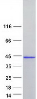
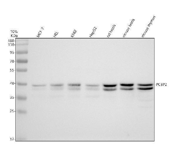
Mass (kDA):
38.58 kDA
Human | |
---|---|
Location: | 12q13.13 |
Sequence: | 12; NC_000012.12 (53452102..53481162) |
Detected in all tissues examined.
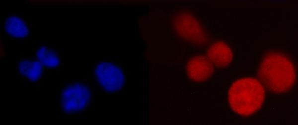
Nucleus. Cytoplasm. Loosely bound in the nucleus. May shuttle between the nucleus and the cytoplasm.
If you're interested in discovering the best uses of the PCBP2 marker, you've come to the right place. This article will introduce you to the Boster Bio line of primary antibodies, the Polypyrimidine tract-binding protein 2, and an ECL chemiluminescent detection system. These are all essential components of the PCBP2 assay. In addition to these components, you'll also learn how to use the Autoradiography film and ECL chemiluminescent detection system.
This primary antibody was raised against the poly(rC) binding protein (PCBP2) and recognizes post translational modifications. It is a versatile tool for protein analysis and detection, allowing researchers to identify proteins that cause disease. Primary antibodies are validated on multiple species and methods, including Western Blotting, Immunohistochemistry, and ELISA. In this article, we'll discuss the properties of PCBP2 and the Boster antibodies used in ELISA and Western Blotting.
The Polypyrimidine tract-binding protein (PTB) is a 58 kDa mRNA-binding protein with a regulatory role in alternative splicing. The PTB binds intronic and exonic splicing silencers by blocking E2AF binding. The PTB has multiple binding sites on mRNAs, with the optimal site being the UCUU.
PTBs are ubiquitous RNA-binding proteins in eukaryotes. They bind to target RNAs to regulate their translation and other metabolic processes. They also function as regulators of translation and stability. Unlike other RNA-binding proteins, PTBs contain four distinct RNA-recognition motifs, which are conserved among plants and animals.
This PTB is an important regulator of pollen germination in Arabidopsis. It is involved in plant cell physiology and negatively regulates reporter splicing. It interacts with the RNAs StBEL5, POTh2, and Xoconostle-Cazares B. It is also associated with plant RNA synthesis and trafficking.
Polypyrimidine tract-binding protein, or PCBP2, is a protein that mediates negative regulation of exon splicing and translation. It is a highly abundant protein in various body tissues, and many biological assays use Ptbp2 antibodies. Boster Bio offers monoclonal and polyclonal antibodies with high affinity. Designed for high-performance use in flow cytometry, Boster's PCBP2 antibodies have received a high number of citations over the past 25 years.
Enhanced chemiluminescent detection (ECL) is a traditional Western blotting method that uses an enzymatic reaction to detect protein concentrations. This system allows researchers to capture the same signal multiple times without the need to spend time optimizing incubation protocols or probing conditions. The C-DiGit(r) Blot Scanner eliminates the need for film and develop reagents, and provides identical performance to Amersham's ECL Western blotting Detection Reagent.
The ECL chemiluminescent detection method offers a higher sensitivity than traditional colorimetric assays. ECL reagents detect medium and high-expression proteins. Moreover, they are scalable, requiring no specialized equipment. This is an essential characteristic for researchers who need to measure the protein concentrations in low-abundance samples.
ECL reagents include HrP and luminol. The latter two reagents are commonly used in chemiluminescent Western detection. Typically, chemiluminescent HRP substrates contain 1,2-dioxetane, luminol, or acridan. Most commonly used reagents contain luminol, which decays to a lower energy state and releases photons.
Enhanced chemiluminescence is a luminol-based substrate commonly used in western blot applications. It produces a signal proportional to the amount of HRP-labeled antibodies on a blot. Luminescence can be measured indirectly, using imaging instruments or photographic film. However, the reagent must be protected from prolonged exposure to light to ensure a high-quality signal.
The DAB chromogenic detection system combines an enhanced HRP chromogen with a diaminobenzidine (DAB) counterstain to produce a blue color shift. This method uses the DAB chromogen stains as a substrate and includes a buffer system to make the reagents more stable. The solution contains 0.125% VWR brand powder, pH 4.2-4.3. The tissue is mounted on slides the night before using distilled water and acetone. The slides are then processed using ethanol dehydration steps. The Methyl Green counterstain is more nuclear in the cortex, and the DAB chromogen detection system enables scientists to see whether a protein is nuclear or not.
If you are looking for an antibody that will recognize target proteins in a Western blot, Boster Bio has the solution. The company's high-affinity primary antibodies have been widely cited for the last 25 years. Their antibodies are trusted by the research community, and have been tested and validated on Western Blotting, Immunohistochemistry, and ELISA. Using their primary antibodies, you will be able to determine the target proteins and normalize the target protein levels in your samples.
To determine the antibodies' specificity, the first step is to prepare the samples. This includes adding the antibodies and blocking buffer. Several things affect this step of the process. First of all, proteins may migrate differently when they are denatured or glycosylated, which can make them appear shorter or longer than they actually are. Then, there is the problem of nonspecific antibody binding, which results in multiple bands on the blot.
Western blotting is also known as Protein Immunoblotting. It is a technique in which the proteins are separated on a polyacrylamide gel using SDS-polyacrylamide electrophoresis. In addition, protein is transferred to a matrix and stained with antibodies specific to the target protein. The intensity of the specific reaction will provide details about protein expression levels. In addition to protein detection, Western blotting has many applications.
PMID: 7607214 by Leffers H., et al. Characterisation of two major cellular poly(rC)-binding human proteins, each containing three K-homologous (KH) domains.
PMID: 12414943 by Walter B.L., et al. Distinct poly(rC) binding protein KH domain determinants for poliovirus translation initiation and viral RNA replication.