Click image to see more details
Product Info Summary
| SKU: | A02425-3 |
|---|---|
| Size: | 100 µg/vial |
| Reactive Species: | Human, Mouse, Rat |
| Host: | Rabbit |
| Application: | ELISA, Flow Cytometry, IF, ICC, WB |
Customers Who Bought This Also Bought

Product info
Product Name
Anti-hnRNP E2/PCBP2 Antibody Picoband®
SKU/Catalog Number
A02425-3
Size
100 µg/vial
Form
Lyophilized
Description
Boster Bio Anti-hnRNP E2/PCBP2 Antibody Picoband® catalog # A02425-3. Tested in ELISA, Flow Cytometry, IF, ICC, WB applications. This antibody reacts with Human, Mouse, Rat. The brand Picoband indicates this is a premium antibody that guarantees superior quality, high affinity, and strong signals with minimal background in Western blot applications. Only our best-performing antibodies are designated as Picoband, ensuring unmatched performance.
Storage & Handling
At -20°C for one year from date of receipt. After reconstitution, at 4°C for one month. It can also be aliquotted and stored frozen at -20°C for six months. Avoid repeated freezing and thawing.
Cite This Product
Anti-hnRNP E2/PCBP2 Antibody Picoband® (Boster Biological Technology, Pleasanton CA, USA, Catalog # A02425-3)
Host
Rabbit
Contents
Each vial contains 4 mg Trehalose, 0.9 mg NaCl, 0.2 mg Na2HPO4.
Clonality
Polyclonal
Isotype
Rabbit IgG
Immunogen
E.coli-derived human hnRNP E2/PCBP2 recombinant protein (Position: D136-K276).
Cross-reactivity
No cross-reactivity with other proteins.
Reactive Species
A02425-3 is reactive to PCBP2 in Human, Mouse, Rat
Observed Molecular Weight
40 kDa
Calculated molecular weight
38.6 kDa
Background of PCBP2
Poly(rC)-binding protein 2 is a protein that in humans is encoded by the PCBP2 gene. The protein encoded by this gene appears to be multifunctional. Along with PCBP-1 and hnRNPK, it is one of the major cellular poly(rC)-binding proteins. The encoded protein contains three K-homologous (KH) domains which may be involved in RNA binding. Together with PCBP-1, this protein also functions as a translational coactivator of poliovirus RNA via a sequence-specific interaction with stem-loop IV of the IRES, promoting poliovirus RNA replication by binding to its 5'-terminal cloverleaf structure. It has also been implicated in translational control of the 15-lipoxygenase mRNA, human papillomavirus type 16 L2 mRNA, and hepatitis A virus RNA. The encoded protein is also suggested to play a part in formation of a sequence-specific alpha-globin mRNP complex which is associated with alpha-globin mRNA stability. This multiexon structural mRNA is thought to be retrotransposed to generate PCBP-1, an intronless gene with functions similar to that of PCBP2. This gene and PCBP-1 have paralogous genes (PCBP3 and PCBP4) which are thought to have arisen as a result of duplication events of entire genes. This gene also has two processed pseudogenes (PCBP2P1 and PCBP2P2). Multiple transcript variants encoding different isoforms have been found for this gene.
Antibody Validation
Boster validates all antibodies on WB, IHC, ICC, Immunofluorescence, and ELISA with known positive control and negative samples to ensure specificity and high affinity, including thorough antibody incubations.
Application & Images
Applications
A02425-3 is guaranteed for ELISA, Flow Cytometry, IF, ICC, WB Boster Guarantee
Recommend Dilution
| Application | Dilution | Species |
|---|---|---|
| Western blot | 0.1-0.25 μg/ml | Human, Mouse, Rat |
| Immunocytochemistry/Immunofluorescence | 5 μg/ml | Human |
| Flow Cytometry (Fixed) | 1-3 μg/1x106 cells | Human |
| ELISA | 0.1-0.5 μg/ml | - |
Tested application
Suggested blocking solution with 5% non-fat milk or BSA; (*)Recommended protein loading: 20-40 µg per lane
Validation Images & Assay Conditions
Click image to see more details
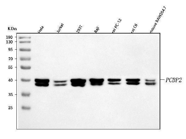
Western blot analysis of hnRNP E2/PCBP2 using anti-hnRNP E2/PCBP2 antibody (A02425-3).
Electrophoresis was performed on a 5-20% SDS-PAGE gel at 70V (Stacking gel) / 90V (Resolving gel) for 2-3 hours. The sample well of each lane was loaded with 30 ug of sample under reducing conditions.
Lane 1: human Hela whole cell lysates,
Lane 2: human Jurkat whole cell lysates,
Lane 3: human 293T whole cell lysates,
Lane 4: human Raji whole cell lysates,
Lane 5: rat PC-12 whole cell lysates,
Lane 6: rat C6 whole cell lysates,
Lane 7: mouse RAW264.7 whole cell lysates.
After electrophoresis, proteins were transferred to a nitrocellulose membrane at 150 mA for 50-90 minutes. Blocked the membrane with 5% non-fat milk/TBS for 1.5 hour at RT. The membrane was incubated with rabbit anti-hnRNP E2/PCBP2 antigen affinity purified polyclonal antibody (Catalog # A02425-3) at 0.25 μg/mL overnight at 4°C, then washed with TBS-0.1%Tween 3 times with 5 minutes each and probed with a goat anti-rabbit IgG-HRP secondary antibody at a dilution of 1:5000 for 1.5 hour at RT. The signal is developed using an Enhanced Chemiluminescent detection (ECL) kit (Catalog # EK1002) with Tanon 5200 system. A specific band was detected for hnRNP E2/PCBP2 at approximately 40 kDa. The expected band size for hnRNP E2/PCBP2 is at 39 kDa.

Click image to see more details
IF analysis of hnRNP E2/PCBP2 using anti-hnRNP E2/PCBP2 antibody (A02425-3) and anti-Tubulin Alpha antibody (M03989-3).
hnRNP E2/PCBP2 was detected in immunocytochemical section of PC-3 cell. Enzyme antigen retrieval was performed using IHC enzyme antigen retrieval reagent (AR0022) for 15 mins. The cells were blocked with 10% goat serum. And then incubated with 5 μg/mL rabbit anti-hnRNP E2/PCBP2 Antibody (A02425-3) and mouse anti-Tubulin Alpha antibody (M03989-3) overnight at 4°C. Cy3 Conjugated Goat Anti-Rabbit IgG (BA1032) and DyLight®488 Conjugated Goat Anti-Mouse IgG (BA1126) were used as secondary antibody at 1:500 dilution and incubated for 30 minutes at 37°C. Visualize using a fluorescence microscope and filter sets appropriate for the label used.

Click image to see more details
Flow Cytometry analysis of Hela cells using anti-hnRNP E2/PCBP2 antibody (A02425-3).
Overlay histogram showing Hela cells stained with A02425-3 (Blue line). To facilitate intracellular staining, cells were fixed with 4% paraformaldehyde and permeabilized with permeabilization buffer. The cells were blocked with 10% normal goat serum. And then incubated with rabbit anti-hnRNP E2/PCBP2 Antibody (A02425-3, 1 μg/1x106 cells) for 30 min at 20°C. DyLight®488 conjugated goat anti-rabbit IgG (BA1127, 5-10 μg/1x106 cells) was used as secondary antibody for 30 minutes at 20°C. Isotype control antibody (Green line) was rabbit IgG (1 μg/1x106) used under the same conditions. Unlabelled sample without incubation with primary antibody and secondary antibody (Red line) was used as a blank control.
Specific Publications For Anti-hnRNP E2/PCBP2 Antibody Picoband® (A02425-3)
Loading publications
Recommended Resources
Here are featured tools and databases that you might find useful.
- Boster's Pathways Library
- Protein Databases
- Bioscience Research Protocol Resources
- Data Processing & Analysis Software
- Photo Editing Software
- Scientific Literature Resources
- Research Paper Management Tools
- Molecular Biology Software
- Primer Design Tools
- Bioinformatics Tools
- Phylogenetic Tree Analysis
Customer Reviews
Have you used Anti-hnRNP E2/PCBP2 Antibody Picoband®?
Share your experimental results or join a short interview to earn up to $1,000 in product credits or other rewards.
0 Reviews For Anti-hnRNP E2/PCBP2 Antibody Picoband®
Customer Q&As
Have a question?
Find answers in Q&As, reviews.
Can't find your answer?
Submit your question
