This website uses cookies to ensure you get the best experience on our website.


- Table of Contents
Plan PDCD5 IHC in paraffin sections using the catalog antibody A02613-1 (datasheet A02613-1). Use HPA’s cytoplasmic tissue staining pattern to select controls, and interpret unexpected staining with its reported off-target binding in mind (HPA tissue IHC).

Expected localisation, validated protocols, controls and antibodies — the at-a-glance facts below, then the full design guide.
| Expected localisation | Cytoplasm in tissue sections (HPA tissue IHC) | |
| Staining pattern | Ubiquitous cytoplasmic staining across cell types (HPA tissue IHC) | |
| Antigen retrieval | EDTA pH 8.0 HIER, heat-mediated (datasheet A02613-1) | |
| Positive control | Placenta+4 more · see all | |
| Negative control | Caudate+4 more · see all |
| Fixation | Keep fixation consistent across sections (standard IHC practice; not target-specific) | |
| Caveat | Presumed off-target staining may mimic PDCD5 (HPA tissue IHC) | |
| Regulation | Genotoxic stress: nuclear accumulation (UniProt) | |
| Isoform / epitope | 2 isoforms; epitope impact unspecified (UniProt) |
Compare the catalog antibody’s IHC-P protocol with four published PDCD5 paraffin-section IHC protocols (datasheet A02613-1; PMC4937001; PMC3939472; PMC13344223; PMC4832950).
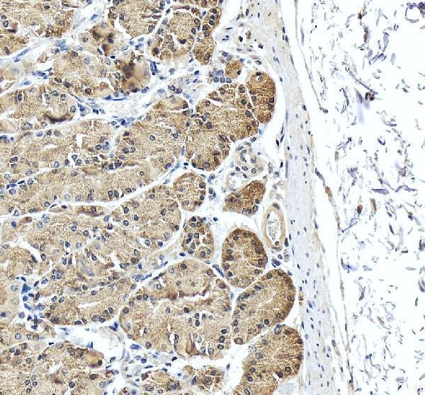
| Sample | Paraffin-embedded human stomach tissue; fixative not specified (datasheet A02613-1) |
| Fixation | Image fixative and duration unreported (datasheet A02613-1); verify before use. |
| Sectioning | 4–5 µm sections on charged slides (standard) |
| Deparaffinisation | Xylene, graded ethanol series to water (standard) |
| Antigen retrieval | Heat retrieval: EDTA pH 8.0 (datasheet A02613-1); 20 min, 95–100 °C (standard) |
| Peroxidase block | 3% H2O2, 10 min, room temperature (standard) |
| Blocking | 10% goat serum (datasheet A02613-1) |
| Primary antibody | Rabbit anti-PDCD5, 1:50 recommended; image 2 μg/ml (datasheet A02613-1) |
| Primary incubation | Overnight at 4 °C (datasheet A02613-1) |
| Detection | HRP-conjugated secondary, DAB chromogen (datasheet A02613-1) |
| Counterstain | Hematoxylin, blue, dehydrate and mount (standard) |
| Expected result | PDCD5-positive staining in cytotrophoblasts of placenta (HPA tissue IHC: High). HPA tissue profile: Ubiquitous cytoplasmic expression. No signal in the no-primary control. |
PDCD5 is mainly cytosolic, with nuclear localisation also reported; UniProt notes nuclear accumulation after genotoxic stress (HPA subcellular; UniProt O14737). In tissue IHC, expect broad cytoplasmic staining, including high staining in placental cytotrophoblasts, skin endothelial cells and late spermatids (HPA tissue IHC). Interpret exceptions cautiously: HPA rates tissue evidence Enhanced but reports medium RNA–staining consistency and disregarded presumed off-target binding (HPA tissue IHC). PDCD5 has no transmembrane segment (UniProt O14737 topology).
| Cytoplasmic staining in the expected cells, especially strong staining in placental cytotrophoblasts, skin endothelial cells or late spermatids. | This fits the reported ubiquitous cytoplasmic tissue pattern and the high levels assigned to those cell populations (HPA tissue IHC). Score the identified cell population and its compartment, rather than treating every stained structure in the section as equivalent (general IHC practice). |
| Predominantly nuclear staining, or a sharp membrane rim, with little cytoplasmic signal. | A membrane-only pattern conflicts with the reported cytosolic localisation and lack of a transmembrane segment (HPA subcellular; UniProt O14737 topology). Nuclear signal can be plausible because nucleoplasmic localisation is approved and nuclear accumulation follows genotoxic stress; assess context and controls before calling it artefact (HPA subcellular; UniProt O14737). |
| Strong staining in a cell population reported as not detected, such as heart cardiomyocytes or prostate glandular cells. | Treat an isolated unexpected result as a specificity or detection warning, not proof of expression: HPA reports those populations as not detected and notes presumed off-target binding in its tissue assessment (HPA tissue IHC). Check morphology, controls and whether the stain follows a plausible cellular compartment (general IHC practice). |
| Uniform colour across cells and empty spaces, or granular deposits that obscure cellular boundaries. | This is difficult to score as PDCD5 because the reported tissue pattern is cellular and cytoplasmic (HPA tissue IHC). Broad haze or deposits can arise from background or chromogen handling; compare a no-primary control and inspect the section for discrete intracellular signal (general IHC practice). |
| No convincing staining in placental cytotrophoblasts, skin endothelial cells or late spermatids. | These are high-staining populations in the HPA tissue record, so absence warrants a technical check before interpreting the specimen as negative (HPA tissue IHC). Verify the expected cells are present, then review section quality, retrieval, antibody incubation and detection with a suitable positive control (general IHC practice). |
| Cell population and tissue context | HPA reports high staining in placental cytotrophoblasts, skin endothelial cells and late spermatids, but no detection in heart cardiomyocytes (HPA tissue IHC). UniProt describes broad expression with high heart levels; that tissue-level statement does not override the cell-specific IHC observation (UniProt O14737; HPA tissue IHC). |
| Subcellular interpretation | Cytosol is the approved main IF location, with nucleoplasm approved additionally; UniProt reports nuclear accumulation after genotoxic stress (HPA subcellular; UniProt O14737). Record cytoplasmic and nuclear staining separately when scoring, and interpret nuclear predominance in its experimental context (general IHC practice). |
| Antibody evidence and limits | The listed antibody HPA018471 has Enhanced IHC validation, while the tissue summary reports medium consistency with RNA and disregarded presumed off-target binding (HPA antibodies; HPA tissue IHC). Validation supports the reference pattern but does not make every stained cell or compartment specific (HPA tissue IHC). |
| Molecular features | PDCD5 has no annotated transmembrane segment or signal peptide and has two listed isoforms (UniProt O14737). These annotations support caution about a membrane-only interpretation, but the supplied record gives no isoform-specific staining pattern or epitope location (UniProt O14737); do not infer one. |
| IF/ICC Q: What localisation should be expected? | A: Mainly cytosolic signal with additional nucleoplasmic signal; HPA approves both locations and lists A-431, U-251MG and U2OS among cell lines with ICC-IF images (HPA subcellular). This is a localisation reference for the separate IF/ICC guide, not a prediction of relative intensity in a new sample (HPA subcellular). |
| Situation | Likely cause | Next action |
|---|---|---|
| Expected high-staining cells are present, but the IHC slide is blank. | The result conflicts with HPA's high staining in placental cytotrophoblasts, skin endothelial cells or late spermatids; the supplied sources do not identify a PDCD5-specific failure mechanism (HPA tissue IHC). | Run a known-positive section alongside the sample and check antibody incubation, retrieval, detector and chromogen steps one at a time (general IHC practice). Interpret a negative specimen only after the control gives interpretable cellular staining (general IHC practice). |
| The whole section has brown haze or staining beyond cell boundaries. | Widespread colour is inconsistent with the reported cellular cytoplasmic pattern (HPA tissue IHC). Background may reflect incomplete blocking, residual detection activity or excess reagent in a chromogenic workflow (general IHC practice). | Compare a no-primary control; review blocking, washes and chromogen development, then rescore only discrete staining in identifiable cells (general IHC practice). Do not use background colour to assign PDCD5-positive cell populations (HPA tissue IHC; general IHC practice). |
| A sharp membrane outline dominates the signal. | That localisation conflicts with cytosolic predominance and the absence of a transmembrane segment (HPA subcellular; UniProt O14737 topology). | Inspect an expected high-staining control and a no-primary control, then check whether intracellular cytoplasmic staining appears under controlled detection conditions (HPA tissue IHC; general IHC practice). Treat a persistent membrane-only pattern as unverified (HPA subcellular; UniProt O14737 topology). |
| Nuclear staining exceeds cytoplasmic staining. | Nucleoplasmic localisation is supported, and UniProt reports nuclear accumulation after genotoxic stress; nuclear predominance alone does not establish a technical failure (HPA subcellular; UniProt O14737). | Score nuclear and cytoplasmic compartments separately, compare matched controls and document any genotoxic treatment (general IHC practice; UniProt O14737). Investigate if the pattern is confined to implausible cells or accompanies diffuse background (HPA tissue IHC; general IHC practice). |
| Strong staining appears in HPA-listed negative cells. | HPA reports no detection in examples including cardiomyocytes and prostate glandular cells, and its tissue assessment acknowledges presumed off-target binding (HPA tissue IHC). | Confirm cell identity on the counterstained section and compare positive and no-primary controls (general IHC practice). If the unexpected signal persists, describe it as discordant with the HPA reference instead of assigning PDCD5 expression from colour alone (HPA tissue IHC). |
| An apparently negative heart section conflicts with UniProt's high heart expression. | UniProt gives a tissue-level expression summary, whereas HPA specifically reports cardiomyocytes as not detected by tissue IHC (UniProt O14737; HPA tissue IHC). | Identify which heart cell population is being scored and use a HPA high-staining cell population as the positive reference (HPA tissue IHC; general IHC practice). Report the cell-specific observation without converting the UniProt tissue summary into an expected cardiomyocyte stain (UniProt O14737; HPA tissue IHC). |


Comprehensive Human Protein Atlas IHC scoring per tissue (reliability: Enhanced — Medium consistency between antibody staining and RNA expression data. Presumed off target binding observed and disregarded.). Rows are taken directly from the HPA tissue chart — click any row's HPA link to view the source.
| Tissue | Cell type | Level | Evidence | Source |
|---|---|---|---|---|
| Placenta | Cytotrophoblasts | High | Protein (IHC) | HPA → |
| Skin | Endothelial cells | High | Protein (IHC) | HPA → |
| Testis | Elongated or late spermatids | High | Protein (IHC) | HPA → |
| Adrenal gland | Glandular cells | Medium | Protein (IHC) | HPA → |
| Appendix | Glandular cells | Medium | Protein (IHC) | HPA → |
| Tissue | Cell type | Level | Evidence | Source |
|---|---|---|---|---|
| Caudate | Glial cells | Not detected | Protein (IHC) | HPA → |
| Cerebellum | Cells in granular layer | Not detected | Protein (IHC) | HPA → |
| Cervix | Glandular cells | Not detected | Protein (IHC) | HPA → |
| Epididymis | Glandular cells | Not detected | Protein (IHC) | HPA → |
| Heart muscle | Cardiomyocytes | Not detected | Protein (IHC) | HPA → |
Troubleshoot PDCD5 staining in paraffin sections by checking retrieval, staining controls, cellular location, and scoring before interpreting biological differences.
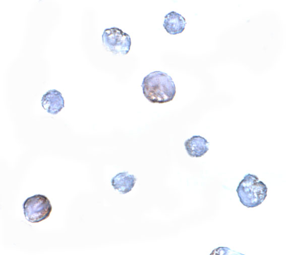
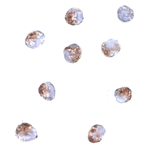
The catalog shows PDCD5 IHC in human paraffin stomach sections and ICC/IF in Jurkat cells (catalog image captions); mouse and rat reactivity is listed for the cell-imaging antibodies (catalog reactivity).
A02613-1 has an IHC image from a human paraffin stomach section (A02613-1 image caption). A02516-2 has a Jurkat ICC image at 5 μg/mL, while A02613 has Jurkat ICC and IF images at 2 μg/mL and 10 μg/mL, respectively (catalog image captions).
Which to pick: Choose A02613-1 for human tissue IHC: it is listed as polyclonal and IHC-reactive, and its image documents a paraffin stomach section with EDTA retrieval; the fixative is unreported (A02613-1 catalog entry and image caption). Choose A02516-2 for ICC or A02613 when IF is needed, based on their application lists and Jurkat images (catalog entries and image captions). For mouse or rat samples, A02516-2 and A02613 list those species as reactive, but the supplied imaging examples are in Jurkat cells, so confirm staining in the intended species (catalog reactivity and image captions).