Click image to see more details
-

-
-
-
-
+1
Product Info Summary
| SKU: | A02613-1 |
|---|---|
| Size: | 100 μl/vial |
| Reactive Species: | Human |
| Host: | Rabbit |
| Application: | ELISA, IHC, WB |
Customers Who Bought This Also Bought

Product info
Product Name
Anti-PDCD5 Antibody
SKU/Catalog Number
A02613-1
Size
100 μl/vial
Form
Liquid
Description
Boster Bio Anti-PDCD5 Antibody catalog # A02613-1. Tested in WB, IHC, ELISA applications. This antibody reacts with Human.
Storage & Handling
12 months from date of receipt,-20℃ as supplied. 6 months 2 to 8℃ after reconstitution. Avoid repeated freezing and thawing.
Cite This Product
Anti-PDCD5 Antibody (Boster Biological Technology, Pleasanton CA, USA, Catalog # A02613-1)
Host
Rabbit
Contents
500 μg/ml antibody with PBS, 0.02% NaN3, 1 mg stabilizing protein and 50% glycerol
*This antibody is supplied in a stabilized formulation.
Compatibility with conjugation reactions depends on the chemistry of the conjugation method used.
For conjugation methods that are not compatible with the stabilizing components present in this formulation, a carrier-free antibody format is required.
Clonality
Polyclonal
Immunogen
E.coli-derived human PDCD5 recombinant protein (Position: M1-D120).
Reactive Species
A02613-1 is reactive to PDCD5 in Human
Calculated molecular weight
14.3 kDa
Background of PDCD5
Programmed cell death 5 (PDCD5), a human apoptosis-related protein, is thought to play an early and universal role in apoptosis. PDCD5 is widely expressed and is upregulated in cells undergoing apoptosis, where it translocates rapidly from the cytoplasm to the nucleus. PDCD5 has a compact core structure of low flexibility with two mobile alpha-helices at N-terminal and a flexible unstructured C-terminal region. The charged residues are crucial for the ability of apoptosis-promoting and cell translocation of the protein. PDCD5 can facilitate apoptosis and enhance TAJ/TROY-induced paraptosis-like cell death. PDCD5 may play a dual role in the Tip60 pathway. It interacts with Tip60 and functions as a Tip60 co-activator to promote apoptosis. The nucleotide polymorphisms in the 5'-upstream region ofPDCD5 affect promoter activity and the susceptibility of a Chinese population to develop chronic myelogenous leukemia and may represent a novel tumor suppressor gene influencing lung cancer.
Antibody Validation
Boster validates all antibodies on WB, IHC, ICC, Immunofluorescence, and ELISA with known positive control and negative samples to ensure specificity and high affinity, including thorough antibody incubations.
Application & Images
Applications
A02613-1 is guaranteed for ELISA, IHC, WB Boster Guarantee
Recommend Dilution
| Application | Dilution | Species |
|---|---|---|
| Western blot | 1:500-2000 | |
| Immunohistochemistry | 1:50-400 | |
| ELISA | 1:100-1000 |
Validation Images & Assay Conditions
Click image to see more details
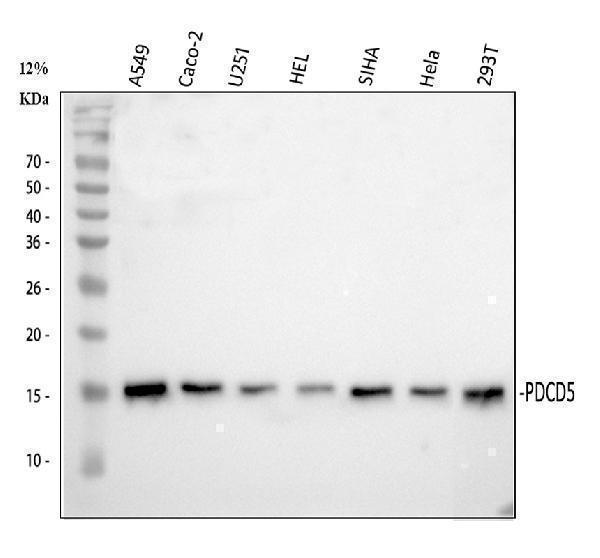
Western blot analysis of PDCD5 using anti-PDCD5 antibody (A02613-1).
Electrophoresis was performed on a 10% SDS-PAGE gel at 80V (Stacking gel) / 120V (Resolving gel) for 2 hours. The sample well of each lane was loaded with 30 ug of sample under reducing conditions.
Lane 1: human A549 whole cell lysates,
Lane 2: human Caco-2 whole cell lysates,
Lane 3: human U251 whole cell lysates,
Lane 4: human HEL whole cell lysates,
Lane 5: human SiHa whole cell lysates,
Lane 6: human Hela whole cell lysates,
Lane 7: human 293T whole cell lysates.
After electrophoresis, proteins were transferred to a nitrocellulose membrane at 150 mA for 50-90 minutes. Blocked the membrane with 5% non-fat milk/TBS for 1.5 hour at RT. The membrane was incubated with rabbit anti-PDCD5 antigen affinity purified polyclonal antibody (A02613-1) at 1:1000 overnight at 4°C, then washed with TBS-0.1%Tween 3 times with 5 minutes each and probed with a goat anti-rabbit IgG-HRP secondary antibody at a dilution of 1:5000 for 1.5 hour at RT. The signal is developed using an ECL Plus Western Blotting Substrate (Catalog # AR1196-200) with Tanon 5200 system. A specific band was detected for PDCD5 at approximately 14 kDa. The expected band size for PDCD5 is at 14 kDa.

Click image to see more details
IHC analysis of PDCD5 using anti-PDCD5 antibody (A02613-1).
PDCD5 was detected in a paraffin-embedded section of human stomach tissue. Heat mediated antigen retrieval was performed in EDTA buffer (pH 8.0, epitope retrieval solution). The tissue section was blocked with 10% goat serum. The tissue section was then incubated with 2 μg/ml rabbit anti-PDCD5 Antibody (A02613-1) overnight at 4°C. Peroxidase Conjugated Goat Anti-rabbit IgG was used as secondary antibody and incubated for 30 minutes at 37°C. The tissue section was developed using HRP Conjugated Rabbit IgG Super Vision Assay Kit (Catalog # SV0002) with DAB as the chromogen.

Click image to see more details
Methylating effect of daphnetin and 5-aza-dc on DR3, PDCD5, FasL and p53 in CIA rat synovial cells. Four experimental groups consisting of untreated cell control, or treated with 5-aza-dc at 20 μM, daphnetin at 40 μg/mL, or combination of 5-aza-dc (20 μM) and daphnetin (40 μg/mL). Cells were cultured, treated and harvested as described in Materials and Methods. Total DNA was extracted and used for MSP after bisulphite modification. M: methylated amplification products, U: hypomethylated amplification products.
Index in PubMed under a CC BY license. PMID: 25311560

Click image to see more details
Effect of daphnetin and 5-aza-dc on expression of DR3, PDCD5, FasL, p53 (A) and DNMT1, DNMT3a, DNMT3b (B) in CIA rat synovial cells. Four experimental groups consisting of untreated cell control, or treated with 5-aza-dc at 20 μM, daphnetin at 40 μg/mL, or combination of 5-aza-dc (20 μM) and daphnetin (40 μg/mL). Cells were cultured, treated and harvested as described in Matreials and Methods. Total RNA was extracted and cDNA was synthesized. After reverse transcription, cDNA was used for real time-PCR. Relative quantification of gene expression was performed by the 2-ΔΔCt method. The results show the mean ± S.D. of six independent experiments. ▲P < 0.05 compared with control, ●P < 0.05 compared with 5-aza-dc, ☆P > 0.05 compared with 5-aza-dc, ★P > 0.05 compared with daphnetin, *P < 0.05 compared with daphnetin.
Index in PubMed under a CC BY license. PMID: 25311560

Click image to see more details
Effect of daphnetin on DR3, PDCD5, FasL and p53 protein expression in CIA rats synovial cells. Cells were cultured, treated and harvested as described Materials and Methods. Four experimental groups consisting of untreated cell control, 5-aza-dc at 20 μM, daphnetin at 40 μg/mL, or combination of 5-aza-dc (20 μM) and daphnetin (40 μg/mL). The results show the mean ± S.D. of six independent experiments. ▲P < 0.05 compared with control, ●P < 0.05 compared with 5-aza-dc, ★P > 0.05 compared with 5-aza-dc, *P < 0.05 compared with daphnetin.
Index in PubMed under a CC BY license. PMID: 25311560
Specific Publications For Anti-PDCD5 Antibody (A02613-1)
Loading publications
Recommended Resources
Here are featured tools and databases that you might find useful.
- Boster's Pathways Library
- Protein Databases
- Bioscience Research Protocol Resources
- Data Processing & Analysis Software
- Photo Editing Software
- Scientific Literature Resources
- Research Paper Management Tools
- Molecular Biology Software
- Primer Design Tools
- Bioinformatics Tools
- Phylogenetic Tree Analysis
Customer Reviews
Have you used Anti-PDCD5 Antibody?
Share your experimental results or join a short interview to earn up to $1,000 in product credits or other rewards.
0 Reviews For Anti-PDCD5 Antibody
Customer Q&As
Have a question?
Find answers in Q&As, reviews.
Can't find your answer?
Submit your question
