This website uses cookies to ensure you get the best experience on our website.
- Table of Contents
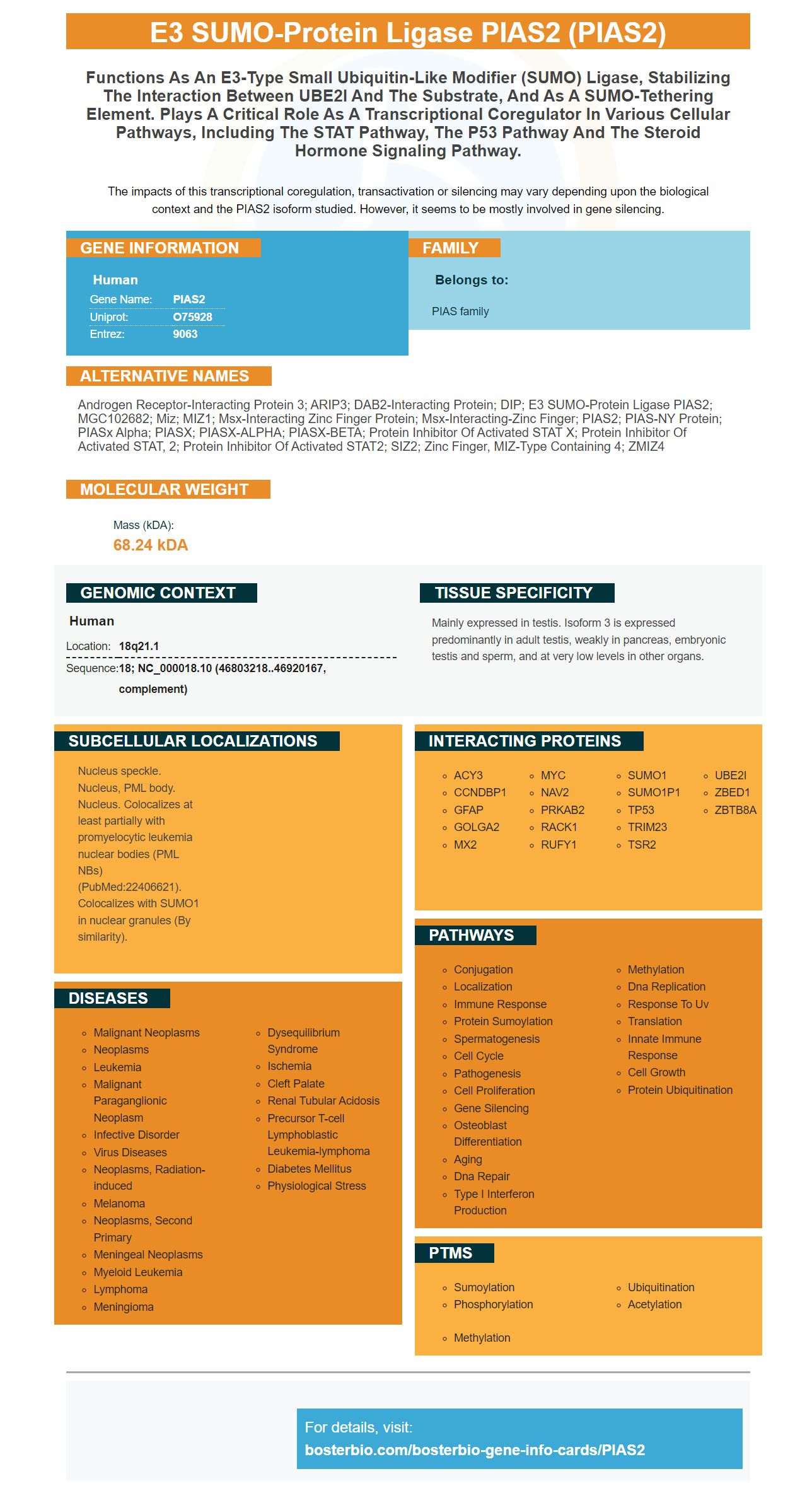
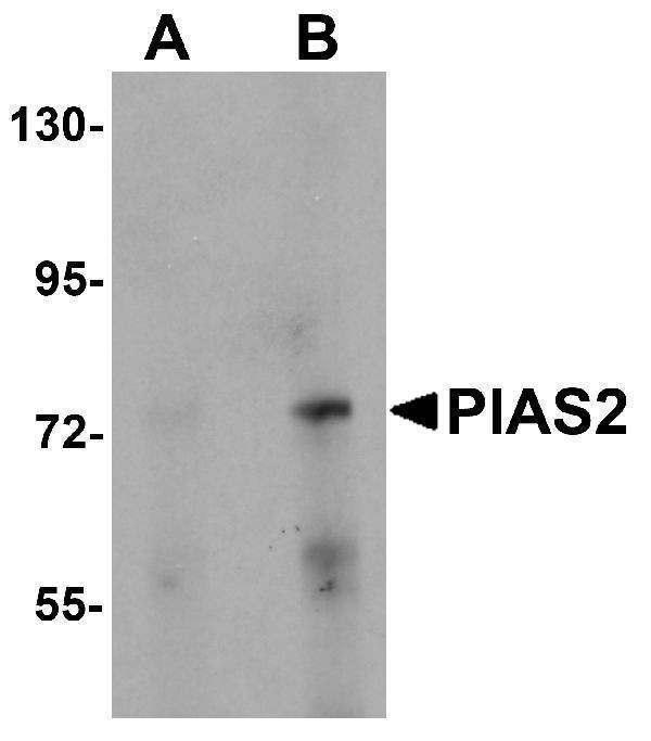
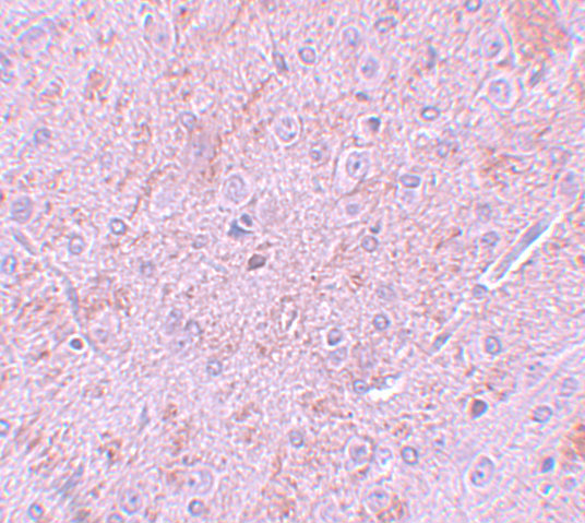
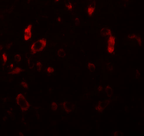
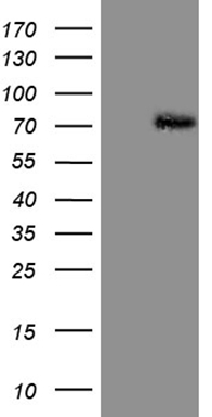
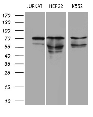
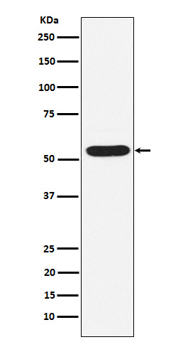
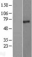
Facts about E3 SUMO-protein ligase PIAS2.
The impacts of this transcriptional coregulation, transactivation or silencing may vary depending upon the biological context and the PIAS2 isoform studied. However, it seems to be mostly involved in gene silencing.
Human | |
---|---|
Gene Name: | PIAS2 |
Uniprot: | O75928 |
Entrez: | 9063 |
Belongs to: |
---|
PIAS family |
Androgen receptor-interacting protein 3; ARIP3; DAB2-interacting protein; DIP; E3 SUMO-protein ligase PIAS2; MGC102682; miz; MIZ1; Msx-interacting zinc finger protein; Msx-interacting-zinc finger; PIAS2; PIAS-NY protein; PIASx alpha; PIASX; PIASX-ALPHA; PIASX-BETA; Protein inhibitor of activated STAT x; protein inhibitor of activated STAT, 2; Protein inhibitor of activated STAT2; SIZ2; zinc finger, MIZ-type containing 4; ZMIZ4
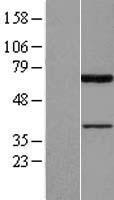
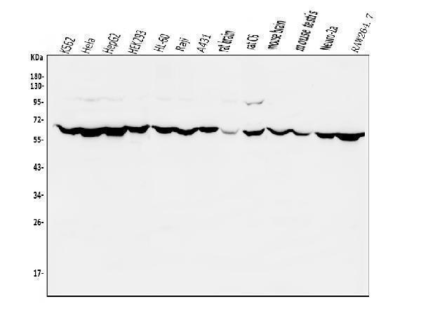
Mass (kDA):
68.24 kDA
Human | |
---|---|
Location: | 18q21.1 |
Sequence: | 18; NC_000018.10 (46803218..46920167, complement) |
Mainly expressed in testis. Isoform 3 is expressed predominantly in adult testis, weakly in pancreas, embryonic testis and sperm, and at very low levels in other organs.
Nucleus speckle. Nucleus, PML body. Nucleus. Colocalizes at least partially with promyelocytic leukemia nuclear bodies (PML NBs) (PubMed:22406621). Colocalizes with SUMO1 in nuclear granules (By similarity).
This is the place to look if you are searching for a protein indicator for PIAS2. PIAS2 stands for PIas-2-associated splicing 2 and is a member of the microtubule-associated protein family. This article will demonstrate how to use PIAS2 as a research tool. Continue reading to learn about this marker and the other PIas proteins.
PIAS2 is an HCV protein-restricting plasmid. Huh7 cells were transfected using a siRNA, pHCVIRES reporter plasmid or both to determine if PIAS2 is involved with regulation of HCV gene transcription. 48 hours later, the cells were fully transfected.
In mouse experiments, PIAS2 knockdown mice showed a greater percentage of time in zone 1 on day one of the probe. They also showed improved spatial memory short-term as well as long-term. PIAS2-knocked mice also retained TH+, NeuN+, and dopamine producing neurons in their SN. However, PIAS2-knocked mice showed a decrease in intracellular pa-syn levels.
PIAS1 & PIAS2 can be orthologous. Pias2, an ortholog for Pias1, is similar in its structural features. Both Pias1 as well as PIAS2 have SP-RING domains and SIM domains. They should be able SUMO protein, which alters transcription factor activity, to transfer between them. The PIAS2 genes are expressed in human cells in a manner similar to Pias1 gene in zebrafish.
The overexpression of PIAS2 in neurons causes oxidative stress and damage to the mitochondria. DJ1, a nuclear protein that blocks the ERK1/2 and P53 signaling pathways, is affected by increased oxidative stress. In PIAS2-knocked mouse models, PIAS2 knockdown restores mitochondrial equilibrium. These studies suggest that PIAS2 knockdown improves cognition.
Ifnb-/ mice were cultured in vitro. PIAS2 was overexpressed in these cells with AAV6 pAAV-SYN1-mCherry-mouse PIAS2 or knocked down with siRNA. The CNs were maintained in artificial cerebrospinal fluid (ACF) containing 125 NaCl, 2.5 KCl, and glucose. Likewise, mutPIAS2-knocked-down mice did not display any neurotoxicity and had minimal effects on SUMO1 levels.
In contrast to neurons expressing only mCherry, PIAS2-mCherry-mCherry AAVs infect primary CNs. Compared with mCherry AAVs, the thresholds and upstrokes of PIAS2-overexpressing neurons are lower than those of mCherry. In addition, PIAS2-mCherry-expressing neurons have lower mean firing rates when stimulated with stimulation pulse intensities.
Motor and cognitive dysfunction can be caused by PIAS2 neuronal overexpression. PIAS2 knockdown mice had significantly improved compared to control siRNA-injected Ifnb-/ mice on the Barnes maze test. Ifnb/ mice showed lower levels of the TH mRNA. When the protein is overexpressed in neurons, PDD is the result. Also, mice with hSNCA deficient in hnb are more likely to have elevated levels.
PIAS2 is a regulator of STAT signaling and IFN-b signaling. It is involved the stabilization of a syn. This protein plays a role in a variety cellular pathways including the inflammatory response. The apparent molecular weight of the PIAS2 proteins is 68 kDa. It is an essential transcriptional co-regulator in many cellular pathways.
The PIAS2 marker has a crucial function in neurons. It is a downstream part of IFNb/IFNAR signaling that is essential for maintaining the homeostasis and excitability of neurons. Overexpression of PIAS2 can cause neuronal survival, neurite growth, and the inhibition of action potential. The PIAS2 marker may be useful in studies that target this particular protein.
The relationship between GFAP biomarker and PIAS2 is still a subject of debate. The GFAP genes encode a protein that is acetylated through its entire length. Although it is not known how acetylation occurs exactly, it is likely that it affects the protein’s function. Further research is needed to determine how acetylation affects GFAP and PIAS2.
There are many therapeutic agents that can be used to suppress glial activity. One of these agents is Withaferin-A, which is derived from the N-terminus of neurotrophic prosaposin. It inhibits the IF function (integrin–fibrillary junction), of GFAP, thereby causing filament destruction. This drug is BBB permeabile and attenuates the activation of glial cells in a retinalgliosis model. Withaferin A is also an anti-inflammatory and asthma medicine.
Some gliosis is beneficial for brain healing, but too much gliosis can hinder recovery. Understanding the role of GFAP as well as the PIAS2 Biomarkers in brain healing is important. Axonal regeneration in GFAP/ mice seems to be unaffected by injury. However, GFAP knockout mice had impaired neural regeneration and a delayed regeneration of nerve fibers.
In a study of patients with PPMS, the biomarkers glial fibrillary acidic prot (GFAP), and neurofilament lighting chain (NfL), was examined. The results from this study indicate that GFAP and NfL serum levels are correlated by age. Gender, age, duration of the disease and treatment status were not associated GFAP and NfL blood levels.
GFAP is a biomarker that may be helpful in the detection of both ischemic and hemorrhagic stroke. Following stroke, GFAP from the brain is released and the CSF flows are followed by circulating levels. They then diffuse over the blood-brain border. An elevated level of GFAP could indicate a higher risk of ischemic stroke. This may provide useful information in the development of new therapeutic strategies.
While GFAP, the PIAS2 Biomarker and GFAP are independent biomarkers but they do correlate well with age. A strong correlation between GFAP levels with age is an indication of a possible confounding factor. Although GFAP and PIAS2 appear to be related, they have different clinical meanings. These results also indicate a possible link between GFAP levels and sTREM2 levels.
A crucial step in diagnosing astroglia is the interaction of GFAP with PIAS2. Astroglia can be described as glial cells in the brain. They provide structural support and functional support for neurons. They also release neurotransmitters (trophic factors) and trophic compounds. Dr. Eng isolated GFAP by nonmyelinating Schwann Cells of the PNS.
CNS injury is currently not covered by FDA-approved treatments. However, biomarkers of GFAP may be useful in drug development. Monitoring biofluid levels GFAP could be a useful neurotoxicity biomarker tool. GFAP may not only be used to assess tumors but also indicate tumor growth and expansion. This could allow doctors to decide whether or not to use certain neuropathologies.
Microtubule-associated protein 2 (MAP2) is a proven marker for human glioma. It is essential for the differentiation of gliomas or astrocytomas. Its expression in different gliomas is evaluated using immunohistochemistry. Recent research compared the immunolabeling intensity for MAP2 in gliomas. We also studied the morphological characteristics and different gliomas.
MAP2 is an 1827-amino-amino acid polypeptide having a mass of 199.5kDa. Its functions include binding of calmodulin and dystroglycan. It is associated with dendrite and neuron development and is involved in various cell types. Its post-translational modifications are phosphorylation. It has been detected in many cell types including human embryonic stem cells, neuronal and other tissues.
MAP2 is a member of the family of microtubule-associated proteins (MAP) and belongs to the MAP2/Tau gene group, which includes MAP4 and MAP3. Four isoforms of map2 have been identified using alternative splicing. The MAP2c and the MAP2d have shorter amino-terminal projection domains and a lower molecular mass.
The regulation and spacing of microtubules has been implicated in the MAP2 family proteins. They are part of KXGS, which binds microtubules. Tau also displays a similar pattern. Numerous experiments have shown that MAP2 binds microtubules from the outside of cells. Some believe that the projection region of MAP2 regulates spacing of microtubules.
F-actin and MAP2 can interact directly. Its microtubulebinding domains are highly conserved in members of Tau family. This means that MAP2 could interact with any protein involved controlling microtubule dynamics. Even in cells that have high levels of stabilizing microtubule proteins, microtubules still show dynamic behavior.
MAP2 interacts directly with microtubules, filamentous (F), and F-actin. Because MAP2 regulates microtubulemediated transport, its interactions may also be implicated in Alzheimer’s disease. The protein may also play a role neuromorphogenic processes such as neurite initiation. These interactions have been associated with neuronal disorders like Alzheimer's disease.
PMID: 9724754 by Liu B., et al. Inhibition of Stat1-mediated gene activation by PIAS1.
PMID: 15301740 by Zheng Y., et al. Molecular cloning and characterization of a novel splicing variant of PIASx.