This website uses cookies to ensure you get the best experience on our website.


- Table of Contents
PIK3R1 shows general cytoplasmic tissue staining, with high signal in lung macrophages and placental trophoblasts (HPA tissue IHC). For paraffin IHC, the catalog antibody A00318-1 has a listed working range of 2–5 μg/ml (datasheet A00318-1).

Expected localisation, validated protocols, controls and antibodies — the at-a-glance facts below, then the full design guide.
| Expected localisation | General cytoplasmic staining (HPA tissue IHC) | |
| Staining pattern | Cytoplasmic staining in lung macrophages (HPA tissue IHC) | |
| Antigen retrieval | EDTA pH 8.0 HIER, heat-mediated (datasheet A00318-1) | |
| Positive control | Bone marrow+4 more · see all | |
| Negative control | None in HPA (detected in all 45 tissues); use no-primary + isotype controls |
| Fixation | Keep fixation consistent across sections (standard IHC practice; not target-specific) | |
| Caveat | Macrophage-rich areas may skew tissue-level scoring (HPA tissue IHC) | |
| Regulation | Expression varies by cell type (HPA tissue IHC) | |
| Isoform / epitope | 5 isoforms; map the antibody epitope for isoform coverage (UniProt) |
The catalog antibody’s IHC-P protocol (datasheet A00318-1) is accompanied by one published human HCC paraffin-section workflow (PMC6264640).
| Sample | Paraffin-embedded human lung cancer tissue; fixative not specified (datasheet A00318-1) |
| Fixation | Image fixative and duration unreported (datasheet A00318-1); verify before use. |
| Sectioning | 4–5 µm sections on charged slides (standard) |
| Deparaffinisation | Xylene, graded ethanol series to water (standard) |
| Antigen retrieval | Heat retrieval: EDTA pH 8.0 (datasheet A00318-1); 20 min, 95–100 °C (standard) |
| Peroxidase block | 3% H2O2, 10 min, room temperature (standard) |
| Blocking | 10% goat serum (datasheet A00318-1) |
| Primary antibody | Rabbit anti-PIK3R1, 2-5 μg/ml (datasheet A00318-1) |
| Primary incubation | Overnight at 4 °C (datasheet A00318-1) |
| Detection | Streptavidin-biotin complex (SABC), DAB chromogen (datasheet A00318-1) |
| Counterstain | Hematoxylin, blue, dehydrate and mount (standard) |
| Expected result | PIK3R1-positive staining in hematopoietic cells of bone marrow (HPA tissue IHC: High). HPA tissue profile: General cytoplasmic expression. No signal in the no-primary control. |
PIK3R1 should appear mainly in the cytoplasm of stained cells (UniProt P27986: cytoplasm; HPA tissue IHC: general cytoplasmic expression). HPA reports high staining in several cell populations, including lung macrophages, heart cardiomyocytes and cerebral cortex neurons (HPA tissue IHC). Its tissue IHC status is Approved, with medium consistency between staining and RNA data (HPA tissue IHC). PIK3R1 has no transmembrane segment (UniProt P27986 topology).
| Clear cytoplasmic staining in lung macrophages or heart cardiomyocytes, with little staining in adjacent unstained structures (HPA tissue IHC: High in both cell populations). | This fits the reported compartment and provides a useful positive comparison (HPA tissue IHC: general cytoplasmic expression; UniProt P27986: cytoplasm). Judge intensity within the named cell population: HPA's High designation describes its observed staining, not a required score for every specimen or staining run. |
| Predominantly nuclear staining, with little cytoplasmic signal in a population expected to stain strongly (HPA tissue IHC: general cytoplasmic expression). | Treat this as a localisation mismatch and investigate staining or interpretation artefact before calling it PIK3R1. HPA reports cytosol in ICC-IF, and UniProt reports cytoplasm (HPA subcellular: approved cytosol; UniProt P27986). PIK3R1 can interact with XBP1 isoform 2, but that interaction alone does not establish a nuclear PIK3R1 staining pattern (UniProt P27986). |
| Strong staining appears mainly in cells expected to show low staining, such as liver cholangiocytes, while a known-positive population is weak (HPA tissue IHC: Low in cholangiocytes; High in lung macrophages). | Check cell identification, antibody specificity and endogenous chromogen-producing activity (general IHC practice). Low HPA staining is not an absolute negative control: HPA reports low tissue RNA specificity and lists no negative cell populations (HPA tissue IHC). A surprising cell pattern therefore needs controls before it is labelled cross-reactivity. |
| Chromogen spreads across cells and surrounding tissue, obscuring cell boundaries and the cytoplasmic pattern. | Interpretation is unreliable when signal cannot be assigned to cells (general IHC practice). Review the no-primary control for detection background, then assess blocking, antibody concentration, washing and chromogen development (general IHC practice). Do not use a uniformly coloured section as evidence that every cell expresses PIK3R1. |
| No convincing staining in a well-preserved positive comparison, such as lung macrophages or heart cardiomyocytes (HPA tissue IHC: High). | Consider an unsuccessful staining run before concluding that the test specimen lacks PIK3R1 (general IHC practice). Check the antibody's documented IHC-P conditions, detection reagents and retrieval trial (general IHC practice). HPA's medium staining-to-RNA consistency limits how confidently any one specimen predicts intensity (HPA tissue IHC). |
| Cell population and comparator (HPA tissue IHC) | HPA reports High staining in bone marrow hematopoietic cells, cerebellar molecular-layer cells, cerebral cortex neurons, fallopian tube glandular cells, cardiomyocytes, lung macrophages, lymph-node germinal-center cells and placental trophoblastic cells. It reports Low staining in cholangiocytes, smooth muscle cells and fibroblasts. Match the cell type when comparing sections; the Low entries are poor absolute-negative controls because HPA lists no negative population (HPA tissue IHC). |
| Antibody validation (HPA antibodies; HPA tissue IHC) | HPA001216 and CAB004268 each have Approved IHC status; HPA001216 also has Approved ICC status (HPA antibodies). The tissue summary has Approved reliability with medium staining-to-RNA consistency (HPA tissue IHC). These labels support use as reference observations but do not prove that every antibody, epitope or specimen will reproduce the same intensity. Confirm the antibody used and compare appropriate controls (general IHC practice). |
| Isoforms and epitope coverage (UniProt P27986) | UniProt lists 5 isoforms; isoforms 2 and 4 are present in skeletal muscle at protein level, and isoform 2 is reported in brain (UniProt P27986). Without an antibody epitope or isoform-coverage record in this payload, do not assign tissue intensity to a particular isoform. Likewise, the listed phosphorylation sites establish modifications, not an effect on IHC retrieval or staining (UniProt P27986). |
| IF/ICC Q&A (HPA subcellular; HPA antibodies) | Q: What localisation should an IF/ICC image show? A: Expect cytosolic signal as the main pattern (HPA subcellular: approved cytosol). HPA lists ICC-IF images and marks HPA001216 Approved for ICC (HPA subcellular; HPA antibodies). This is localisation context for the separate IF/ICC guide, not an IF protocol or a prediction of fluorescence intensity in paraffin sections. |
| Situation | Likely cause | Next action |
|---|---|---|
| Expected cells are unstained in both the test and positive-comparison sections (HPA tissue IHC: High in lung macrophages or cardiomyocytes). | A run-level failure is possible when a documented positive population also lacks signal (general IHC practice). | Check antibody identity and documented IHC-P conditions; verify detection and chromogen steps, then assess retrieval using the antibody's instructions (general IHC practice). No PIK3R1-specific fixation sensitivity is established by the supplied sources. |
| The test section is weak, but expected cells in the positive comparison stain (HPA tissue IHC: High populations). | Biological variation, sampling or staining differences remain possible; HPA reports medium staining-to-RNA consistency (HPA tissue IHC). | Confirm the cell population and section quality, then compare a second suitable specimen or staining run before scoring absence (general IHC practice). Do not infer an isoform or fixation effect from intensity alone. |
| Nuclei dominate the signal while expected cytoplasm is faint (HPA tissue IHC: general cytoplasmic expression). | The pattern conflicts with HPA's approved cytosolic ICC-IF localisation and UniProt's cytoplasm annotation (HPA subcellular; UniProt P27986). | Review the counterstain and no-primary control, then reassess antibody concentration and detection background (general IHC practice). Score PIK3R1 localisation only where cell boundaries and cytoplasm are clear. |
| Broad brown staining persists across tissue, including spaces without identifiable cells. | Nonspecific binding, endogenous detection activity or excessive chromogen development can obscure localisation (general chromogenic IHC practice). | Compare a no-primary control; check blocking appropriate to the detection system, washing and development time (general IHC practice). Reassess the cell-specific pattern only after background is controlled. |
| Strong signal appears in an HPA Low population and little appears in an HPA High comparator (HPA tissue IHC). | Cell misidentification, cross-reactivity or endogenous detection activity may explain the mismatch (general IHC practice). HPA Low does not mean absent (HPA tissue IHC). | Confirm morphology and comparator identity; examine no-primary staining and, if available, an independently validated antibody (general IHC practice). Report the discrepancy rather than treating the Low population as a proven negative. |
| Staining hugs cell edges, with no clear cytoplasmic component. | PIK3R1 can be recruited to the plasma membrane during signalling, but its reported overall localisation is cytoplasmic or cytosolic (UniProt P27986; HPA subcellular). | Inspect higher magnification and a cytoplasmic positive comparison; exclude edge artefact and background with appropriate controls (general IHC practice). Avoid scoring edge-only colour as a definitive membrane pattern. |
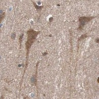


Comprehensive Human Protein Atlas IHC scoring per tissue (reliability: Approved — Medium consistency between antibody staining and RNA expression data.). Rows are taken directly from the HPA tissue chart — click any row's HPA link to view the source.
| Tissue | Cell type | Level | Evidence | Source |
|---|---|---|---|---|
| Bone marrow | Hematopoietic cells | High | Protein (IHC) | HPA → |
| Cerebellum | Cells in molecular layer | High | Protein (IHC) | HPA → |
| Cerebral cortex | Neuronal cells | High | Protein (IHC) | HPA → |
| Fallopian tube | Glandular cells | High | Protein (IHC) | HPA → |
| Heart muscle | Cardiomyocytes | High | Protein (IHC) | HPA → |
| Tissue | Cell type | Level | Evidence | Source |
|---|---|---|---|---|
| None in HPA: PIK3R1 is detected in all 45 scored tissues. Use a no-primary (secondary-only) and an isotype control instead. | ||||
Troubleshoot PIK3R1 staining in paraffin sections by checking retrieval, cell identity, cytoplasmic localisation and controls before interpreting signal intensity.
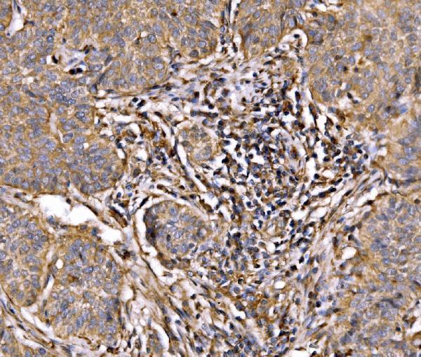
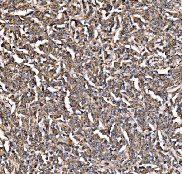
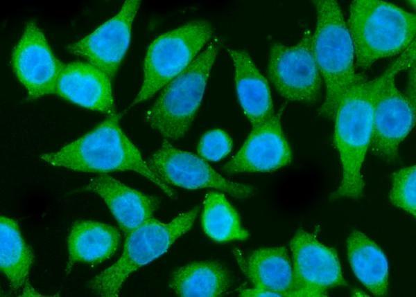
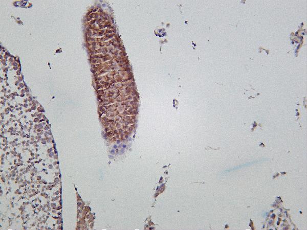
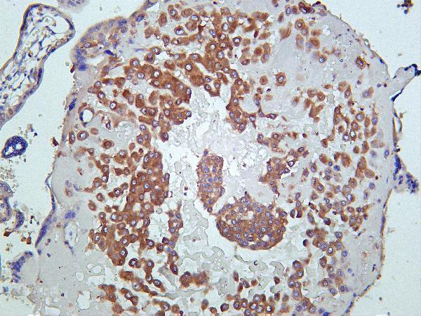
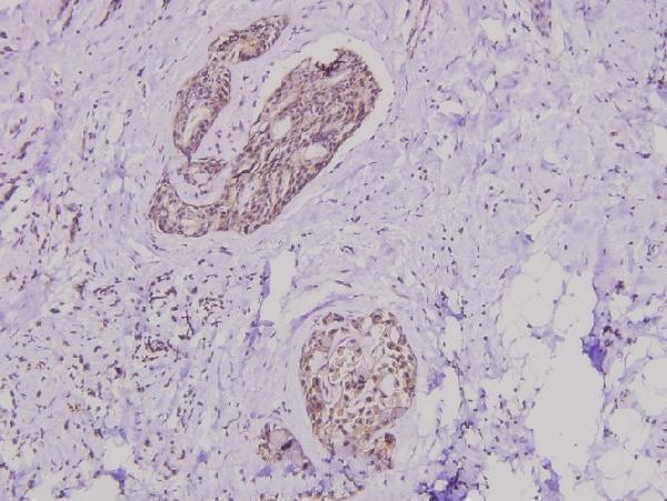
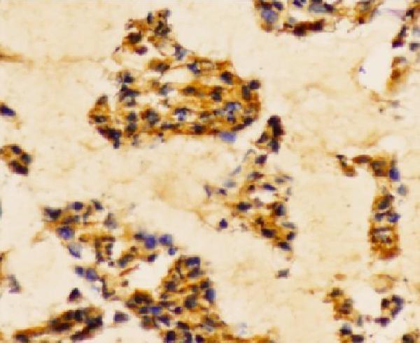
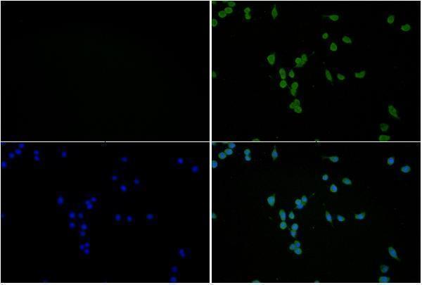
These anti-PIK3R1 reagents have IHC images from human and mouse tissues and IF images from Caco-2 and BV2 cells (catalog image captions); listed reactivity includes human, mouse and rat (catalog reactivity).
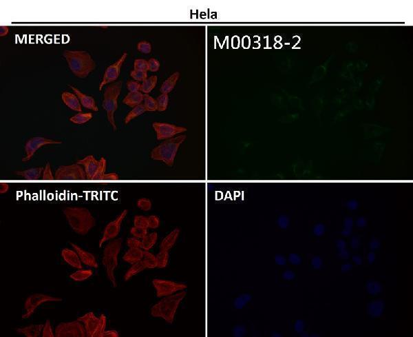
A00318-1 shows IHC in paraffin sections of human lung cancer, lymphoma and testicular cancer and mouse brain (A00318-1 IHC captions); M00318-3 shows IHC in mouse testis and human placenta (M00318-3 IHC captions); A00318Y463 and A00318Y467 show IHC in paraffin sections of human breast carcinoma (respective IHC captions). A00318Y607 shows IF in BV2 cells (A00318Y607 IF caption), while M00318-2 has IF images with no sample identified (M00318-2 IF captions).
Which to pick: For paraffin-section IHC, choose A00318-1: its lung cancer image documents EDTA retrieval at pH 8.0 and 2 μg/ml primary antibody overnight at 4°C; the fixative is unreported (A00318-1 IHC caption). For IF/ICC, choose A00318-1 for the documented Caco-2 cell image at 5 μg/ml (A00318-1 IF caption), or monoclonal M00318-2 for listed IF/ICC use, while its IF captions leave the sample unidentified (M00318-2 catalog applications, clone and IF captions). For cross-species tissue IHC, monoclonal M00318-3 has images from human placenta and mouse testis and lists rat reactivity; the captions do not report the fixative, and rat IHC is not shown (M00318-3 catalog clone, reactivity and IHC captions).