Click image to see more details
Product Info Summary
| SKU: | M00318-2 |
|---|---|
| Size: | 100 μl/vial |
| Reactive Species: | Human, Mouse, Rat |
| Host: | Rabbit |
| Application: | IF, ICC, WB |
Customers Who Bought This Also Bought

Product info
Product Name
Anti-PI3 Kinase p85 alpha Rabbit Monoclonal Antibody
SKU/Catalog Number
M00318-2
BM5187 is an alternative SKU for this antibody, used in previous lots.
Size
100 μl/vial
Form
Liquid
Description
Boster Bio Anti-PI3 Kinase p85 alpha Rabbit Monoclonal Antibody catalog # M00318-2. Tested in WB, ICC/IF applications. This antibody reacts with Human, Mouse, Rat.
Storage & Handling
Store at -20°C for one year. For short term storage and frequent use, store at 4°C for up to one month. Avoid repeated freeze-thaw cycles.
Cite This Product
Anti-PI3 Kinase p85 alpha Rabbit Monoclonal Antibody (Boster Biological Technology, Pleasanton CA, USA, Catalog # M00318-2)
Host
Rabbit
Contents
Rabbit IgG in stabilizing components, phosphate buffered saline, pH 7.4, 150mM NaCl, 0.02% sodium azide and 50% glycerol.
*This antibody is supplied in a stabilized formulation.
Compatibility with conjugation reactions depends on the chemistry of the conjugation method used.
For conjugation methods that are not compatible with the stabilizing components present in this formulation, a carrier-free antibody format is required.
Clonality
Monoclonal
Clone Number
17P38
Isotype
IgG
Immunogen
A synthesized peptide derived from human PI3 Kinase p85 alpha
Reactive Species
M00318-2 is reactive to PIK3R1 in Human, Mouse, Rat
Observed Molecular Weight
84 kDa
Calculated molecular weight
83.6 kDa
Antibody Validation
Boster validates all antibodies on WB, IHC, ICC, Immunofluorescence, and ELISA with known positive control and negative samples to ensure specificity and high affinity, including thorough antibody incubations.
Application & Images
Applications
M00318-2 is guaranteed for IF, ICC, WB Boster Guarantee
Recommend Dilution
WB 1:500-2000
Tested application
Suggested blocking solution with 5% non-fat milk or BSA; (*)Recommended protein loading: 20-40 µg per lane
Validation Images & Assay Conditions
Click image to see more details
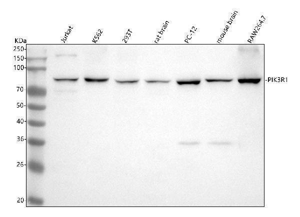
Western blot analysis of PI3 Kinase p85 alpha using anti-PI3 Kinase p85 alpha antibody (M00318-2).
Electrophoresis was performed on a 5-20% SDS-PAGE gel at 70V (Stacking gel) / 90V (Resolving gel) for 2-3 hours. The sample well of each lane was loaded with 30 ug of sample under reducing conditions.
Lane 1: human Jurkat whole cell lysates,
Lane 2: human K562 whole cell lysates,
Lane 3: human 293T whole cell lysates,
Lane 4: rat brain tissue lysates,
Lane 5: rat PC-12 whole cell lysates,
Lane 6: mouse brain tissue lysates,
Lane 7: mouse RAW264.7 whole cell lysates.
After electrophoresis, proteins were transferred to a nitrocellulose membrane at 150 mA for 50-90 minutes. Blocked the membrane with 5% non-fat milk/TBS for 1.5 hour at RT. The membrane was incubated with rabbit anti-PI3 Kinase p85 alpha antigen affinity purified monoclonal antibody (Catalog # M00318-2) at 1:500 overnight at 4°C, then washed with TBS-0.1%Tween 3 times with 5 minutes each and probed with a goat anti-rabbit IgG-HRP secondary antibody at a dilution of 1:500 for 1.5 hour at RT. The signal is developed using an Enhanced Chemiluminescent detection (ECL) kit (Catalog # EK1002) with Tanon 5200 system. A specific band was detected for PI3 Kinase p85 alpha at approximately 84 kDa. The expected band size for PI3 Kinase p85 alpha is at 84 kDa.

Click image to see more details
Immunofluorescent analysis using the Antibody at 1:50 dilution.

Click image to see more details
Immunofluorescent analysis using the Antibody at 1:150 dilution.

Click image to see more details
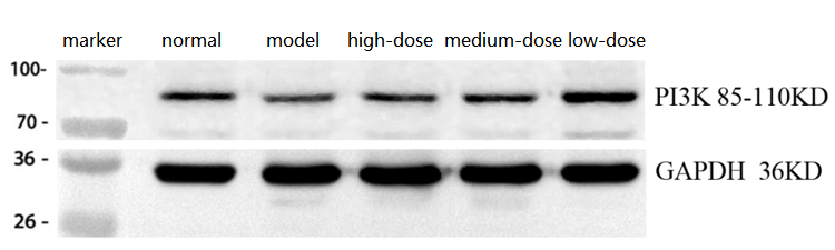
Western blot analysis of PI3 Kinase p85 alpha using anti-PI3 Kinase p85 alpha antibody (M00318-2).
Electrophoresis was performed on a 5-20% SDS-PAGE gel at 70V (Stacking gel) / 90V (Resolving gel) for 2-3 hours. The sample well of each lane was loaded with 30 ug of sample under reducing conditions.
Lane 1: normal group-Rat skeletal muscle tissue lysates,
Lane 2: model group-Rat skeletal muscle tissue lysates,
Lane 3: high-dose group-Rat skeletal muscle tissue lysates,
Lane 4: medium-dose group-Rat skeletal muscle tissue lysates,
Lane 5: low-dose group-Rat skeletal muscle tissue lysates.
After electrophoresis, proteins were transferred to a nitrocellulose membrane at 150 mA for 50-90 minutes. Blocked the membrane with 5% non-fat milk/TBS for 1.5 hour at RT. The membrane was incubated with rabbit anti-PI3 Kinase p85 alpha antigen affinity purified monoclonal antibody (Catalog # M00318-2) at 1:2000 overnight at 4°C, then washed with TBS-0.1%Tween 3 times with 5 minutes each and probed with a goat anti-rabbit IgG-HRP secondary antibody at a dilution of 1:10000 for 1.5 hour at RT. The signal is developed using an Enhanced Chemiluminescent detection (ECL) kit (Catalog # EK1002) with Tanon 5200 system. A specific band was detected for PI3 Kinase p85 alpha at approximately 85-110 kDa. The expected band size for PI3 Kinase p85 alpha is at 84 kDa.
Specific Publications For Anti-PI3 Kinase p85 alpha Rabbit Monoclonal Antibody (M00318-2)
Loading publications
Recommended Resources
Here are featured tools and databases that you might find useful.
- Boster's Pathways Library
- Protein Databases
- Bioscience Research Protocol Resources
- Data Processing & Analysis Software
- Photo Editing Software
- Scientific Literature Resources
- Research Paper Management Tools
- Molecular Biology Software
- Primer Design Tools
- Bioinformatics Tools
- Phylogenetic Tree Analysis
Customer Reviews
Have you used Anti-PI3 Kinase p85 alpha Rabbit Monoclonal Antibody?
Share your experimental results or join a short interview to earn up to $1,000 in product credits or other rewards.
1 Reviews For Anti-PI3 Kinase p85 alpha Rabbit Monoclonal Antibody
The PI3K p85 alpha Antibody (M00318-2) showed clear and specific bands in WB of rat skeletal muscle, with PI3K p85 expression increased in the model group and decreased in a dose-dependent manner in the treatment groups, consistent with expectations.
Excellent
| SKU | M00318-2 |
|---|---|
| Application | Western Blot |
| Sample | Rat skeletal muscle tissue |
| Sample Processing Description | Normal rat skeletal muscle, injury model, and high-, medium-, and low-dose treatment groups, with total protein extracted. |
| Other Reagents | RIPA lysis buffer, Protease inhibitor, Running buffer, Transfer buffer,Blocking buffer |
| Primary Antibody | PI3 Kinase p85 alpha Rabbit Monoclonal Antibody |
| Primary Incubation | 1:2000, overnight at 4 ℃ |
| Secondary Antibody | HRP Conjugated AffiniPure Goat Anti-Rabbit IgG (H+L) (BA1054) |
| Secondary Incubation | 1:10000, 1 h in RT |
| Detection | Substrate: ECL substrate, Image system: ChemiDoc MP |
| Results Summary | Based on the experimental results, the band of PI3K p85 is clear. The PI3K p85 level in the model group is significantly higher than in the normal group, whereas in the treatment group, the PI3K p85 level decreases with increasing drug dose, which is consistent with expectations. |
Guangtian Yu, Ningxia Medical University
Verified customer
Submitted 2026-03-26
Customer Q&As
Have a question?
Find answers in Q&As, reviews.
Can't find your answer?
Submit your question
