Click image to see more details
-

-
-
-
-
+6
Product Info Summary
| SKU: | A00318-1 |
|---|---|
| Size: | 100 μg/vial |
| Reactive Species: | Human, Mouse, Rat |
| Host: | Rabbit |
| Application: | ELISA, Flow Cytometry, IF, IHC, ICC, WB |
Customers Who Bought This Also Bought

Product info
Product Name
Anti-PI 3 Kinase p85 alpha/PIK3R1 Antibody Picoband®
SKU/Catalog Number
A00318-1
Size
100 μg/vial
Form
Lyophilized
Description
Boster Bio Anti-PI 3 Kinase p85 alpha/PIK3R1 Antibody Picoband® catalog # A00318-1. Tested in ELISA, Flow Cytometry, IF, IHC, ICC, WB applications. This antibody reacts with Human, Mouse, Rat. The brand Picoband indicates this is a premium antibody that guarantees superior quality, high affinity, and strong signals with minimal background in Western blot applications. Only our best-performing antibodies are designated as Picoband, ensuring unmatched performance.
Storage & Handling
At -20°C for one year from date of receipt. After reconstitution, at 4°C for one month. It can also be aliquotted and stored frozen at -20°C for six months. Avoid repeated freezing and thawing.
Cite This Product
Anti-PI 3 Kinase p85 alpha/PIK3R1 Antibody Picoband® (Boster Biological Technology, Pleasanton CA, USA, Catalog # A00318-1)
Host
Rabbit
Contents
Each vial contains 4 mg Trehalose, 0.9 mg NaCl, 0.2 mg Na2HPO4.
Clonality
Polyclonal
Isotype
Rabbit IgG
Immunogen
E.coli-derived human PI 3 Kinase p85 alpha/PIK3R1 recombinant protein (Position: D117-Q153).
Cross-reactivity
No cross-reactivity with other proteins
Reactive Species
A00318-1 is reactive to PIK3R1 in Human, Mouse, Rat
Observed Molecular Weight
85 kDa
Calculated molecular weight
83.6 kDa
Background of PIK3R1
Phosphatidylinositol 3-kinase regulatory subunit alpha is an enzyme that in humans is encoded by the PIK3R1 gene. Its gene is mapped to 5q13. the bovine PI3K p85 subunit consists of 2 closely related proteins, p85-alpha and p85-beta. They cloned cDNAs encoding both p85 subunits, each of which is a 724-amino acid polypeptide. Phosphatidylinositol 3-kinase plays an important role in the metabolic actions of insulin, and a mutation in this gene has been associated with insulin resistance.
Antibody Validation
Boster validates all antibodies on WB, IHC, ICC, Immunofluorescence, and ELISA with known positive control and negative samples to ensure specificity and high affinity, including thorough antibody incubations.
Application & Images
Applications
A00318-1 is guaranteed for ELISA, Flow Cytometry, IF, IHC, ICC, WB Boster Guarantee
Recommend Dilution
| Application | Dilution | Species |
|---|---|---|
| Western blot | 0.25-0.5 μg/ml | Human, Mouse, Rat |
| Immunohistochemistry(Paraffin-embedded Section) | 2-5 μg/ml | Human, Mouse, Rat |
| Immunocytochemistry/Immunofluorescence | 5 μg/ml | Human |
| Flow Cytometry (Fixed) | 1-3 μg/1x106 cells | Human |
| ELISA | 0.1-0.5 μg/ml | - |
Tested application
Suggested blocking solution with 5% non-fat milk or BSA; (*)Recommended protein loading: 20-40 µg per lane
Use TE buffer pH 9.0 for antigen retrieval; (*) citrate buffer pH 6.0 is an alternative.
Validation Images & Assay Conditions
Click image to see more details
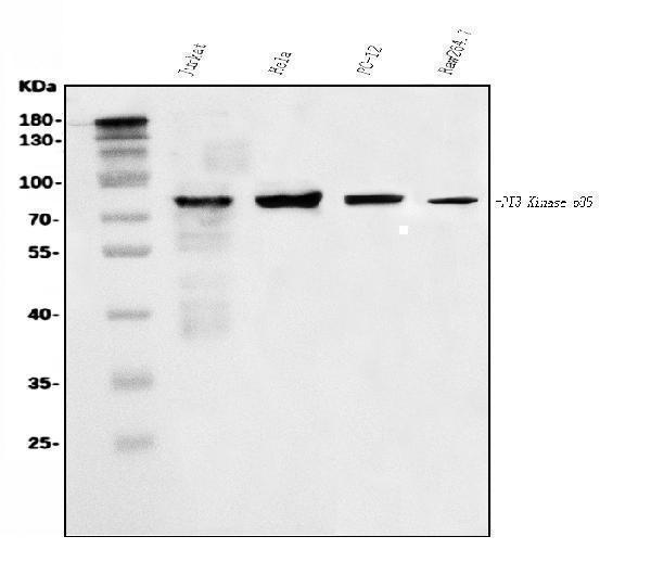
Western blot analysis of PI 3 Kinase p85 alpha/PIK3R1 using anti-PI 3 Kinase p85 alpha/PIK3R1 antibody (A00318-1).
Electrophoresis was performed on a 5-20% SDS-PAGE gel at 70V (Stacking gel) / 90V (Resolving gel) for 2-3 hours. The sample well of each lane was loaded with 30 ug of sample under reducing conditions.
Lane 1: human Jurkat whole cell lysates,
Lane 2: human Hela whole cell lysates,
Lane 3: rat PC-12 whole cell lysates,
Lane 4: mouse RAW264.7 whole cell lysates.
After electrophoresis, proteins were transferred to a nitrocellulose membrane at 150 mA for 50-90 minutes. Blocked the membrane with 5% non-fat milk/TBS for 1.5 hour at RT. The membrane was incubated with rabbit anti-PI 3 Kinase p85 alpha/PIK3R1 antigen affinity purified polyclonal antibody (Catalog # A00318-1) at 0.5 μg/mL overnight at 4°C, then washed with TBS-0.1%Tween 3 times with 5 minutes each and probed with a goat anti-rabbit IgG-HRP secondary antibody at a dilution of 1:5000 for 1.5 hour at RT. The signal is developed using an Enhanced Chemiluminescent detection (ECL) kit (Catalog # EK1002) with Tanon 5200 system. A specific band was detected for PI 3 Kinase p85 alpha/PIK3R1 at approximately 85 kDa. The expected band size for PI 3 Kinase p85 alpha/PIK3R1 is at 85 kDa.

Click image to see more details
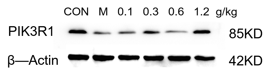
Western blot analysis of PI 3 Kinase p85 alpha/PIK3R1 using anti-PI 3 Kinase p85 alpha/PIK3R1 antibody (A00318-1).
Electrophoresis was performed on a 5-20% SDS-PAGE gel at 80V (Stacking gel) / 120V (Resolving gel) for 2 hours. The sample well of each lane was loaded with 30 ug of sample under reducing conditions.
Lane 1: control group-mouse hippocampus tissue lysates,
Lane 2: model group-mouse hippocampus tissue lysates
Lane 3:Drug treatment (0.1g/kg) – Mouse hippocampus tissue lysates,
Lane 4:Drug treatment (0.3g/kg) – Mouse hippocampus tissue lysates,
Lane 5: Drug treatment (0.6g/kg) – Mouse hippocampus tissue lysates,
Lane 6: Drug treatment (1.2g/kg) – Mouse hippocampus tissue lysates.
After electrophoresis, proteins were transferred to a nitrocellulose membrane at 150 mA for 50-90 minutes. Blocked the membrane with 5% non-fat milk/TBS for 1.5 hour at RT. The membrane was incubated with rabbit anti-PI 3 Kinase p85 alpha/PIK3R1 antigen affinity purified polyclonal antibody (A00318-1) overnight at 4°C, then washed with TBS-0.1%Tween 3 times with 5 minutes each and probed with a goat anti-rabbit IgG-HRP secondary antibody (Catalog # BA1054) at a dilution of 1:5000 for 1 hour at RT. The signal is developed using an ECL Plus Western Blotting Substrate with ChemiDoc MP system. A specific band was detected for PI 3 Kinase p85 alpha/PIK3R1 at approximately 85 kDa. The expected band size for PI 3 Kinase p85 alpha/PIK3R1 is at 85 kDa.

Click image to see more details
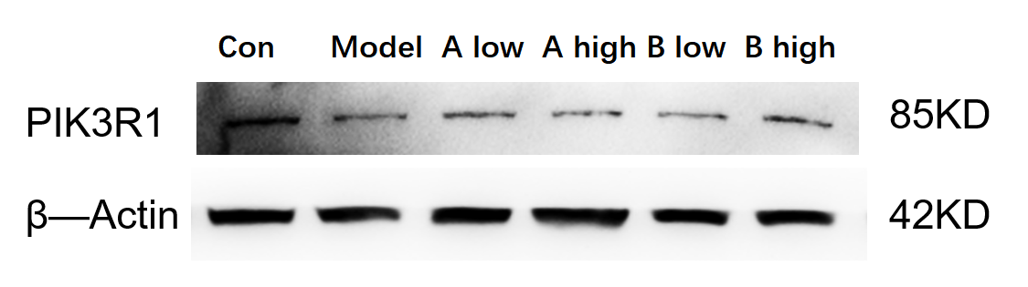
Western blot analysis of PI 3 Kinase p85 alpha/PIK3R1 using anti-PI 3 Kinase p85 alpha/PIK3R1 antibody (A00318-1).
Electrophoresis was performed on a 5-20% SDS-PAGE gel at 80V (Stacking gel) / 120V (Resolving gel) for 2 hours. The sample well of each lane was loaded with 30 ug of sample under reducing conditions.
Lane 1: control group-mouse brain tissue lysates,
Lane 2: model group-mouse brain tissue lysates
Lane 3: A drug low-concentration treatment– Mouse brain tissue lysates,
Lane 4: A drug high-concentration treatment– Mouse brain tissue lysates,
Lane 5: B drug low-concentration treatment– Mouse brain tissue lysates,
Lane 6: B drug high-concentration treatment– Mouse brain tissue lysates.
After electrophoresis, proteins were transferred to a nitrocellulose membrane at 150 mA for 50-90 minutes. Blocked the membrane with 5% non-fat milk/TBS for 1.5 hour at RT. The membrane was incubated with rabbit anti-PI 3 Kinase p85 alpha/PIK3R1 antigen affinity purified polyclonal antibody (A00318-1) overnight at 4°C, then washed with TBS-0.1%Tween 3 times with 5 minutes each and probed with a goat anti-rabbit IgG-HRP secondary antibody (Catalog # BA1054) at a dilution of 1:5000 for 1 hour at RT. The signal is developed using an ECL Plus Western Blotting Substrate with ChemiDoc MP system. A specific band was detected for PI 3 Kinase p85 alpha/PIK3R1 at approximately 85 kDa. The expected band size for PI 3 Kinase p85 alpha/PIK3R1 is at 85 kDa.

Click image to see more details
IHC analysis of PI 3 Kinase p85 alpha/PIK3R1 using anti-PI 3 Kinase p85 alpha/PIK3R1 antibody (A00318-1).
PI 3 Kinase p85 alpha/PIK3R1 was detected in a paraffin-embedded section of human lung cancer tissue. Heat mediated antigen retrieval was performed in EDTA buffer (pH 8.0, epitope retrieval solution). The tissue section was blocked with 10% goat serum. The tissue section was then incubated with 2 μg/ml rabbit anti-PI 3 Kinase p85 alpha/PIK3R1 Antibody (A00318-1) overnight at 4°C. Biotinylated goat anti-rabbit IgG was used as secondary antibody and incubated for 30 minutes at 37°C. The tissue section was developed using Strepavidin-Biotin-Complex (SABC) (Catalog # SA1022) with DAB as the chromogen.

Click image to see more details
IHC analysis of PI 3 Kinase p85 alpha/PIK3R1 using anti-PI 3 Kinase p85 alpha/PIK3R1 antibody (A00318-1).
PI 3 Kinase p85 alpha/PIK3R1 was detected in a paraffin-embedded section of human lymphoma tissue. Heat mediated antigen retrieval was performed in EDTA buffer (pH 8.0, epitope retrieval solution). The tissue section was blocked with 10% goat serum. The tissue section was then incubated with 2 μg/ml rabbit anti-PI 3 Kinase p85 alpha/PIK3R1 Antibody (A00318-1) overnight at 4°C. Biotinylated goat anti-rabbit IgG was used as secondary antibody and incubated for 30 minutes at 37°C. The tissue section was developed using Strepavidin-Biotin-Complex (SABC) (Catalog # SA1022) with DAB as the chromogen.

Click image to see more details
IHC analysis of PI 3 Kinase p85 alpha/PIK3R1 using anti-PI 3 Kinase p85 alpha/PIK3R1 antibody (A00318-1).
PI 3 Kinase p85 alpha/PIK3R1 was detected in a paraffin-embedded section of human testicular cancer tissue. Heat mediated antigen retrieval was performed in EDTA buffer (pH 8.0, epitope retrieval solution). The tissue section was blocked with 10% goat serum. The tissue section was then incubated with 2 μg/ml rabbit anti-PI 3 Kinase p85 alpha/PIK3R1 Antibody (A00318-1) overnight at 4°C. Biotinylated goat anti-rabbit IgG was used as secondary antibody and incubated for 30 minutes at 37°C. The tissue section was developed using Strepavidin-Biotin-Complex (SABC) (Catalog # SA1022) with DAB as the chromogen.

Click image to see more details
IHC analysis of PI 3 Kinase p85 alpha/PIK3R1 using anti-PI 3 Kinase p85 alpha/PIK3R1 antibody (A00318-1).
PI 3 Kinase p85 alpha/PIK3R1 was detected in a paraffin-embedded section of mouse brain tissue. Heat mediated antigen retrieval was performed in EDTA buffer (pH 8.0, epitope retrieval solution). The tissue section was blocked with 10% goat serum. The tissue section was then incubated with 2 μg/ml rabbit anti-PI 3 Kinase p85 alpha/PIK3R1 Antibody (A00318-1) overnight at 4°C. Biotinylated goat anti-rabbit IgG was used as secondary antibody and incubated for 30 minutes at 37°C. The tissue section was developed using Strepavidin-Biotin-Complex (SABC) (Catalog # SA1022) with DAB as the chromogen.

Click image to see more details
IHC analysis of PI 3 Kinase p85 alpha/PIK3R1 using anti-PI 3 Kinase p85 alpha/PIK3R1 antibody (A00318-1).
PI 3 Kinase p85 alpha/PIK3R1 was detected in a paraffin-embedded section of rat brain tissue. Heat mediated antigen retrieval was performed in EDTA buffer (pH 8.0, epitope retrieval solution). The tissue section was blocked with 10% goat serum. The tissue section was then incubated with 2 μg/ml rabbit anti-PI 3 Kinase p85 alpha/PIK3R1 Antibody (A00318-1) overnight at 4°C. Biotinylated goat anti-rabbit IgG was used as secondary antibody and incubated for 30 minutes at 37°C. The tissue section was developed using Strepavidin-Biotin-Complex (SABC) (Catalog # SA1022) with DAB as the chromogen.

Click image to see more details
IF analysis of PI 3 Kinase p85 alpha/PIK3R1 using anti-PI 3 Kinase p85 alpha/PIK3R1 antibody (A00318-1).
PI 3 Kinase p85 alpha/PIK3R1 was detected in an immunocytochemical section of Caco-2 cells. Enzyme antigen retrieval was performed using IHC enzyme antigen retrieval reagent (AR0022) for 15 mins. The cells were blocked with 10% goat serum. And then incubated with 5 μg/mL rabbit anti-PI 3 Kinase p85 alpha/PIK3R1 Antibody (A00318-1) overnight at 4°C. DyLight®488 Conjugated Goat Anti-Rabbit IgG (BA1127) was used as secondary antibody at 1:100 dilution and incubated for 30 minutes at 37°C. The section was counterstained with DAPI. Visualize using a fluorescence microscope and filter sets appropriate for the label used.

Click image to see more details
Flow Cytometry analysis of HL-60 cells using anti-PI 3 Kinase p85 alpha/PIK3R1 antibody (A00318-1).
Overlay histogram showing HL-60 cells stained with A00318-1 (Blue line). To facilitate intracellular staining, cells were fixed with 4% paraformaldehyde and permeabilized with permeabilization buffer. The cells were blocked with 10% normal goat serum. And then incubated with rabbit anti-PI 3 Kinase p85 alpha/PIK3R1 Antibody (A00318-1, 1 μg/1x106 cells) for 30 min at 20°C. DyLight®488 conjugated goat anti-rabbit IgG (BA1127, 5-10 μg/1x106 cells) was used as secondary antibody for 30 minutes at 20°C. Isotype control antibody (Green line) was rabbit IgG (1 μg/1x106) used under the same conditions. Unlabelled sample without incubation with primary antibody and secondary antibody (Red line) was used as a blank control.
Specific Publications For Anti-PI 3 Kinase p85 alpha/PIK3R1 Antibody Picoband® (A00318-1)
Loading publications
Recommended Resources
Here are featured tools and databases that you might find useful.
- Boster's Pathways Library
- Protein Databases
- Bioscience Research Protocol Resources
- Data Processing & Analysis Software
- Photo Editing Software
- Scientific Literature Resources
- Research Paper Management Tools
- Molecular Biology Software
- Primer Design Tools
- Bioinformatics Tools
- Phylogenetic Tree Analysis
Customer Reviews
Have you used Anti-PI 3 Kinase p85 alpha/PIK3R1 Antibody Picoband®?
Share your experimental results or join a short interview to earn up to $1,000 in product credits or other rewards.
2 Reviews For Anti-PI 3 Kinase p85 alpha/PIK3R1 Antibody Picoband®
This antibody is highly specific and efficient, suitable for Western blot detection of Anti-PI3K p85 alpha Antibody protein in mouse hippocampus, with only minor nonspecific bands observed.
Excellent
| SKU | A00318-1 |
|---|---|
| Application | Western Blot |
| Sample | Mouse brain tissue |
| Sample Processing Description | Mouse brain tissue was lysed in RIPA buffer supplemented with protease inhibitors at 4°C for 2 hours. The lysate was centrifuged to collect the supernatant, and protein concentration was determined. After adjusting the concentration, samples were mixed with 5× protein loading buffer and heated at 95–100°C for 10 minutes to denature. Then, 15 μL of each protein sample was loaded per lane for electrophoresis. |
| Other Reagents | Blocking buffer |
| Primary Antibody | PI 3 Kinase p85 alpha/PIK3R1 Antibody Picoband® |
| Primary Incubation | 1:2000, overnight at 4 ℃ |
| Secondary Antibody | HRP Goat Anti-Rabbit IgG |
| Secondary Incubation | 1:10000, 1 hour in room temperature |
| Detection | Substrate: ECL, Imaging system:ChemiDoc MP |
| Results Summary | The figure shows a schematic representation of Western blot results for PIK3R1 and the loading control β-actin in mouse brain tissues from normal mice, model group, and mice treated with high and low doses of drug AB. Although the expression differences between experimental groups are not pronounced, the Western blot results obtained with this antibody are still clear and well-defined. |
Yetao Ju, Liaoning Univ. of TCM
Verified customer
Submitted 2025-12-25
The antibody shows clear bands with no non-specific signals in WB, and is suitable for detecting PIK3R1 protein in mouse hippocampus by Western blot.
Excellent
| SKU | A00318-1 |
|---|---|
| Application | Western Blot |
| Sample | Mouse hippocampus tissue |
| Sample Processing Description | The mouse hippocampus was lysed with RIPA lysis buffer containing protease inhibitors, followed by protein quantification. The samples were then mixed with 5× protein loading buffer and heated for 10 minutes for denaturation. Five microliters of protein sample were loaded into each lane for SDS-PAGE. |
| Primary Antibody | Anti-PI 3 Kinase p85 alpha/PIK3R1 Antibody Picoband® |
| Primary Incubation | overnight at 4 ℃ |
| Secondary Antibody | HRP-conjugated Anti-Rabbit IgG Secondary Antibody |
| Secondary Incubation | 1 hour in room temperature |
| Detection | Substrate: Ultra-sensitive ECL luminescent reagent , Imaging system:ChemiDoc MP |
| Results Summary | The antibody shows clear bands with no non-specific signals in WB, and is suitable for detecting PIK3R1 protein in mouse hippocampus by Western blot. |
Yali Yang, LNUTCM
Verified customer
Submitted 2025-11-10
Customer Q&As
Have a question?
Find answers in Q&As, reviews.
Can't find your answer?
Submit your question
