This website uses cookies to ensure you get the best experience on our website.
- Table of Contents
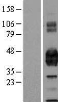
Facts about Endothelial protein C receptor.
.
Human | |
---|---|
Gene Name: | PROCR |
Uniprot: | Q9UNN8 |
Entrez: | 10544 |
Belongs to: |
---|
No superfamily |
APC receptor; CCCA; CCD41; CCD41centrosome-associated protein; CD201 antigen; CD201; centrocyclin; Endothelial cell protein C receptor; endothelial protein C receptor; EPCR; EPCRMGC23024; PROCR; protein C receptor, endothelial
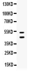
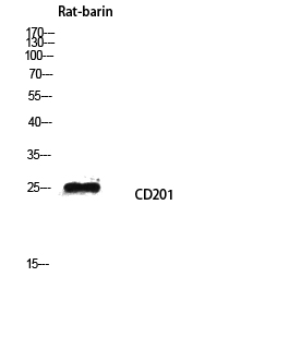
Mass (kDA):
26.671 kDA
Human | |
---|---|
Location: | 20q11.22 |
Sequence: | 20; NC_000020.11 (35172072..35215989) |
Expressed strongly in the endothelial cells of arteries and veins in heart and lung, less intensely in capillaries in the lung and skin, and not at all in the endothelium of small vessels of the liver and kidney.
Membrane; Single-pass type I membrane protein.
This article will discuss the benefits of using the Boster Bio PROCR Marker, the IHC protocol and the use of primary antibodies. For more details, visit Boster Bio's website. For a start with PROCR-based molecular and cellular imaging Download a free trial kit today! The best use for the PROCR marker in molecular biology applications is immunohistochemistry, but the use of the PROCR Marker is not limited to immunohistochemistry and IHC.
A primary antibody bound to p21 during an IHC protocol using the PROCR marker, which binds the antigen found in human colon cancer specimens. It is a distinct marker due to its placement in FFPE sections. The antibody detects p21 through analyzing its signal intensity, allowing a densitometric analysis to compare the reporter's signal with protein expression levels. The secondary antibody binds to the enzyme that transforms the substrate into brown stars.
Once the organ of choice is prepared and the tissue sample is taken, it must be fixed or rapidly frozen to remove blood and other blood components. Tissue can also be washed and dried. Once the tissue has been fixed, the immunostaining procedure can be done on the targeted organ. To avoid false positives fix the tissue as soon as possible prior to IHC. Depending on the antigen that is of interest, the sample could be fixed or not.
Many antibodies recognize small peptides, molecular mimicry and various other molecular peptides. These antibodies can stain tissue and other cell types other than the tissue of the target. The prognostic value of the PROCR marker in IHC is unknown. It is used to track the growth of tumors and their microenvironment. The results of immunostaining can provide information about the immune status of the tumor's microenvironment. There are a myriad of possible pitfalls to applying this marker, such as the possibility of false positive results.
The primary antibody must be specific to the target protein. If it is not be, it shouldn't be used in IHC. The primary antibody should be of high-quality. If the antigen's activity is inactive the antibody will not be able to bind to the protein target. The secondary antibody should be specific to primary antibody's source. To avoid false positives, it's essential to purify the primary antibody prior to using it. A second antibody should be available that is specific to the target protein.
The use of immunohistochemistry is common to identify haptens and antigens in biological tissues. This technique makes use of antibodies to detect proteins while keeping the characteristics of the tissue. For instance, tumors that contain HER2 receptor proteins are frequently identified using IHC. If there are too many of these proteins, cancerous cells could grow out of control. It is essential to understand the expression of HER2 receptors in tumors to determine if they are active.
An IHC test that detects estrogen receptors or progesterone inside breast cancer cells is referred to as hormone receptor-positive (HER2). The results aren't conclusive as different pathologists use different criteria to determine the presence of hormone receptors. However the IHC protocol using the PROCR marker remains effective and widely used in the diagnosis of breast cancer.
The primary-secondary antibody system is the ability to label two different substances, allowing scientists to study the same specimen using two distinct primary antibodies. Dual-labeling allows researchers to ask many more questions and provide concrete answers. This allows them to differentiate between different kinds of antibodies and the different ways to test for a specific antibody.
This involves binding a primary antibody to an enzyme-labeled strainavidin that detects biotinylated antibody. The enzyme-labeled streptavidin is smaller than a biotin-conjugated one which means it is able to penetrate deeper into tissues. The secondary antibodies that are conjugated to LSB are more sensitive than the ABC method and have superior tissue penetration. They also can detect proteins that are not expressed.
Scientists should seek out an antibody that recognizes PROCR protein when conducting tests using immunohistochemicals. Boster's histology laboratory is well-equipped to perform the tests necessary to determine if the primary antibody recognizes the specific antigen. To ensure that the right antibody is used to identify the target, scientists can purchase blocking peptides. It is possible to determine the specific protein in a sample without using an antibody as a primary.
Secondary antibody dilutions must have to be customized for each use. They should be reduced to 1:1000 or less. The type of antigen employed and the specimen being evaluated will determine the best concentration of secondary antibodies. The incubation period should take place at 37°C for one hour or at room temperature for two hours. If overnight incubation is needed the secondary antibody must be kept at 4degC in a refrigerator.
The PROCR marker is a crucial tool to produce primary polyclonal antibodies. It allows scientists to develop polyclonal antibody without having to face somatic mutations. These antibodies are derived from the same VH and VL genes. These genes encode variable regions that detect pathogens or haptens. This allows you to track the changes in these antibodies molecules.
When primary antibodies are made using the PROCR marker the results are more reliable. Using this marker in the production of primary antibodies is more precise than using an antibody that is a primary one in the sandwich assay. The primary antibodies are immobilized onto the proteins that are targeted, and the secondary antibodies are used to identify them. These procedures come with many problems.
It is now simpler to distinguish memory B cells from non-specific ones by using the PROCR marker in primary antibody production. Memory B cells are immune types that have undergone somatic mutations or acquired VH and VL gene segments. These antibodies are not specific because they have higher affinity for the antigen. Although the differences between these two types of antibodies aren't immediately obvious, they can be assessed.
PMID: 7929370 by Fukudome K., et al. Identification, cloning, and regulation of a novel endothelial cell protein C/activated protein C receptor.
PMID: 10397730 by Simmonds R.E., et al. Structural and functional implications of the intron/exon organization of the human endothelial cell protein C/activated protein C receptor (EPCR) gene: comparison with the structure of CD1/major histocompatibility complex alpha1 and alpha2 domains.