PTPN6 / Tyrosine-protein phosphatase non-receptor type 6 · IHC design guide

Design Immunohistochemistry for PTPN6

Plan PTPN6 paraffin IHC with catalog antibody M00938-2 at 2 μg/mL (datasheet M00938-2). Use cytoplasmic staining in lung macrophages or lymphoid cells as a positive reference (HPA tissue IHC), and score cell populations separately (standard IHC practice).

Evidence assembled Oct 2026 · For research use; verify linked source records and product datasheet before use
Immunohistochemistry protocol sheet for PTPN6 (IHC for PTPN6): expected localisation Cytoplasmic staining in immune tissues (HPA tissue IHC), antibody M00938-2, validated IHC image, and IHC protocol steps
Printable PTPN6 IHC protocol sheet — expected localisation Cytoplasmic staining in immune tissues (HPA tissue IHC), antibody M00938-2, controls and protocol steps. Open the full PTPN6 IHC guide →

PTPN6 Immunohistochemistry Experimental Design Guide

Expected localisation, validated protocols, controls and antibodies — the at-a-glance facts below, then the full design guide.

Must know before staining
Expected localisation Cytoplasmic staining in immune tissues (HPA tissue IHC)
Staining pattern Cytoplasmic staining in lung macrophages and lymphoid cells (HPA tissue IHC)
Antigen retrieval EDTA pH 8.0 HIER, heat-mediated (datasheet M00938-2)
Positive control ⓘ Appendix+4 more · see all
Negative control ⓘ Adrenal gland+4 more · see all
Important caveats
Reasons your staining may differ from the expected pattern.
Fixation Keep paraffin-section fixation consistent across samples (standard IHC practice; not target-specific)
Caveat Immune infiltrates can complicate cell-specific scoring (HPA tissue IHC)
Regulation Immune-tissue enrichment (HPA tissue IHC)
Isoform / epitope 4 isoforms; epitope coverage needs validation (UniProt)
Section 1

Recommended PTPN6 IHC & IF Protocols

The catalog antibody uses heat-mediated EDTA retrieval at pH 8.0 (datasheet M00938-2). One published PTPN6 IHC protocol uses citrate retrieval at pH 6.1 (PMC4036221).

Recommended immunohistochemistry (IHC-P) protocol parameters
SampleParaffin-embedded human laryngeal squamous cell carcinomas tissue; fixative not specified (datasheet M00938-2)
FixationImage fixative and duration unreported (datasheet M00938-2); verify before use.
Sectioning4–5 µm sections on charged slides (standard)
DeparaffinisationXylene, graded ethanol series to water (standard)
Antigen retrievalHeat retrieval: EDTA pH 8.0 (datasheet M00938-2); 20 min, 95–100 °C (standard)
Peroxidase block3% H2O2, 10 min, room temperature (standard)
Blocking10% goat serum (datasheet M00938-2)
Primary antibodyMouse monoclonal (clone 8H11B10) anti-PTPN6, 2 μg/ml (datasheet M00938-2)
Primary incubationOvernight at 4 °C (datasheet M00938-2)
DetectionHRP-conjugated secondary, DAB chromogen (datasheet M00938-2)
CounterstainHematoxylin, blue, dehydrate and mount (standard)
Expected resultPTPN6-positive staining in lymphoid tissue of appendix (HPA tissue IHC: High). HPA tissue profile: Distinct cytoplasmic expression in immune tissues. No signal in the no-primary control.
💡Decision noteStart with EDTA at pH 8.0 for the catalog antibody (datasheet M00938-2); evaluate citrate at pH 6.1 as a published alternative (PMC4036221).
Section 2

What Is the Expected PTPN6 Staining Pattern?

PTPN6 is a soluble protein found in the cytoplasm and nucleus, with no transmembrane segment (UniProt P29350 topology; subcellular location). In tissue IHC, expect distinct cytoplasmic staining in immune cells, including lung macrophages and lymphoid cells (HPA: tissue IHC). HPA rates the tissue profile Enhanced, while describing antibody staining versus RNA expression as having medium consistency (HPA: tissue IHC reliability).

What am I looking at on my slide?
Distinct cytoplasmic staining in lung macrophages or lymph node germinal center cells.This matches cell types scored High by HPA (HPA: lung macrophages; lymph node germinal center cells). Score the identified cells, rather than assigning the whole tissue one intensity; nearby cells may differ (standard IHC interpretation).
Staining is confined to cell borders, luminal surfaces, or extracellular material.That distribution does not match HPA's distinct cytoplasmic tissue profile or UniProt's soluble topology (HPA: tissue IHC profile; UniProt P29350 topology). Review morphology and controls before calling it PTPN6.
Strong signal appears in cells HPA scores Not detected, such as bronchial respiratory epithelium.Treat this as discordant with the reference cell type, not proof of absence across the entire section (HPA: bronchus respiratory epithelial cells, Not detected). Check antibody specificity and endogenous chromogen activity (standard IHC practice).
Diffuse color covers both cells and empty tissue areas.A noncellular haze obscures the distinct cytoplasmic pattern expected in immune tissue (HPA: tissue IHC profile). Compare a control without primary antibody and review blocking, washes, and detection conditions (standard IHC practice).
No staining is visible in lung macrophages or lymph node germinal center cells.These are HPA High reference cells, so a blank result is uninformative until the run is checked (HPA: lung; lymph node). Confirm cell identification and assess the antibody and detection controls (standard IHC practice).
💡Expected PTPN6 appearanceCall a section positive when identifiable immune cells show distinct cytoplasmic signal, potentially High in lung macrophages or lymph node germinal center cells; isolated border or extracellular color is discordant (HPA: tissue IHC profile, lung, lymph node; UniProt P29350 topology).
How each factor affects the staining
Cell type and tissueHPA scores appendix lymphoid tissue, spleen white pulp cells, and tonsil non-germinal center cells High, but bone marrow hematopoietic cells Low (HPA: tissue IHC). Choose controls by the scored cell type; do not infer uniform staining from a tissue name.
Antibody validationHPA lists IHC as Enhanced for HPA001466, CAB004572, and CAB072845 (HPA: antibody validation). Its tissue profile still reports medium consistency with RNA expression, so interpret unexpected staining against morphology and controls (HPA: tissue IHC reliability).
Soluble topology and processingPTPN6 has no transmembrane segment, signal peptide, or propeptide; its annotated chain spans residues 1–595 (UniProt P29350 topology; processing). Surface-only or extracellular staining therefore warrants scrutiny; the record supplies no shedding-based pattern.
Isoforms and compartmentUniProt describes isoform 1 in hematopoietic cells and isoform 2 in non-hematopoietic cells, and lists both cytoplasm and nucleus (UniProt P29350 tissue specificity; subcellular location). These annotations alone do not establish which isoform an IHC antibody detects.
IF/ICC question: can nuclear signal be expected?Yes. HPA reports supported nucleoplasmic and additional nucleolar ICC-IF localization, while UniProt also lists the nucleus (HPA: subcellular ICC-IF; UniProt P29350 subcellular location). Judge tissue IHC against its cytoplasmic reference profile; IF/ICC conditions belong in the separate guide.
Why is my staining missing, weak or wrong?
SituationLikely causeNext action
High reference cells are blank.The run may have failed, or the scored cells may be absent from the field (standard IHC practice; HPA: lung macrophages, High).Locate the reference cells, then check positive control tissue, antibody application, and detection reagents (standard IHC practice).
Signal is weak in a putative positive control.Bone marrow hematopoietic cells are scored Low and may be a poor strong-signal benchmark (HPA: bone marrow hematopoietic cells, Low).Use an HPA High cell population for comparison and optimize the established IHC-P workflow against controls (HPA: lung macrophages, High; standard IHC practice).
Respiratory epithelium stains strongly.HPA scores bronchial respiratory epithelial cells Not detected; nonspecific binding or endogenous detection activity is possible (HPA: bronchus; standard IHC practice).Inspect a control without primary antibody and reassess blocking and detection chemistry (standard IHC practice).
Staining follows cell membranes or lumens.That pattern conflicts with the cytoplasmic tissue profile and lack of a transmembrane segment (HPA: tissue IHC profile; UniProt P29350 topology).Compare with cellular morphology and a known-positive section before scoring (standard IHC practice).
Diffuse background hides cell boundaries.Background from detection or insufficient washing can obscure a cellular pattern (standard IHC practice).Compare the control without primary antibody; review blocking, wash steps, and detection exposure (standard IHC practice).
Nuclear staining accompanies cytoplasmic staining.A nuclear pool is biologically plausible, although HPA tissue IHC emphasizes cytoplasm (UniProt P29350 subcellular location; HPA: tissue IHC profile).Score tissue cytoplasm separately and check nuclear staining against controls; consult the separate IF/ICC guide for that application's pattern (standard IHC interpretation; HPA: subcellular ICC-IF).

Sample controls for PTPN6 IHC & IF

🧪Run lung first and confirm staining in macrophages, where PTPN6 is reported as High (HPA: High in lung macrophages). Use bronchus respiratory epithelial cells as the negative tissue (HPA: Not detected in bronchus respiratory epithelial cells); on the lung slide, score neighboring nonmacrophage cells as internal negatives only if they are unstained, since their PTPN6 status is not specified by the supplied HPA row.
Positive control tissue: Appendix (Lymphoid tissue, HPA High)
Negative control tissue: Adrenal gland (HPA Not detected)
ICC-IF cell lines (HPA subcellular resource): HPA ICC-IF images show PTPN6 in A-431, MCF-7, U2OS, with annotated localisation: Nucleoplasm (supported) (HPA subcellular).
Technical controls: Include a no-primary, secondary-only control and a mouse-host isotype control matched to the primary antibody’s Ig class and, where known, clonality (caption: mouse primary antibody; standard IHC practice). Use a PTPN6 knockout sample as a biological negative, or immunizing-peptide competition if the peptide is available; check lung macrophages for endogenous peroxidase activity before interpreting DAB signal (standard IHC practice).
⚠️Feasibility: No target-specific fixation window or fixation effect is reported, and the selected M00938-2 tissue-IHC caption does not state the fixative (caption: fixative unreported). The paraffin-section example used heat-mediated retrieval in EDTA at pH 8.0, but it does not establish that retrieval is required for every specimen (caption: EDTA pH 8.0 retrieval). The supplied evidence does not establish whether frozen sections or IF are easier; lung macrophage endogenous peroxidase can complicate chromogenic scoring (standard IHC practice).

HPA tissue IHC evidence for PTPN6

Comprehensive Human Protein Atlas IHC scoring per tissue (reliability: Enhanced — Medium consistency between antibody staining and RNA expression data.). Rows are taken directly from the HPA tissue chart — click any row's HPA link to view the source.

Positive expression · recommended positive controls

TissueCell typeLevelEvidenceSource
Appendix Lymphoid tissue High Protein (IHC) HPA →
Lung Macrophages High Protein (IHC) HPA →
Lymph node Germinal center cells High Protein (IHC) HPA →
Skin Cells in granular layer High Protein (IHC) HPA →
Spleen Cells in white pulp High Protein (IHC) HPA →

Undetected expression · recommended negative controls

TissueCell typeLevelEvidenceSource
Adrenal gland Glandular cells Not detected Protein (IHC) HPA →
Bronchus Respiratory epithelial cells Not detected Protein (IHC) HPA →
Caudate Glial cells Not detected Protein (IHC) HPA →
Cerebellum Cells in granular layer Not detected Protein (IHC) HPA →
Cerebral cortex Endothelial cells Not detected Protein (IHC) HPA →
Section 3

Advanced PTPN6 IHC Tips

Troubleshoot PTPN6 staining by checking retrieval, cell type and compartment before comparing signal across paraffin sections (datasheet M00938-2; HPA tissue IHC; UniProt P29350 localisation).

What should I change when PTPN6 staining is weak after antigen retrieval?
Start with heat-mediated retrieval in EDTA at pH 8.0 for paraffin sections (datasheet M00938-2). The selected tissue image used this retrieval before applying the catalog antibody at 2 μg/ml overnight at 4°C; its caption does not identify the fixative (datasheet M00938-2). If staining remains weak, compare a small set of heating and cooling conditions while keeping antibody concentration and detection constant, and inspect section integrity (standard IHC practice). Include a lymphoid tissue control with expected cytoplasmic signal, then judge any gain against background in adjacent cells (HPA: distinct cytoplasmic expression in immune tissues; standard IHC practice).
Can I attribute poor PTPN6 staining to the tissue fixative?
The selected PTPN6 image identifies a paraffin-embedded section but does not report its fixative, so target-specific fixation sensitivity is unknown (datasheet M00938-2). Record the fixative, fixation duration and processing history for each specimen, and compare sections processed alike before changing retrieval or antibody concentration (standard IHC practice). Use the reported EDTA pH 8.0 retrieval and 2 μg/ml antibody condition as a reproducible starting point, while treating the unreported fixation step as an unresolved variable (datasheet M00938-2). If matched control tissue stains and a test section does not, inspect preservation and processing alongside cell composition before attributing the difference to PTPN6 expression (standard IHC practice).
How should I assess cytoplasmic versus nuclear PTPN6 staining?
Score cytoplasmic and nuclear staining separately because UniProt lists both compartments for PTPN6, while tissue IHC shows distinct cytoplasmic expression in immune tissues (UniProt P29350 localisation; HPA tissue IHC). Supported ICC/IF localisation is mainly nucleoplasmic, with additional nucleolar signal, so nuclear staining deserves review rather than automatic rejection (HPA subcellular). Compare each compartment within identified cells and against a no-primary control, and check whether staining follows intact cell boundaries (standard IHC practice). PTPN6 has no transmembrane segment, so a continuous surface-only DAB rim is not its expected topology and warrants scrutiny for background or misidentified cells (UniProt P29350 topology; standard IHC practice).
Can this stain distinguish PTPN6 isoforms or modified epitopes?
PTPN6 has 4 listed isoforms; isoform 1 is expressed in hematopoietic cells and isoform 2 in non-hematopoietic cells (UniProt P29350 isoforms and tissue specificity). The supplied caption does not map the catalog antibody’s epitope, so staining alone cannot establish which isoform it detects (datasheet M00938-2). PTPN6 contains two SH2 domains and a phosphatase domain, and its record lists modified residues including phosphotyrosines at positions 64 and 377 (UniProt P29350 domains and modified residues). Treat differences between cell populations as descriptive IHC findings until epitope mapping or an independent isoform-specific assay supports a stronger interpretation (standard IHC practice).
How can IF help resolve ambiguous PTPN6 localisation in tissue?
For a separate IF study, multiplex PTPN6 with a marker identifying the expected cell population, such as macrophages, to assign signal to cells rather than nearby structures (HPA: high in lung macrophages; standard IF practice). Select a fluorophore whose emission is separated from measured tissue autofluorescence, and assess single-channel controls before interpreting overlap (standard IF practice). PTPN6 is listed in the cytoplasm and nucleus and has no transmembrane segment, so intracellular epitopes require access through the cell membrane; choose permeabilisation to preserve the compartment being assessed (UniProt P29350 localisation and topology; standard IF practice). Compare cytoplasmic and nuclear signal independently because supported ICC/IF data place PTPN6 mainly in the nucleoplasm (HPA subcellular).
What controls help explain diffuse DAB background in PTPN6 IHC?
Start with a no-primary control and inspect whether diffuse DAB remains in the same cells or tissue regions (standard IHC practice). The selected image used 10% goat serum blocking, a mouse primary at 2 μg/ml, a peroxidase-conjugated secondary and DAB development (datasheet M00938-2). If background persists, check blocking, wash stringency, primary concentration and secondary specificity one variable at a time; include a peroxidase block as a general chromogenic IHC control (standard IHC practice). Compare suspected background with expected cytoplasmic immune-cell staining, while remembering that some non-immune cells also show reported PTPN6 staining (HPA tissue IHC).
How should I quantify PTPN6 across mixed-cell tissue sections? ⚠ ANSWER MARKED FOR VERIFICATION
Define the cell population and scoring compartment before quantification, because tissue IHC reports cytoplasmic immune-tissue staining and differing levels among named cell types (HPA tissue IHC). For identified cells, report the percentage positive and an intensity-weighted H-score from 0–300; for spatial questions, report positive-cell density per mm² of evaluable tissue (standard IHC scoring practice). Normalise counts to the number of relevant cells or their evaluable area, and keep retrieval, imaging and threshold settings consistent across specimens (standard IHC practice). Record nuclear and cytoplasmic results separately so a shift in cell composition or compartment does not masquerade as a change in PTPN6 abundance (UniProt P29350 localisation; standard IHC practice).
When is a PTPN6-positive DAB signal likely to be artefactual?
A convincing result follows intact cells and fits the sampled population: HPA reports high staining in lung macrophages and lymphoid populations, alongside some other positive cell types (HPA tissue IHC). Treat staining confined to section edges, necrotic areas or no-primary controls as suspect, and check endogenous peroxidase when DAB appears without specific primary staining (standard IHC practice). Strong surface-only outlines also merit review because PTPN6 has no transmembrane segment; cytoplasmic and nuclear locations are listed, although tissue and ICC/IF profiles differ (UniProt P29350 topology and localisation; HPA tissue IHC; HPA subcellular). Confirm an unexpected pattern in preserved, identifiable cells using appropriate controls before assigning biological meaning (standard IHC practice).
Boster reagents

Best PTPN6 / Tyrosine-protein phosphatase non-receptor type 6 IHC Antibodies

Anti-PTPN6 antibodies have IHC images from human and rat paraffin sections and an IF/ICC image from human cells (catalog image captions). Catalog reactivity includes human, mouse, and rat (catalog applications and reactivity).

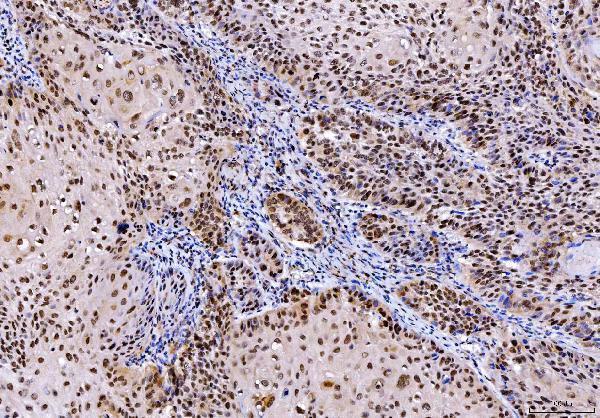
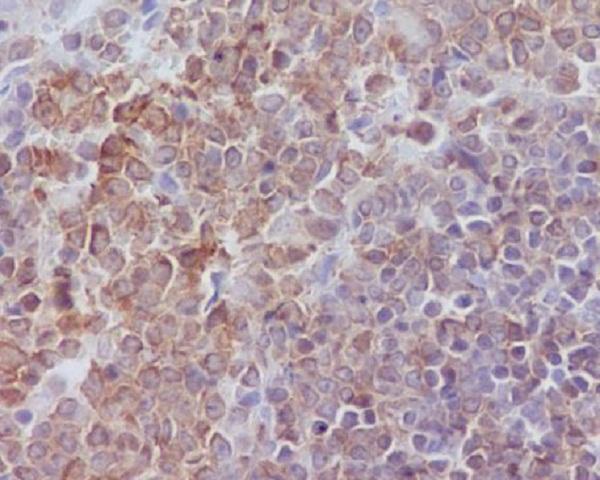
Real IHC data IHC analysis of SHP1/PTPN6 using anti-SHP1/PTPN6 antibody (M00938-2). SHP1/PTPN6 was detected in a paraffin-embedded section of human laryngeal squamous cell carcinomas tissue. Heat mediated antigen retrieval was performed in EDTA buffer (pH 8.0, epitope retrieval solution). The tissue section was blocked with 10% goat serum. The tissue section was then incubated with 2 μg/ml mouse anti-SHP1/PTPN6 Antibody (M00938-2) overnight at 4°C. Peroxidase Conjugated Goat Anti-mouse IgG was used as secondary antibody and incubated for 30 minutes at 37°C. The tissue section was developed using HRP Conjugated Mouse IgG Super Vision Assay Kit (Catalog # SV0001) with DAB as the chromogen.
Anti-SHP1/PTPN6 Antibody ® (monoclonal, 8H11B10)
Cat # M00938-2
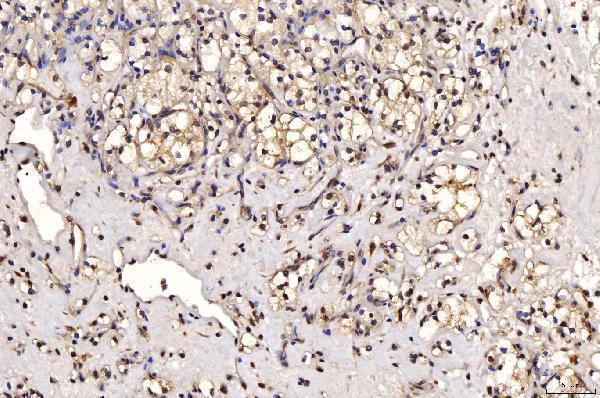
Real IHC data Immunohistochemistry (IHC) analyzes of SHP-1 (phospho-Y536) polyclonal antibody in paraffin-embedded human breast carcinoma tissue at 1:100.
Anti-SHP-1 (phospho-Y536) PTPN6 Antibody
Cat # A00938Y536
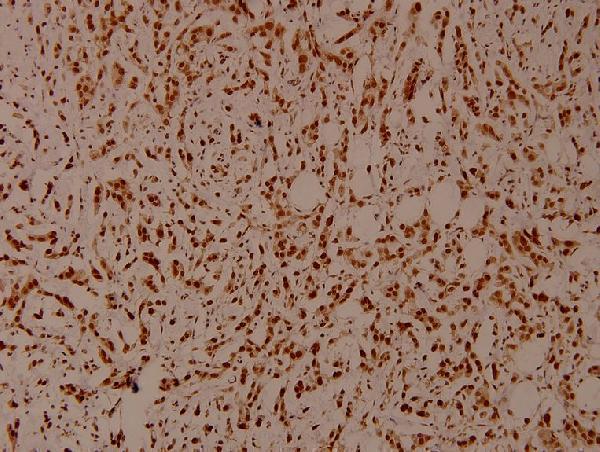
Real IHC data Immunohistochemical analysis of paraffin-embedded rat spleen, using SHP1 Antibody.
Anti-SHP1 PTPN6 Rabbit Monoclonal Antibody
Cat # M00938
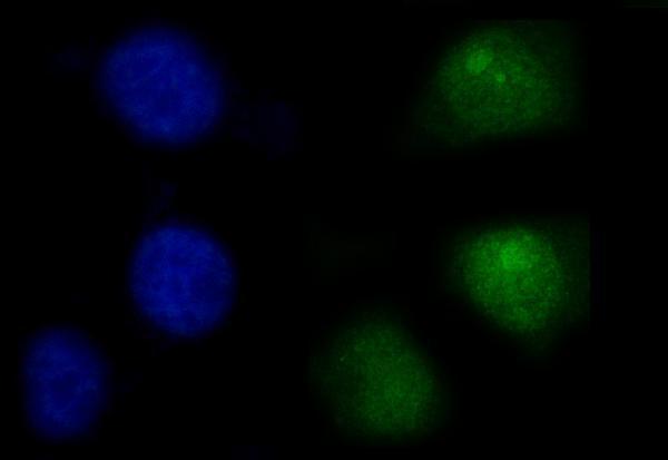
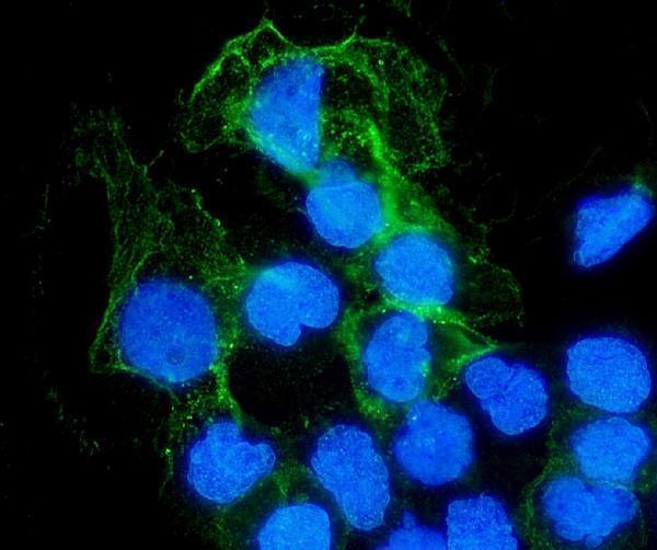
Real IF data IF analysis of SHP1/PTPN6 using anti-SHP1/PTPN6 antibody (A00938-3). SHP1/PTPN6 was detected in immunocytochemical section of A431 cells. Enzyme antigen retrieval was performed using IHC enzyme antigen retrieval reagent (AR0022) for 15 mins. The cells were blocked with 10% goat serum. And then incubated with 5μg/mL rabbit anti-SHP1/PTPN6 Antibody (A00938-3) overnight at 4°C. DyLight®488 Conjugated Goat Anti-Rabbit IgG (BA1127) was used as secondary antibody at 1:100 dilution and incubated for 30 minutes at 37°C. The section was counterstained with DAPI. Visualize using a fluorescence microscope and filter sets appropriate for the label used.
Anti-SHP1/PTPN6 Antibody ®
Cat # A00938-3

M00938-2 shows IHC in human laryngeal squamous cell carcinoma, A00938Y536 shows phospho-Y536 IHC in human breast carcinoma, and M00938 shows IHC in rat spleen (each SKU’s IHC image caption). A00938-3 shows IF/ICC in A431 cells (A00938-3 IF image caption).

Which to pick: For tissue IHC, choose M00938-2 for its documented paraffin-section protocol using 2 μg/mL primary antibody; its caption does not report the fixative (M00938-2 IHC image caption). Choose A00938-3 for IF/ICC at 5 μg/mL in the pictured cell preparation (A00938-3 IF image caption). For a human, mouse, or rat project, M00938-2 lists all three species and IHC among its applications, but its pictured IHC evidence is human tissue only (M00938-2 catalog reactivity and applications; M00938-2 IHC image caption).

Each figure is that product's own IHC / IF validation image from its datasheet.

References

  1. UniProt Consortium. UniProt entry P29350 (PTN6_HUMAN, Tyrosine-protein phosphatase non-receptor type 6).
  2. Human Protein Atlas. PTPN6 tissue IHC expression (reliability: Enhanced).
  3. Human Protein Atlas. PTPN6 subcellular location (ICC-IF): Mainly localized to the nucleoplasm. In addition localized to the nucleoli..
  4. Human Protein Atlas. PTPN6 antibody validation summary (3 antibodies).
  5. Epigenetic mechanisms of protein tyrosine phosphatase 6 suppression in diffuse large B-cell lymphoma: implications for epigenetic therapy. Leukemia 2014 — PMC4036221.
  6. Pan-cancer analysis of PTPN6: prognostic significance and functional implications in tumor progression. Discover oncology 2025 — PMC12378822.
  7. PTPN6-EGFR Protein Complex: A Novel Target for Colon Cancer Metastasis. Journal of oncology 2022 — PMC8856819.
  8. Multi-omic analyses reveal PTPN6's impact on tumor immunity across various cancers. Scientific reports 2025 — PMC11958644.
  9. PubMed PMID:1732748 — UniProt-cited evidence.
  10. PubMed PMID:1652101 — UniProt-cited evidence.
  11. PubMed PMID:1736296 — UniProt-cited evidence.