This website uses cookies to ensure you get the best experience on our website.


- Table of Contents
Real validated RETN Western blot protocols, expected-band and isoform facts, troubleshooting for weak or shifted signal, and recommended anti-RETN WB antibodies. Everything you need to plan the experiment before you commit precious samples.
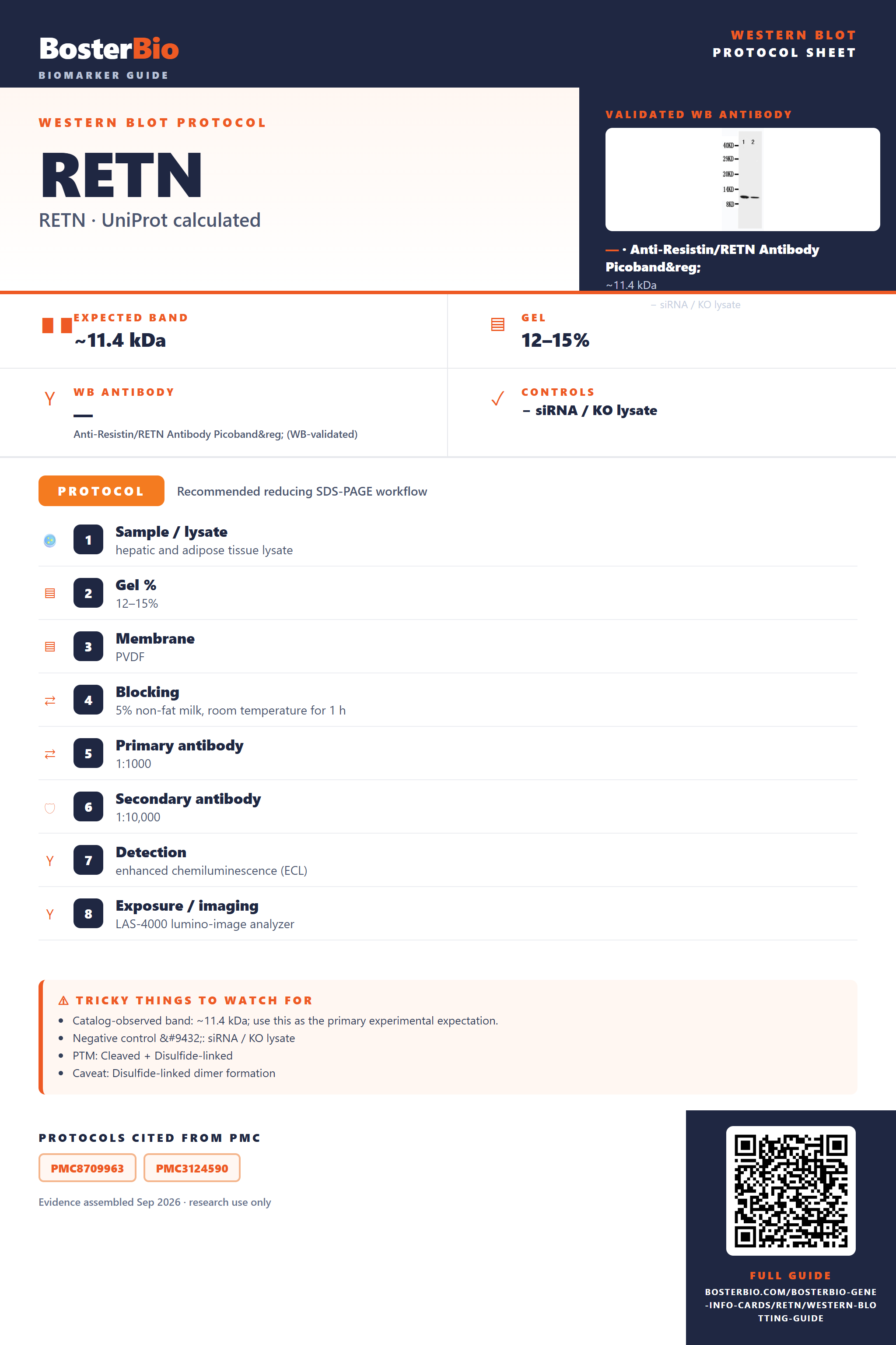
Expected bands, validated protocols, controls and antibodies — the at-a-glance facts below, then the full design guide.
| Expected band | ~11.4 kDa | |
| Observed band | ~11 kDa | |
| Gel | 12–15% | |
| Negative control | siRNA / KO lysate |
| PTM | Cleaved + Disulfide-linked | |
| Caveat | Disulfide-linked dimer formation | |
| Regulation | Adipogenesis | |
| Isoform | 2 isoform(s) |
Literature-validated Western blot parameters for RETN — gel percentage, transfer, blocking, antibody incubation and detection, extracted from published methods.
| Sample / lysate | hepatic and adipose tissue lysate |
| Gel % | 12–15% |
| Membrane | PVDF |
| Blocking | 5% non-fat milk, room temperature for 1 h |
| Primary antibody | 1:1000 |
| Secondary antibody | 1:10,000 |
| Detection | enhanced chemiluminescence (ECL) |
| Exposure / imaging | LAS-4000 lumino-image analyzer |
Resistin's 11.4 kDa precursor runs as the mature, signal-cleaved monomer at the empirically observed ~11 kDa, with a disulfide-linked ~22 kDa homodimer possible if incompletely reduced.
| single sharp band at ~11 kDa | matches the mature, signal-cleaved resistin monomer, consistent with the empirically observed ~11 kDa band and the 11.4 kDa predicted mass |
| band near ~22 kDa, roughly double the monomer | disulfide-linked homodimer formed via the inter-chain Cys22-Cys22 bond, seen when reduction is incomplete or samples are run non-reducing |
| band slightly smaller than the 11.4 kDa full-length precursor | loss of the 18-residue signal peptide upon secretion yields a mature protein lighter than the unprocessed precursor |
| little or no band in whole-cell lysate | resistin is a secreted protein, so most of the mature protein leaves the cell rather than accumulating intracellularly |
| two closely spaced bands or a faint doublet | the two annotated splice isoforms (1 and 2) can differ slightly in length and migrate as separate species |
| Predicted mass (UniProt, 11.4 kDa) | sets the baseline monomer size, matching the ~11 kDa band typically observed |
| Signal peptide cleavage (residues 1-18) | removal during secretion yields a mature protein modestly smaller than the 11.4 kDa full-length precursor |
| Inter-chain disulfide bond (Cys22) forming a homodimer | produces a band at roughly double the monomer mass (~22 kDa) unless samples are fully reduced |
| Intrachain disulfide bonds (Cys51-104, Cys63-103, Cys72-89, Cys74-91, Cys78-93) | stabilize a compact C-terminal domain that can shift mobility slightly if these bonds are not fully reduced |
| Alternative splicing (isoforms 1 and 2) | named isoforms may differ in length, potentially yielding an extra or shifted band relative to the canonical monomer |
| Secreted subcellular localization | protein is exported from cells, so whole-cell lysates show weaker signal than conditioned media, serum, or plasma |
| Situation | Likely cause | Next action |
|---|---|---|
| No band in lysate | resistin is secreted and largely exits the cell rather than accumulating intracellularly | probe conditioned media, serum, plasma, or extracellular fractions instead of relying solely on whole-cell lysate |
| Band higher than expected | incomplete reduction leaves the inter-chain Cys22 disulfide intact, preserving the ~22 kDa homodimer | increase reducing agent concentration and boiling time to fully dissociate the homodimer into the ~11 kDa monomer |
| Band lower than expected | the mature, signal-cleaved protein runs below the 11.4 kDa full-length precursor mass used for reference | compare against mature-form standards rather than the unprocessed precursor mass |
| Multiple bands | the two annotated splice isoforms can co-migrate as distinct species | confirm isoform identity by mass spectrometry or an isoform-specific antibody |
| Weak or no signal | low secreted abundance or degradation of the small, disulfide-stabilized protein outside a stabilizing buffer | use fresh samples with protease inhibitors and gentle handling to preserve the disulfide-stabilized structure before detection |
| Fragments below expected size | disruption of the stabilizing disulfide bonds can expose the small protein to proteolytic cleavage | add protease inhibitors and avoid premature reduction or denaturation prior to the intended electrophoresis step |
Comprehensive Human Protein Atlas IHC scoring per tissue. Rows are taken directly from the HPA tissue chart — click any row's HPA link to view the source.
| Tissue | Cell type | Level | Evidence | Source |
|---|
| Tissue | Cell type | Level | Evidence | Source |
|---|
Deeper troubleshooting and optimisation questions for RETN, answered from its protein features.
BosterBio's RETN antibodies are among the best-performing WB antibodies on the market — well cited, thoroughly validated, and orthogonally cross-validated against negative tissues and complementary methods.
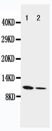

These Boster anti-Resistin antibodies are top-performing, widely cited Western blot reagents, thoroughly validated with recombinant protein standards and cross-validated against negative-tissue controls and complementary detection methods for confident, reproducible RETN detection.
Which to pick: Both PA1473 and PB9412 include real Western blot validation images. PA1473 was tested against recombinant mouse Resistin protein at two loading amounts; PB9412 shows a full SDS-PAGE western blot analysis of Resistin. Either is a solid choice from this list.