This website uses cookies to ensure you get the best experience on our website.


- Table of Contents
Plan chromogenic RIMS2 IHC in paraffin sections using adrenal zona fasciculata as a high-staining reference (HPA tissue IHC). Assess cytoplasmic adrenal staining (HPA tissue IHC) while accounting for reported off-target binding and splice or transcript discrepancies (HPA tissue IHC).

Expected localisation, validated protocols, controls and antibodies — the at-a-glance facts below, then the full design guide.
| Expected localisation | Adrenal cortical cytoplasm (HPA tissue IHC); presynaptic membrane (UniProt) | |
| Staining pattern | Zona fasciculata cells: strong cytoplasmic staining (HPA tissue IHC) | |
| Antigen retrieval | Citrate pH 6.0 HIER, 95–98 °C, 20 min (rule: cytoplasmic / membrane antigen) | |
| Positive control | Adrenal gland+2 more · see all | |
| Negative control | Adipose tissue+4 more · see all |
| Fixation | Keep fixation consistent across sections. (standard IHC practice; not target-specific) | |
| Caveat | Presumed off-target binding; assess staining specificity (HPA tissue IHC) | |
| Regulation | Fetal brain predominance (UniProt) | |
| Isoform / epitope | 8 isoforms; no transmembrane segment; map the epitope (UniProt) |
The catalog antibody’s datasheet-derived IHC-P protocol is paired with 1 published human-tissue RIMS2 IHC protocol (PMC7273530).
| Sample | Tissue sections; selected-image fixative not specified (standard IHC workflow) |
| Fixation | Image fixative and duration unreported (datasheet A09915); verify before use. |
| Sectioning | 4–5 µm sections on charged slides (standard) |
| Deparaffinisation | Xylene, graded ethanol series to water (standard) |
| Antigen retrieval | Heat-induced epitope retrieval in citrate buffer, pH 6.0, 20 min at 95–98 °C (standard rule: cytoplasmic / membrane antigen) |
| Peroxidase block | 3% H2O2, 10 min, room temperature (standard) |
| Blocking | 10% normal serum of the secondary host, 30 min, room temperature (standard) |
| Primary antibody | Rabbit anti-RIMS2, 5 μg/mL (datasheet A09915) |
| Primary incubation | Overnight at 4 °C (standard) |
| Detection | HRP-polymer secondary, DAB chromogen 5–10 min (standard) |
| Counterstain | Hematoxylin, blue, dehydrate and mount (standard) |
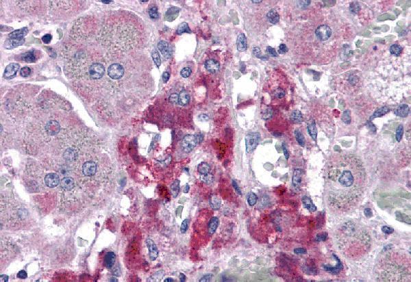
| Expected result | RIMS2-positive staining in cells in zona fasciculata of adrenal gland (HPA tissue IHC: High). HPA tissue profile: Cytoplasmic expression in adrenal cortex. No signal in the no-primary control. |
RIMS2 is associated with synapses and the presynaptic cell membrane, with no transmembrane segment (UniProt Q9UQ26 topology). In tissue IHC, expect cytoplasmic staining in adrenal zona fasciculata cells, with staining also reported in ciliated cell bodies (HPA tissue IHC). HPA rates the tissue profile Enhanced, while noting presumed off-target staining and a splice or transcript discrepancy (HPA tissue IHC).
| Strong cytoplasmic staining in adrenal zona fasciculata cells. | This matches HPA’s reported high staining and adrenal cortex cytoplasmic profile (HPA tissue IHC). Judge the result by both cell identity and compartment. |
| Predominantly nuclear staining, or staining confined to extracellular material. | Neither is a reported RIMS2 location (UniProt Q9UQ26; HPA subcellular). Treat it as suspect; check background and staining controls before scoring it as positive (general IHC practice). |
| Strong staining in an HPA-listed negative cell population. | Possible causes include cross-reactivity or endogenous detection activity (general IHC practice). Compare the specific cell type, since an HPA negative call does not describe every cell in that tissue (HPA tissue IHC). |
| Diffuse chromogen across cells and surrounding tissue, without a distinct cellular pattern. | This limits interpretation even if the slide is dark (general IHC practice). Reassess blocking, washes, detection controls and counterstain before assigning RIMS2 staining. |
| No staining in adrenal zona fasciculata cells. | The expected high-staining reference population is absent (HPA tissue IHC). Check section quality, primary antibody conditions and detection with a known-positive section (general IHC practice). |
| Reference tissue and cell type | HPA reports high staining in adrenal zona fasciculata and nasopharyngeal ciliated cell bodies, medium in bronchial ciliated cell bodies, and low in fallopian tube ciliated cell bodies (HPA tissue IHC). Compare like cells when assessing intensity. |
| Compartment | The adrenal IHC profile is cytoplasmic (HPA tissue IHC). UniProt places RIMS2 at synapses and the presynaptic cell membrane (UniProt Q9UQ26); those annotations do not require every positive paraffin section to show a crisp membrane rim. |
| Antibody evidence | HPA066498 has Enhanced IHC validation; CAB079049 is Supported for IHC (HPA antibodies). HPA also flags presumed off-target staining and a splice or transcript discrepancy, so resolve ambiguous patterns against cell-specific controls (HPA tissue IHC). |
| Isoforms and epitope coverage | UniProt lists 8 RIMS2 isoforms (UniProt Q9UQ26). Epitope coverage for these antibodies is not supplied here; do not treat a negative cell population as proof that every RIMS2 isoform is absent. |
| IF/ICC: what location is reported? | HPA reports approved plasma membrane and cytosol localization in ICC-IF, with images in HeLa, RT-4 and U2OS cells (HPA subcellular). Its ICC-approved antibody is HPA046538; that approval does not establish IHC performance (HPA antibodies). |
| Situation | Likely cause | Next action |
|---|---|---|
| Adrenal reference section has no zona fasciculata signal. | The section misses an HPA high-staining cell population, or the staining run failed (HPA tissue IHC; general IHC practice). | Confirm cell identity and section quality; review the primary antibody conditions and a positive detection control (general IHC practice). |
| A suspected negative control stains strongly. | HPA negative calls are cell-specific; alternatively, nonspecific binding or endogenous detection activity may contribute (HPA tissue IHC; general IHC practice). | Check the named cell population and include an appropriate primary-omission or detection control (general IHC practice). |
| Cerebellar granular layer is negative despite reported cerebellar RIMS2. | HPA reports granular-layer cells as not detected, while UniProt reports protein in Purkinje cells (HPA tissue IHC; UniProt Q9UQ26). | Score the identified cell populations separately; do not use granular-layer negativity to dismiss a Purkinje-cell result. |
| Signal is mainly nuclear. | A nuclear pattern lacks support from the supplied localization records (UniProt Q9UQ26; HPA subcellular). | Review controls and compartment boundaries, then repeat with an IHC-validated antibody if the pattern persists (HPA antibodies; general IHC practice). |
| Chromogen appears broadly diffuse. | Background may reflect nonspecific binding, incomplete washing or detection activity (general IHC practice). | Review blocking, washes and detection controls; score RIMS2 only where cell and compartment remain interpretable (general IHC practice). |
| Two antibodies give different cell patterns. | HPA flags presumed off-target binding and a splice or transcript discrepancy; its IHC antibody validation levels also differ (HPA tissue IHC; HPA antibodies). | Compare staining in the same named cells and reference section, document each antibody separately, and avoid combining discordant scores (general IHC practice). |


Comprehensive Human Protein Atlas IHC scoring per tissue (reliability: Enhanced — Medium consistency between antibody staining and RNA expression data. Presumed off target binding observed and disregarded. Caution, Splice and/or transcript discrepancy exists.). Rows are taken directly from the HPA tissue chart — click any row's HPA link to view the source.
| Tissue | Cell type | Level | Evidence | Source |
|---|---|---|---|---|
| Adrenal gland | Cells in zona fasciculata | High | Protein (IHC) | HPA → |
| Nasopharynx | Ciliated cells (cell body) | High | Protein (IHC) | HPA → |
| Bronchus | Ciliated cells (cell body) | Medium | Protein (IHC) | HPA → |
| Tissue | Cell type | Level | Evidence | Source |
|---|---|---|---|---|
| Adipose tissue | Adipocytes | Not detected | Protein (IHC) | HPA → |
| Appendix | Glandular cells | Not detected | Protein (IHC) | HPA → |
| Bone marrow | Hematopoietic cells | Not detected | Protein (IHC) | HPA → |
| Breast | Adipocytes | Not detected | Protein (IHC) | HPA → |
| Caudate | Glial cells | Not detected | Protein (IHC) | HPA → |
Troubleshoot RIMS2 staining in paraffin sections by comparing cellular pattern, compartment, and controls with the available IHC evidence.
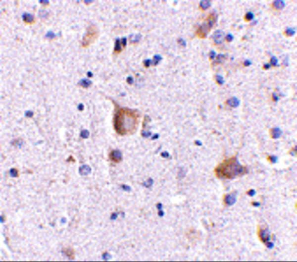
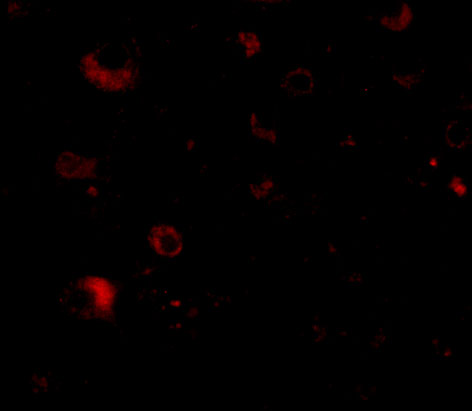
Two anti-RIMS2 antibodies have human tissue IHC images: adrenal gland for A09915 and brain for A09915-1; A09915-1 also has a human brain IF image (catalog image captions).
A09915 has a human adrenal gland IHC image at 5 μg/mL (A09915 IHC image caption). A09915-1 has human brain IHC and IF images at 5 μg/mL and 20 μg/mL, respectively (A09915-1 image captions).
Which to pick: For human paraffin section IHC, either SKU lists IHC-P; choose A09915 for the adrenal gland image or A09915-1 for the brain image (catalog applications; each SKU’s IHC image caption). Choose A09915-1 for tissue IF or work involving mouse or rat because it lists IF and those species; ICC is not listed (A09915-1 applications and reactivity). A09915 is described as polyclonal, while A09915-1 has no clone designation; neither IHC caption reports a fixative (A09915 dilution text; A09915-1 clone field; IHC image captions).