This website uses cookies to ensure you get the best experience on our website.


- Table of Contents
Plan chromogenic SCARB1 IHC in paraffin sections with fixation, retrieval, and staining controls. Compare the expected membranous pattern with staining in adrenal zona fasciculata and testis Leydig cells (HPA tissue IHC).

Expected localisation, validated protocols, controls and antibodies — the at-a-glance facts below, then the full design guide.
| Expected localisation | Membranous in a subset of tissues (HPA tissue IHC) | |
| Staining pattern | Membranous in adrenal zona fasciculata and testis Leydig cells (HPA tissue IHC) | |
| Antigen retrieval | Citrate pH 6.0 HIER, 95–98 °C, 20 min (rule: cytoplasmic / membrane antigen) | |
| Positive control | Adrenal gland+4 more · see all | |
| Negative control | Adipose tissue+4 more · see all |
| Fixation | Keep fixation consistent across sections (standard IHC practice; not target-specific) | |
| Caveat | Presumed off-target GI staining can mislead (HPA tissue IHC) | |
| Regulation | No specific expression regulator given (UniProt) | |
| Isoform / epitope | 5 isoforms; verify extracellular vs cytoplasmic epitope (UniProt) |
The catalog antibody’s IHC-P protocol is followed by three published SCARB1 chromogenic IHC protocols (PMC8741905; PMC12010815; PMC9907373).
| Sample | Tissue sections; selected-image fixative not specified (standard IHC workflow) |
| Fixation | Image fixative and duration unreported (datasheet A01093); verify before use. |
| Sectioning | 4–5 µm sections on charged slides (standard) |
| Deparaffinisation | Xylene, graded ethanol series to water (standard) |
| Antigen retrieval | Heat-induced epitope retrieval in citrate buffer, pH 6.0, 20 min at 95–98 °C (standard rule: cytoplasmic / membrane antigen) |
| Peroxidase block | 3% H2O2, 10 min, room temperature (standard) |
| Blocking | 10% normal serum of the secondary host, 30 min, room temperature (standard) |
| Primary antibody | Rabbit anti-SCARB1, 2.5 μg/mL (datasheet A01093) |
| Primary incubation | Overnight at 4 °C (standard) |
| Detection | HRP-polymer secondary, DAB chromogen 5–10 min (standard) |
| Counterstain | Hematoxylin, blue, dehydrate and mount (standard) |
| Expected result | SCARB1-positive staining in cells in zona fasciculata of adrenal gland (HPA tissue IHC: High). HPA tissue profile: Membranous expression in a subset of tissues, including adrenal gland, Leydig cells, sinusoids in liver, vessels in placenta and salivary ducts. No signal in the no-primary control. |
SCARB1 is a membrane receptor with two transmembrane segments and a large extracellular region (UniProt Q8WTV0 topology). Expect chiefly membranous staining in selected tissues, especially adrenal zona fasciculata cells and testicular Leydig cells (HPA: High in both). HPA rates tissue IHC reliability Enhanced, while noting medium consistency with RNA expression and presumed off-target binding in the GI tract (HPA tissue IHC).
| Distinct membranous staining in adrenal zona fasciculata cells or Leydig cells. | This matches two HPA High cell populations and the reported membranous tissue pattern (HPA tissue IHC). Compare the stained cells with tissue morphology when scoring (general IHC practice). |
| Strong staining confined to nuclei, with no corresponding membranous pattern. | An exclusively nuclear result does not match SCARB1's reported cell-membrane location (UniProt Q8WTV0) or HPA tissue IHC. Review the negative control and antibody specificity (general IHC practice). |
| Strong staining in duodenal glandular cells or colonic endothelial cells. | HPA reports these cells as Not detected and flags presumed off-target GI binding (HPA tissue IHC). Treat this pattern cautiously; compare it with an HPA High cell population and appropriate controls. |
| Diffuse chromogen across cells and surrounding tissue, obscuring cell borders. | This is difficult to score as the reported membranous pattern (HPA tissue IHC). Background can arise from nonspecific antibody binding or detection activity; inspect a negative control (general IHC practice). |
| No staining in adrenal zona fasciculata cells or Leydig cells. | These are HPA High populations, so a blank result needs a technical check (HPA tissue IHC). Verify tissue identity, controls, retrieval, primary-antibody incubation and detection (general IHC practice). |
| Membrane topology and epitope location (UniProt Q8WTV0 topology). | SCARB1 has extracellular residues 33–443 and cytoplasmic ends (UniProt Q8WTV0 topology). Interpret staining against the antibody's stated epitope, if available; this record does not locate that epitope. |
| Uneven expression across tissues (HPA tissue IHC). | HPA reports High staining in adrenal zona fasciculata cells and Leydig cells, Medium in kidney proximal-tubule cell bodies and placental trophoblasts, and Not detected in several listed populations (HPA tissue IHC). |
| Antibody validation and GI caveat (HPA antibodies; HPA tissue IHC). | Two listed antibodies have Enhanced IHC validation, yet HPA notes medium consistency with RNA expression and disregarded presumed off-target GI staining (HPA tissue IHC). Assess unexpected GI signal against this caveat. |
| IF/ICC Q: Should an IF vesicle signal be scored as the expected IHC pattern? | A: HPA reports vesicles as the main approved ICC-IF location, with additional lysosome and rods-and-rings locations; tissue IHC is described as membranous (HPA subcellular; HPA tissue IHC). Interpret each application in its own context. |
| Target-specific fixation sensitivity. | The supplied UniProt and HPA records do not report a SCARB1-specific fixation effect. Evaluate retrieval and processing with routine IHC controls without assigning a SCARB1-specific cause (general IHC practice). |
| Situation | Likely cause | Next action |
|---|---|---|
| Expected-positive adrenal or testis section is blank (HPA: High). | The result conflicts with HPA High staining; the cause is unresolved by these records (HPA tissue IHC). | Confirm the expected cell population, then check section quality, retrieval, primary-antibody step and detection controls (general IHC practice). |
| Membrane staining is weak in an HPA High population. | A weak result differs from HPA's High category, but HPA does not identify the cause in this specimen (HPA tissue IHC). | Compare a matched positive control and review retrieval, antibody incubation and detection settings (general IHC practice). |
| Only nuclear staining appears. | This conflicts with the reported membrane location (UniProt Q8WTV0; HPA tissue IHC). | Check the negative control, examine the correct cell population and reassess antibody specificity before scoring positive (general IHC practice). |
| Strong GI staining appears in cells listed as Not detected. | HPA notes presumed off-target GI binding and lists duodenal glandular cells as Not detected (HPA tissue IHC). | Record the cell type and compare negative controls and an HPA High tissue; do not assign that GI signal to SCARB1 without corroboration. |
| Chromogen covers the section diffusely. | Diffuse background obscures the cell-specific, membranous pattern reported by HPA (HPA tissue IHC). | Inspect a no-primary control and review blocking, washes, antibody concentration and chromogen development (general IHC practice). |
| Placental or kidney staining seems weaker than adrenal staining. | HPA rates placental trophoblasts and kidney proximal-tubule cell bodies Medium, versus High in adrenal zona fasciculata cells (HPA tissue IHC). | Identify the scored cell population first; compare like tissues and controls before treating the intensity difference as a failed run (general IHC practice). |
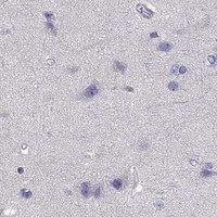


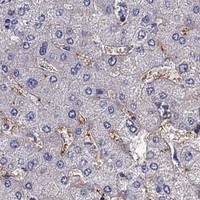


Comprehensive Human Protein Atlas IHC scoring per tissue (reliability: Enhanced — Medium consistency between antibody staining and RNA expression data. Presumed off target binding in GI tract observed and disregarded.). Rows are taken directly from the HPA tissue chart — click any row's HPA link to view the source.
| Tissue | Cell type | Level | Evidence | Source |
|---|---|---|---|---|
| Adrenal gland | Cells in zona fasciculata | High | Protein (IHC) | HPA → |
| Bronchus | Ciliated cells (cilia axoneme) | High | Protein (IHC) | HPA → |
| Fallopian tube | Ciliated cells (cilia axoneme) | High | Protein (IHC) | HPA → |
| Testis | Leydig cells | High | Protein (IHC) | HPA → |
| Kidney | Proximal tubules (cell body) | Medium | Protein (IHC) | HPA → |
| Tissue | Cell type | Level | Evidence | Source |
|---|---|---|---|---|
| Adipose tissue | Adipocytes | Not detected | Protein (IHC) | HPA → |
| Appendix | Glandular cells | Not detected | Protein (IHC) | HPA → |
| Bone marrow | Hematopoietic cells | Not detected | Protein (IHC) | HPA → |
| Breast | Adipocytes | Not detected | Protein (IHC) | HPA → |
| Cerebellum | Cells in granular layer | Not detected | Protein (IHC) | HPA → |
Troubleshoot SCARB1 chromogenic IHC in paraffin sections using matched controls, expected cell types and compartment patterns; IF considerations appear in one separate entry.
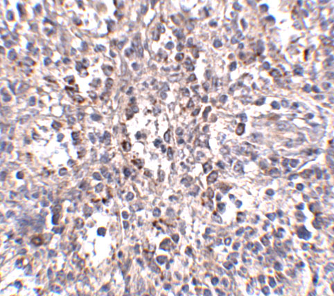
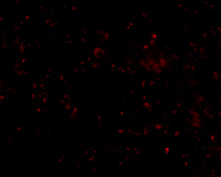
Two anti-SCARB1 antibodies have human tissue IHC images; one also has a human spleen IF image (catalog image captions). Both list Human, Mouse and Rat reactivity (catalog applications/reactivity).
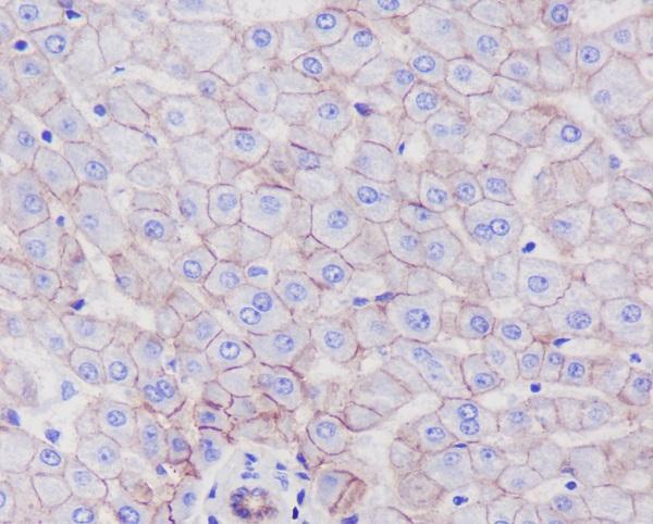
A01093 has IHC and IF images from human spleen tissue (A01093 image captions). M01093 has an IHC image from paraffin-embedded human liver and lists ICC/IF among its applications (M01093 image caption; catalog applications).
Which to pick: For tissue IHC, choose A01093 for the demonstrated human spleen sample at 2.5 μg/mL (A01093 IHC image caption), or M01093 for the demonstrated paraffin-embedded human liver sample at 1:50–1:200; the latter caption does not report the fixative (M01093 IHC image caption; catalog dilution). For IF, A01093 has a human spleen image at 20 μg/mL; for ICC/IF, M01093 lists both applications and a 1:50–1:200 dilution, but has no IF image in the payload (A01093 IF image caption; M01093 catalog applications/dilution/image captions). Both list Human, Mouse and Rat reactivity; M01093 is monoclonal, while A01093 has no clone listed, and the supplied tissue images show only human samples (catalog reactivity/clone; image captions).