This website uses cookies to ensure you get the best experience on our website.


- Table of Contents
Use the IHC-validated antibody at 0.5–1 µg/mL for paraffin sections (datasheet A06559-1). This guide helps interpret granular cytoplasmic and additional plasma staining using glandular and glial cells as reference patterns (HPA tissue IHC).

Expected localisation, validated protocols, controls and antibodies — the at-a-glance facts below, then the full design guide.
| Expected localisation | Granular cytoplasm; additional plasma staining (HPA tissue IHC) | |
| Staining pattern | Granular cytoplasm in glandular and glial cells; plasma positivity (HPA tissue IHC) | |
| Antigen retrieval | EDTA pH 8.0 HIER, heat-mediated (datasheet A06559-1) | |
| Positive control | Adrenal gland+4 more · see all | |
| Negative control | None in HPA (detected in all 44 tissues); use no-primary + isotype controls |
| Fixation | Keep fixation consistent across paraffin sections. (standard IHC practice; not target-specific) | |
| Caveat | Secreted variants may uncouple tissue RNA and protein staining (HPA tissue IHC) | |
| Regulation | Enhanced RNA in adrenal gland and brain (HPA tissue RNA) | |
| Isoform / epitope | 2 isoforms; mature chain 25–749 lacks a cytoplasmic domain (UniProt) |
The catalog antibody’s IHC-P protocol (datasheet: A06559-1) is followed by three published SEMA3B IHC protocols (PMC6598462; PMC8864000; PMC11371361).
| Sample | Paraffin-embedded human mammary cancer tissue; fixative not specified (datasheet A06559-1) |
| Fixation | Image fixative and duration unreported (datasheet A06559-1); verify before use. |
| Sectioning | 4–5 µm sections on charged slides (standard) |
| Deparaffinisation | Xylene, graded ethanol series to water (standard) |
| Antigen retrieval | Heat retrieval: EDTA pH 8.0 (datasheet A06559-1); 20 min, 95–100 °C (standard) |
| Peroxidase block | 3% H2O2, 10 min, room temperature (standard) |
| Blocking | 10% goat serum (datasheet A06559-1) |
| Primary antibody | Rabbit anti-SEMA3B, 0.5-1μg/ml (datasheet A06559-1) |
| Primary incubation | Overnight at 4 °C (datasheet A06559-1) |
| Detection | Streptavidin-biotin complex (SABC), DAB chromogen (datasheet A06559-1) |
| Counterstain | Hematoxylin, blue, dehydrate and mount (standard) |
| Expected result | SEMA3B-positive staining in glandular cells of adrenal gland (HPA tissue IHC: High). HPA tissue profile: General cytoplasmic expression with a granular pattern. Additional plasma positivity. No signal in the no-primary control. |
SEMA3B is secreted and can accumulate in the endoplasmic reticulum; it has no transmembrane segment (UniProt Q13214). In tissue sections, expect granular cytoplasmic staining, with additional plasma positivity (HPA tissue IHC). HPA reports high staining in several glandular and neural cell populations, but rates its tissue IHC evidence Uncertain because secretion complicates comparison of RNA and protein locations (HPA tissue IHC).
| Granular cytoplasmic staining in adrenal or duodenal glandular cells. | This fits the reported general pattern and High staining in those cells (HPA tissue IHC). Judge the signal against nearby tissue and the assay controls (standard IHC practice). |
| Plasma staining accompanies cellular staining. | Additional plasma positivity is reported (HPA tissue IHC). Secreted SEMA3B makes extracellular signal plausible (UniProt Q13214); plasma staining alone does not identify its producing cell. |
| Strong nuclear or sharply membrane restricted staining dominates. | That differs from HPA's granular cytoplasmic pattern and UniProt's secreted, nontransmembrane annotation (HPA tissue IHC; UniProt Q13214). Check for artefact before scoring it as SEMA3B. |
| Strong signal appears mainly in a cell type outside the reported pattern. | Possible causes include cross reactivity or endogenous detection activity (standard IHC practice). HPA lists no negative tissue, so an unlisted cell type alone cannot establish a false positive (HPA tissue IHC). |
| A reported High tissue has no specific staining. | This conflicts with the reported result but does not prove biological absence (HPA tissue IHC). Check tissue identity, positive control and detection performance (standard IHC practice). |
| Cellular versus extracellular location | SEMA3B is secreted and accumulates in the endoplasmic reticulum (UniProt Q13214). HPA reports granular cytoplasm plus plasma positivity; extracellular signal need not mark the source cell (HPA tissue IHC). |
| Choice of comparison tissue | HPA reports High staining in adrenal and duodenal glandular cells, cortical glia and cardiomyocytes, among others; ovarian stroma is Low, not negative (HPA tissue IHC). |
| Strength of the tissue evidence | HPA rates tissue IHC Uncertain and HPA030651 IHC Uncertain (HPA tissue IHC; HPA antibodies). Treat a matching pattern as supportive rather than definitive identification. |
| Processing and isoforms | The signal peptide spans residues 1–24; the annotated chain spans 25–749, with 2 isoforms (UniProt Q13214). Epitope location is unspecified, so isoform recognition cannot be inferred. |
| Glycosylation | UniProt lists glycosylation sites at 82, 124 and 427 (UniProt Q13214). Their effect on this antibody's staining or antigen retrieval is unreported. |
| IF/ICC Q: Is the same pattern established in cultured cells? | A: HPA calls SEMA3B secreted but lists no ICC-IF image cell lines or main location; HPA030651 has no ICC validation entry (HPA subcellular; HPA antibodies). An IF pattern remains unverified here. |
| Situation | Likely cause | Next action |
|---|---|---|
| Reported High tissue is blank. | Tissue mix-up or a failed staining run is possible (standard IHC practice); a blank slide alone cannot overturn HPA's reported High staining (HPA tissue IHC). | Verify the tissue and review the run's positive control, antibody application and detection steps before interpreting SEMA3B as absent (standard IHC practice). |
| Most of the section has diffuse brown color. | Nonspecific background or endogenous detection activity may obscure specific staining (standard IHC practice). | Compare a reagent omission control, review blocking and washing, then score only a distinguishable granular cellular pattern (standard IHC practice; HPA tissue IHC). |
| Nuclei are the strongest stained compartment. | This conflicts with the reported granular cytoplasmic pattern and secreted annotation (HPA tissue IHC; UniProt Q13214). | Inspect controls and counterstain, then reassess whether the nuclear color is specific before assigning a positive score (standard IHC practice). |
| A vessel or plasma rich area stains without clear cellular signal. | Additional plasma positivity is reported for secreted SEMA3B (HPA tissue IHC; UniProt Q13214). It cannot localize production to adjacent cells. | Record plasma and cellular signals separately; use the reported cell pattern when assessing cellular positivity (HPA tissue IHC). |
| Unexpected cells stain strongly. | Cross reactivity or endogenous activity is possible (standard IHC practice). HPA supplies no negative tissue designation for a decisive comparison (HPA tissue IHC). | Review morphology and detection controls, and avoid calling the cells SEMA3B positive from location alone (standard IHC practice). |
| Staining is confined to ovarian stroma at low intensity. | HPA reports ovarian stroma as Low and rates its tissue IHC evidence Uncertain (HPA tissue IHC). | Compare a reported High tissue in the same run before treating the weak signal as either a positive control or proof of assay failure (HPA tissue IHC; standard IHC practice). |




Comprehensive Human Protein Atlas IHC scoring per tissue (reliability: Uncertain — At least one protein variant secreted, tissue location of RNA and protein might differ and correlation is complex.). Rows are taken directly from the HPA tissue chart — click any row's HPA link to view the source.
| Tissue | Cell type | Level | Evidence | Source |
|---|---|---|---|---|
| Adrenal gland | Glandular cells | High | Protein (IHC) | HPA → |
| Appendix | Glandular cells | High | Protein (IHC) | HPA → |
| Caudate | Glial cells | High | Protein (IHC) | HPA → |
| Cerebellum | Cells in molecular layer | High | Protein (IHC) | HPA → |
| Cerebral cortex | Glial cells | High | Protein (IHC) | HPA → |
| Tissue | Cell type | Level | Evidence | Source |
|---|---|---|---|---|
| None in HPA: SEMA3B is detected in all 44 scored tissues. Use a no-primary (secondary-only) and an isotype control instead. | ||||
Use the catalog antibody’s paraffin-section example as the starting point, then assess SEMA3B staining by compartment, cell type and controls.
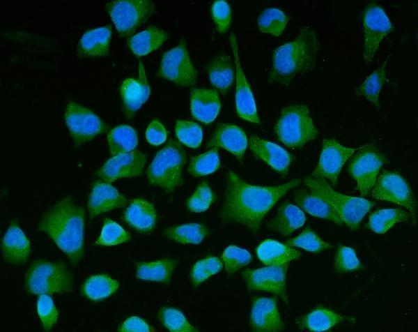
SEMA3B antibodies have IHC data from human cancer and mouse brain paraffin sections (A06559-1 image captions), plus IF/ICC data from human A431 and HeLa cells (A06559-1 and M06559 image captions).
A06559-1 is listed for IHC and IF/ICC in human, mouse and rat (catalog applications and reactivity); its images show paraffin-section IHC in human mammary and colon cancer and mouse brain, plus IF/ICC in A431 cells (A06559-1 image captions). M06559 is listed for IF/ICC, but not IHC, with human, mouse and rat reactivity (catalog applications and reactivity); its IF image shows HeLa cells (M06559 image caption).
Which to pick: Choose rabbit A06559-1 for tissue IHC because its own images document human and mouse paraffin sections; the fixative is unreported (A06559-1 image captions). For IF/ICC, both A06559-1 and mouse monoclonal M06559 have cell images, in A431 and HeLa respectively (catalog host and clone; A06559-1 and M06559 image captions). For cross-species tissue IHC, A06559-1 has human and mouse images and listed rat reactivity, though no rat IHC image is supplied (A06559-1 image captions; catalog reactivity).