SNAP25 / Synaptosomal-associated protein 25 · IHC design guide

Design Immunohistochemistry for SNAP25

Plan paraffin-section SNAP25 IHC around high neuropil staining in cerebellum and cerebral cortex (HPA tissue IHC). Keep fixation consistent and start with the catalog antibody’s 0.5–1 μg/mL IHC range (datasheet A01625).

Evidence assembled Oct 2026 · For research use; verify linked source records and product datasheet before use
Immunohistochemistry protocol sheet for SNAP25 (IHC for SNAP25): expected localisation Neuropil in cerebellum and cerebral cortex (HPA tissue IHC), antibody A01625, validated IHC image, and IHC protocol steps
Printable SNAP25 IHC protocol sheet — expected localisation Neuropil in cerebellum and cerebral cortex (HPA tissue IHC), antibody A01625, controls and protocol steps. Open the full SNAP25 IHC guide →

SNAP25 Immunohistochemistry Experimental Design Guide

Expected localisation, validated protocols, controls and antibodies — the at-a-glance facts below, then the full design guide.

Must know before staining
Expected localisation Neuropil in cerebellum and cerebral cortex (HPA tissue IHC)
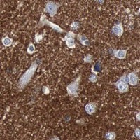
Staining pattern High neuropil staining in cerebellum and cerebral cortex (HPA tissue IHC)
Antigen retrieval Citrate pH 6 HIER, heat-mediated (datasheet A01625)
Positive control ⓘ Cerebellum+1 more · see all
Negative control ⓘ Adipose tissue+4 more · see all
Important caveats
Reasons your staining may differ from the expected pattern.
Fixation Keep fixation conditions consistent across sections. (standard IHC practice; not target-specific)
Caveat Hippocampal and caudate neuronal staining is low (HPA tissue IHC)
Regulation No expression regulator specified (UniProt)
Isoform / epitope Two isoforms; verify antibody epitope coverage (UniProt)
Section 1

Recommended SNAP25 IHC & IF Protocols

Compare the catalog antibody’s IHC-P protocol (datasheet A01625) with three published chromogenic SNAP25 IHC protocols (PMC8502944; PMC11125604; PMC9179158).

Recommended immunohistochemistry (IHC-P) protocol parameters
SampleParaffin-embedded rat brain tissue; fixative not specified (datasheet A01625)
FixationImage fixative and duration unreported (datasheet A01625); verify before use.
Sectioning4–5 µm sections on charged slides (standard)
DeparaffinisationXylene, graded ethanol series to water (standard)
Antigen retrievalHeat retrieval: Citrate pH 6, 20 min (datasheet A01625)
Peroxidase block3% H2O2, 10 min, room temperature (standard)
Blocking10% goat serum (datasheet A01625)
Primary antibodyRabbit anti-SNAP25, 0.5-1μg/ml recommended; image 2μg/ml (datasheet A01625)
Primary incubationOvernight at 4 °C (datasheet A01625)
DetectionStreptavidin-biotin complex (SABC), DAB chromogen (datasheet A01625)
CounterstainHematoxylin, blue, dehydrate and mount (standard)
Expected resultSNAP25-positive staining in molecular layer - neuropil of cerebellum (HPA tissue IHC: High). HPA tissue profile: Distinct expression CNS. No signal in the no-primary control.
💡Decision noteStart with citrate pH 6 heat-induced retrieval (datasheet A01625); the published protocols specify heat-induced retrieval but do not give a retrieval buffer (PMC8502944; PMC11125604; PMC9179158).
Section 2

What Is the Expected SNAP25 Staining Pattern?

SNAP25 is a t-SNARE associated with the cell membrane and synaptosomes; its membrane association requires palmitoylation, and it has no transmembrane segment (UniProt P60880). In paraffin-section IHC, expect the strongest signal in CNS neuropil, especially cerebellar molecular layer and cerebral cortex neuropil (HPA: High). HPA rates its tissue staining reliability Enhanced, with high consistency between antibody staining and RNA expression (HPA: Enhanced).

What am I looking at on my slide?
Strong staining outlines the cerebellar molecular layer or fills cerebral cortex neuropil.This matches the reported high SNAP25 signal in those neuropil compartments (HPA: High). Assess the neuropil pattern separately from neuronal cell bodies: HPA reports low staining in hippocampal and caudate neuronal cells (HPA: Low). A weak soma signal therefore does not, by itself, overturn a clear positive neuropil result.
The main signal is confined to nuclei or another compartment without a plausible membrane or neuronal-process pattern.Treat this as a localization mismatch requiring review, rather than an expected SNAP25 result: UniProt places SNAP25 at the cell membrane, synaptosome and in cytoplasm with perinuclear concentration (UniProt P60880). Check the counterstain and compare with a known-positive neuropil section before assigning the signal to SNAP25 (general IHC practice).
Adipocytes or glandular cells stain strongly while the expected CNS neuropil pattern is absent.Consider cross-reactivity or endogenous chromogenic activity (general IHC practice). HPA reports no detection in adipose adipocytes and adrenal glandular cells (HPA: Not detected); those findings concern the named cells, not every cell in either tissue. Review the staining distribution and detection controls before treating unexpected positive cells as biological expression.
Brown precipitate is widespread across tissue and obscures cell boundaries or neuropil.Diffuse staining is difficult to score as specific SNAP25 signal (general IHC practice). Compare a section processed without primary antibody, then inspect blocking, washes and chromogen development (general IHC practice). A convincing result should still show the tissue and compartment contrast reported by HPA, including high CNS neuropil signal (HPA: High).
Cerebellar molecular layer and cerebral cortex neuropil show no convincing signal.First treat the run as uninformative: both are reported high-staining positive compartments (HPA: High). Check section integrity, antibody incubation and detection controls, then optimize the IHC workflow if needed (general IHC practice). The supplied sources do not establish SNAP25-specific fixation sensitivity; a negative run alone cannot identify fixation as its cause.
💡Expected SNAP25 appearanceCall a section positive when neuropil in cerebellar molecular layer or cerebral cortex shows strong, spatially coherent signal (HPA: High); isolated nuclear staining or widespread precipitate is a localization or background concern (UniProt P60880; general IHC practice).
How each factor affects the staining
Membrane association and epitope interpretationSNAP25 has no transmembrane segment, and its membrane association requires palmitoylation (UniProt P60880). A membrane-associated pattern is plausible, but the record gives no antibody epitope; topology alone cannot predict how this antibody will stain an individual section.
Tissue and cell comparisonCerebellar molecular layer and cortical neuropil are high, whereas hippocampal and caudate neuronal cells are low (HPA: High; HPA: Low). Use the specified compartments when judging a positive control; do not equate low neuronal-cell staining with failure of the entire brain section.
Antibody validationHPA lists three antibodies with Enhanced IHC staining status: HPA001830, HPA071013 and CAB000360 (HPA: Enhanced). Enhanced reflects agreement between antibodies or orthogonal data (HPA: IHC validation); it supports the reported tissue pattern but does not validate every new antibody or staining run.
Isoforms and modificationsUniProt lists two SNAP25 isoforms and phosphorylation sites at residues 138, 154 and 187 (UniProt P60880). The supplied records do not map the IHC antibody epitope or show that isoform choice or phosphorylation changes the observed staining; interpret such effects only with antibody-specific evidence.
IF/ICC Q: Where should SNAP25 appear?A: HPA reports mainly plasma-membrane signal, with additional vesicle localization in ICC-IF (HPA: approved plasma membrane and vesicles). This answers the localization question for IF/ICC; the reported cell-line images do not replace the paraffin-section IHC tissue pattern (HPA: ICC-IF; HPA: tissue IHC).
Why is my staining missing, weak or wrong?
SituationLikely causeNext action
No signal in a known-positive CNS sectionAn absent high-neuropil pattern may reflect an unsuccessful staining run (HPA: High; general IHC practice).Verify section quality, primary-antibody incubation, detection reagents and a positive control; optimize retrieval only as a general IHC variable (general IHC practice). The supplied sources identify no SNAP25-specific retrieval condition.
Signal appears mainly nuclearNuclear-only staining conflicts with the reported membrane, synaptosomal and cytoplasmic locations (UniProt P60880).Check the counterstain, examine a no-primary control and compare neuropil in the same run (general IHC practice; HPA: High). Avoid scoring nuclei as positive SNAP25 solely because they are colored.
Strong staining in HPA-negative cell typesCross-reactivity or endogenous chromogenic activity is possible (general IHC practice); HPA reports no detection in the specified adipocytes and glandular cells (HPA: Not detected).Use a no-primary control and inspect whether the signal follows those specific cells or nonspecific deposits (general IHC practice). Do not generalize the HPA finding to every cell in the tissue.
High background masks neuropilIncomplete blocking, washing or excessive chromogen development can obscure the pattern (general IHC practice).Review control-section background, blocking, washes and development time (general IHC practice). Score SNAP25 only when neuropil contrast remains interpretable against surrounding tissue (HPA: High).
Weak neuronal cell bodies but clear neuropilHPA distinguishes high cerebellar and cortical neuropil staining from low hippocampal and caudate neuronal-cell staining (HPA: High; HPA: Low).Record the tissue, named compartment and relative intensity separately (general IHC practice). Use the high-neuropil compartments to judge whether the run detected the expected pattern (HPA: High).
IF/ICC vesicle signal differs from IHC tissue stainingHPA reports additional vesicle localization in ICC-IF and high CNS neuropil staining in tissue IHC (HPA: approved vesicles; HPA: High).Interpret each preparation against its own reported pattern (HPA: ICC-IF; HPA: tissue IHC). For this paraffin-section guide, assess the IHC neuropil result; consult the separate IF/ICC guide for that application.

Sample controls for SNAP25 IHC & IF

🧪Run cerebellum first: molecular-layer neuropil should stain strongly (HPA: High in cerebellar molecular-layer neuropil). Use adipose tissue as the negative tissue; adipocytes should lack detectable staining (HPA: Not detected in adipocytes). Within the cerebellar section, nuclei should remain unstained while neuropil stains (UniProt P60880 subcellular localization; HPA: High in molecular-layer neuropil).
Positive control tissue: Cerebellum (Molecular layer - neuropil, HPA High)
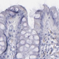
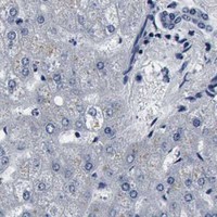
Negative control tissue: Adipose tissue (HPA Not detected)
ICC-IF cell lines (HPA subcellular resource): HPA ICC-IF images show SNAP25 in HeLa, SH-SY5Y, U2OS, ASC52telo, RPTEC/TERT1, hTERT-RPE1 (serum starved), with annotated localisation: Plasma membrane (approved) (HPA subcellular).
Technical controls: Include a no-primary, secondary-only control and a concentration-matched rabbit IgG isotype control for the rabbit catalog antibody (selected-SKU caption: rabbit anti-SNAP25; standard IHC practice); use SNAP25 knockout tissue or immunizing-peptide blocking, if available, as a biological specificity control (standard IHC practice). For DAB detection in brain, block endogenous peroxidase and check for endogenous biotin when using the caption’s streptavidin–biotin system (selected-SKU caption: SABC with DAB; standard IHC practice).
⚠️Feasibility: A target-specific fixation window or fixation effect is unreported, and the selected-SKU paraffin-section caption does not state the fixative (selected-SKU caption: fixative not stated). The caption reports heat retrieval in citrate buffer at pH 6 for 20 minutes; whether SNAP25 staining depends on that retrieval condition is unreported (selected-SKU caption). The supplied evidence does not establish that frozen sections or IF/ICC are easier than paraffin IHC; brain autofluorescence requires a background control if IF is used (supplied application evidence; standard IF practice).

HPA tissue IHC evidence for SNAP25

Comprehensive Human Protein Atlas IHC scoring per tissue (reliability: Enhanced — High consistency between antibody staining and RNA expression data.). Rows are taken directly from the HPA tissue chart — click any row's HPA link to view the source.

Positive expression · recommended positive controls

TissueCell typeLevelEvidenceSource
Cerebellum Molecular layer - neuropil High Protein (IHC) HPA →
Cerebral cortex Neuropil High Protein (IHC) HPA →

Undetected expression · recommended negative controls

TissueCell typeLevelEvidenceSource
Adipose tissue Adipocytes Not detected Protein (IHC) HPA →
Adrenal gland Glandular cells Not detected Protein (IHC) HPA →
Appendix Endocrine cells Not detected Protein (IHC) HPA →
Bone marrow Hematopoietic cells Not detected Protein (IHC) HPA →
Breast Adipocytes Not detected Protein (IHC) HPA →
Section 3

Advanced SNAP25 IHC Tips

Troubleshooting chromogenic SNAP25 IHC in paraffin sections, with one companion question on IF multiplexing.

Which retrieval conditions should I try first when SNAP25 staining is weak?
Begin with heat-mediated citrate retrieval at pH 6 for 20 minutes on paraffin sections (datasheet A01625). The selected rat brain image used this treatment before incubation with 2 µg/ml antibody overnight at 4°C (A01625 tissue-IHC caption). If staining is weak, compare a longer citrate exposure on adjacent sections while keeping antibody concentration and detection constant (standard IHC practice). Include a cerebellar or cortical section with expected neuropil staining to judge whether retrieval improved the signal (HPA: high in cerebellar molecular-layer neuropil and cortical neuropil). Excess retrieval can damage morphology, so assess tissue preservation alongside staining intensity (standard IHC practice).
Could fixation explain weak or uneven SNAP25 staining?
The selected paraffin-section caption does not state a fixative, and target-specific fixation sensitivity is unknown (A01625 tissue-IHC caption). Record the fixative, fixation duration, section thickness, and processing history for each specimen before attributing weak staining to the antibody (standard IHC practice). Compare similarly processed sections using the documented citrate retrieval at pH 6 for 20 minutes (datasheet A01625). Run a known-positive brain section in the same staining batch to distinguish a batch failure from a specimen-specific problem (HPA: high cortical and cerebellar neuropil staining). Assess morphology and regional staining together; neither alone establishes a fixation effect on SNAP25 (standard IHC practice).
What staining pattern should I expect in a positive brain section?
Look first for neuropil staining in the cerebellar molecular layer and cerebral cortex (HPA: high in both neuropil compartments). SNAP25 is associated with synapses and the cell membrane, and can also appear in cytoplasm with perinuclear concentration (UniProt P60880 localisation). It has no transmembrane segment, while membrane association requires palmitoylation (UniProt P60880 topology and localisation). Compare the stained compartment with nearby neuronal structures on the counterstained section before scoring an isolated cell-body signal as convincing (standard IHC practice). Low neuronal-cell staining in hippocampus or caudate does not, by itself, invalidate strong neuropil staining elsewhere (HPA: low in hippocampal and caudate neuronal cells).
Can this stain distinguish SNAP25 isoforms or modification states?
Treat staining as total antibody-accessible SNAP25 unless the antibody epitope and isoform specificity have been established independently (standard IHC interpretation). The record lists 2 isoforms and t-SNARE coiled-coil regions at residues 19–81 and 140–202 (UniProt P60880 isoforms and domains). It also lists phosphorylation at residues 138, 154, and 187, but the supplied caption does not map the antibody epitope (UniProt P60880 modified residues; A01625 tissue-IHC caption). Compare staining across identically processed sections before inferring biological differences from intensity (standard IHC practice). An isoform-specific or phospho-specific conclusion requires an antibody with documented specificity and matching controls (standard IHC practice).
How should I check SNAP25 localisation in a companion IF experiment?
Use a neuronal marker such as NeuN in a separate fluorescence channel to relate SNAP25 signal to the expected cell population (UniProt P60880: neuronal tissue specificity; standard IF practice). Choose a far-red fluorophore when tissue autofluorescence obscures shorter-wavelength channels, and inspect unstained and single-label controls (standard IF practice). SNAP25 has no transmembrane segment and is membrane-associated through palmitoylation, so choose permeabilisation according to whether the antibody epitope is accessible on the cytoplasmic side (UniProt P60880 topology and localisation). Compare membrane or vesicular signal with the reported IF localisation while checking for bleed-through (HPA subcellular: approved plasma-membrane and vesicle localisation; standard IF practice).
How can I reduce diffuse DAB signal without losing neuropil staining?
Start with the documented 10% goat-serum block and 2 µg/ml primary-antibody incubation overnight at 4°C (A01625 tissue-IHC caption). Include a no-primary section to reveal signal from the biotin-based detection system or tissue background (A01625 tissue-IHC caption; standard IHC practice). For peroxidase-based DAB detection, block endogenous peroxidase and compare shorter chromogen development on adjacent sections (standard IHC practice). Titrate primary antibody and detection reagents while holding retrieval at citrate pH 6 constant (datasheet A01625; standard IHC practice). Preserve the expected cortical or cerebellar neuropil pattern when selecting the lowest-background condition (HPA: high neuropil staining in both regions).
What should I quantify when SNAP25 staining is mainly neuropil? ⚠ ANSWER MARKED FOR VERIFICATION
Define comparable cortical neuropil or cerebellar molecular-layer regions before measuring staining (HPA: high in both neuropil compartments; standard image-analysis practice). For chromogenic sections, report DAB-positive area fraction or optical density within each region, normalised to the analysed neuropil area in mm² (standard IHC quantification practice). Keep threshold, illumination, section thickness, retrieval, and development settings consistent across groups (standard IHC quantification practice). An H-score or percentage of positive cells answers a different question and can miss diffuse neuropil signal (HPA: neuropil localisation; standard IHC quantification practice). Exclude damaged tissue and document region selection before comparing biological groups (standard IHC quantification practice).
How do I distinguish convincing SNAP25 staining from an artefact?
Give greatest weight to reproducible cortical or cerebellar neuropil staining with preserved morphology (HPA: high neuropil staining; standard IHC interpretation). Membrane-associated and cytoplasmic signal can fit SNAP25 biology, but an isolated nuclear pattern warrants scrutiny (UniProt P60880 localisation; standard IHC interpretation). Examine section edges, folds, and necrotic areas for abrupt staining unrelated to anatomy (standard IHC practice). Compare no-primary controls and endogenous-peroxidase blocking to identify detection-related DAB deposits (standard IHC practice). Widespread strong staining in adipocytes or unrelated glandular cells would conflict with the listed tissue profile and should prompt a specificity review (HPA: adipocytes and listed glandular cells not detected).
Boster reagents

Best SNAP25 / Synaptosomal-associated protein 25 IHC Antibodies

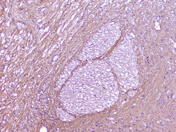
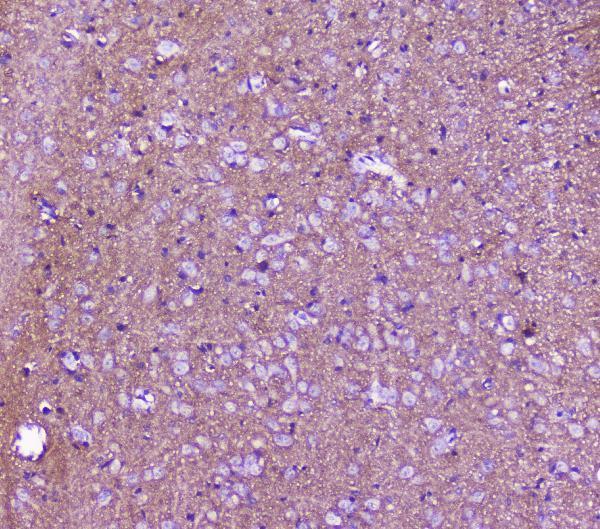
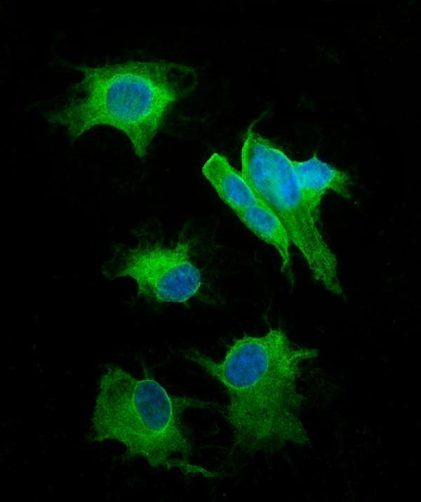
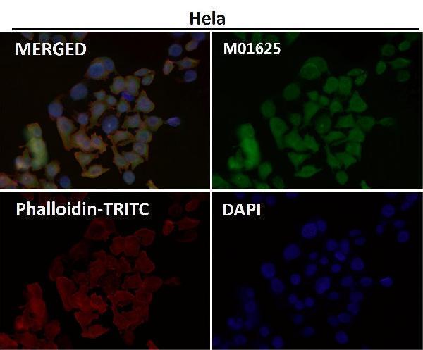
The catalog includes SNAP25 IHC images from rat and mouse brain and human glioma (A01625 image captions), plus an IF image with no sample identified (M01625 image caption).

Real IHC data IHC analysis of SNAP25 using anti-SNAP25 antibody (A01625). SNAP25 was detected in paraffin-embedded section of rat brain tissue. Heat mediated antigen retrieval was performed in citrate buffer (pH6, epitope retrieval solution) for 20 mins. The tissue section was blocked with 10% goat serum. The tissue section was then incubated with 2μg/ml rabbit anti-SNAP25 Antibody (A01625) overnight at 4°C. Biotinylated goat anti-rabbit IgG was used as secondary antibody and incubated for 30 minutes at 37°C. The tissue section was developed using Strepavidin-Biotin-Complex (SABC)(Catalog # SA1022) with DAB as the chromogen.
Anti-SNAP25 Antibody ®
Cat # A01625
Real IF data Immunofluorescent analysis using the Antibody at 1:50 dilution.
Anti-SNAP25 Rabbit Monoclonal Antibody
Cat # M01625

A01625 has IHC images from paraffin sections of rat and mouse brain and human glioma, and an IF image from SH-SY5Y cells (A01625 image captions). M01625 is listed for IF/ICC, with an IF image whose caption gives no sample or species (M01625 applications and image caption).

Which to pick: Choose A01625 for paraffin-section IHC: its rat brain caption documents citrate retrieval at pH 6 for 20 minutes, and the fixative is unreported (A01625 IHC image caption). Choose M01625 for IF/ICC when a rabbit monoclonal is preferred, while noting that its IF caption identifies neither sample nor species (M01625 catalog and image caption). For human, mouse, or rat work, both SKUs list reactivity with all three species; A01625 provides species-identified tissue IHC images, whereas M01625 does not (catalog reactivity; A01625 and M01625 image captions).

Each figure is that product's own IHC / IF validation image from its datasheet.

References

  1. UniProt Consortium. UniProt entry P60880 (SNP25_HUMAN, Synaptosomal-associated protein 25).
  2. Human Protein Atlas. SNAP25 tissue IHC expression (reliability: Enhanced).
  3. Human Protein Atlas. SNAP25 subcellular location (ICC-IF): Mainly localized to the plasma membrane. In addition localized to vesicles..
  4. Human Protein Atlas. SNAP25 antibody validation summary (3 antibodies).
  5. Recombinant botulinum neurotoxin serotype A1 in vivo characterization. Pharmacology research & perspectives 2021 — PMC8502944.
  6. Intramuscular Botulinum Neurotoxin Serotypes E and A Elicit Distinct Effects on SNAP25 Protein Fragments, Muscular Histology, Spread and Neuronal Transport: An Integrated Histology-Based Study in the Rat. Toxins 2024 — PMC11125604.
  7. SNAP25-induced MYC upregulation promotes high-grade neuroendocrine lung carcinoma progression. Frontiers in immunology 2024 — PMC11486671.
  8. Evaluation of Recombinant Botulinum Neurotoxin Type A1 Efficacy in Peripheral Inflammatory Pain in Mice. Frontiers in molecular neuroscience 2022 — PMC9179158.
  9. PubMed PMID:8112622 — UniProt-cited evidence.
  10. PubMed PMID:8056350 — UniProt-cited evidence.
  11. PubMed PMID:8760387 — UniProt-cited evidence.