SREBF1 / Sterol regulatory element-binding protein 1 · IHC design guide

Design Immunohistochemistry for SREBF1

Plan SREBF1 chromogenic IHC around cytoplasmic tissue staining, with adrenal glandular cells as a high-staining reference (HPA tissue IHC). Interpret nuclear staining in light of sterol-dependent processing and epitope position (UniProt).

Evidence assembled Oct 2026 · For research use; verify linked source records and product datasheet before use
Immunohistochemistry protocol sheet for SREBF1 (IHC for SREBF1): expected localisation Cytoplasm in tissue; ER/Golgi precursor, nuclear product (HPA tissue IHC; UniProt), antibody A00282-2, validated IHC image, and IHC protocol steps
Printable SREBF1 IHC protocol sheet — expected localisation Cytoplasm in tissue; ER/Golgi precursor, nuclear product (HPA tissue IHC; UniProt), antibody A00282-2, controls and protocol steps. Open the full SREBF1 IHC guide →

SREBF1 Immunohistochemistry Experimental Design Guide

Expected localisation, validated protocols, controls and antibodies — the at-a-glance facts below, then the full design guide.

Must know before staining
Expected localisation Cytoplasm in tissue; ER/Golgi precursor, nuclear product (HPA tissue IHC; UniProt)
Staining pattern Cytoplasmic staining, highest in adrenal glandular cells (HPA tissue IHC)
Antigen retrieval EDTA pH 8.0 HIER, heat-mediated (datasheet A00282-2)
Positive control ⓘ Adrenal gland+4 more · see all
Negative control ⓘ Adipose tissue+4 more · see all
Important caveats
Reasons your staining may differ from the expected pattern.
Fixation Keep paraffin-section fixation consistent across samples (standard IHC practice; not target-specific)
Caveat Sterol-dependent processing can shift staining to nuclei (UniProt)
Regulation Low sterols promote nuclear entry (UniProt)
Isoform / epitope Six isoforms; epitope position affects processed-form detection (UniProt)
Section 1

Recommended SREBF1 IHC & IF Protocols

The catalog antibody’s IHC-P protocol (datasheet A00282-2) is followed by published SREBF1 IHC methods for prostate tumors, breast tumors, and mouse aorta (PMC10256812; PMC8605000; PMC13337896).

Recommended immunohistochemistry (IHC-P) protocol parameters
SampleParaffin-embedded human melanoma tissue; fixative not specified (datasheet A00282-2)
FixationImage fixative and duration unreported (datasheet A00282-2); verify before use.
Sectioning4–5 µm sections on charged slides (standard)
DeparaffinisationXylene, graded ethanol series to water (standard)
Antigen retrievalHeat retrieval: EDTA pH 8.0 (datasheet A00282-2); 20 min, 95–100 °C (standard)
Peroxidase block3% H2O2, 10 min, room temperature (standard)
Blocking10% goat serum (datasheet A00282-2)
Primary antibodyRabbit anti-SREBF1, 1:50 recommended; image 1:100 (datasheet A00282-2)
Primary incubationOvernight at 4 °C (datasheet A00282-2)
DetectionHRP-conjugated secondary, DAB chromogen (datasheet A00282-2)
CounterstainHematoxylin, blue, dehydrate and mount (standard)
Expected resultSREBF1-positive staining in glandular cells of adrenal gland (HPA tissue IHC: High). HPA tissue profile: Cytoplasmic expression in several tissues, most abundant in adrenal gland. No signal in the no-primary control.
💡Decision noteStart with heat retrieval in EDTA pH 8.0 for the catalog antibody (datasheet A00282-2). The prostate study used citrate pH 6.0 (PMC10256812).
Section 2

What Is the Expected SREBF1 Staining Pattern?

SREBF1 staining may appear in the cytoplasm, including membrane-associated precursor, or in nuclei after processing (UniProt P36956). In paraffin-section IHC, HPA reports mainly cytoplasmic staining, strongest in adrenal glandular cells (HPA: High in adrenal glandular cells). Treat this as a reference pattern, not a definitive specificity test: HPA rates its tissue IHC evidence Uncertain because staining and RNA expression show low consistency (HPA: tissue IHC reliability Uncertain).

What am I looking at on my slide?
Adrenal glandular cells show clear cytoplasmic chromogen above nearby background (HPA: High in adrenal glandular cells).This matches HPA’s strongest reported tissue IHC pattern; score the glandular-cell staining separately from background (HPA: High in adrenal glandular cells; standard IHC practice). Nuclear staining can also be biologically plausible for processed SREBF1 (UniProt P36956).
Lung macrophages or placental trophoblastic cells show moderate cellular staining (HPA: Medium in both cell types).These are reported positive populations, though weaker than adrenal glandular cells (HPA: tissue IHC). Check cell identity and intracellular localisation before scoring; HPA’s Uncertain tissue reliability limits how strongly any single field supports specificity (HPA: tissue IHC reliability Uncertain).
Chromogen collects outside cells or follows tissue edges instead of cytoplasm or nuclei (UniProt P36956 localisation; standard IHC practice).An extracellular or edge-bound pattern does not fit SREBF1’s reported compartments (UniProt P36956). Consider section artefact or detection background, then compare with a matched negative control before interpreting the deposit as target staining (standard IHC practice).
A population reported as not detected, such as adipocytes in adipose tissue, stains strongly (HPA: Not detected in adipocytes).Unexpected staining raises cross-reactivity or endogenous detection activity as possibilities (standard IHC practice). It is not proof of either: HPA’s tissue calls are Uncertain, so inspect the compartment and controls before rejecting a biological signal (HPA: tissue IHC reliability Uncertain).
Adrenal glandular cells show no discernible staining above the negative control (HPA: High in adrenal glandular cells).The reported positive population is missing, which makes the run difficult to interpret (HPA: tissue IHC; standard IHC practice). Review antibody and detection performance alongside section quality; one negative specimen alone cannot establish absence of SREBF1 (standard IHC practice).
💡Expected SREBF1 appearanceA convincing positive is cell-associated cytoplasmic staining, strongest in adrenal glandular cells, with possible nuclear signal; extracellular or edge-only chromogen is suspect (HPA: tissue IHC Uncertain; UniProt P36956 localisation; standard IHC practice).
How each factor affects the staining
Membrane precursor and nuclear product (UniProt P36956).SREBF1 occupies ER and Golgi membranes before processing; its released transcription-factor form enters nuclei (UniProt P36956). Interpret cytoplasmic and nuclear signals in that context rather than requiring one compartment in every cell.
Epitope location and processing (UniProt P36956 topology and chains).UniProt places two transmembrane segments at residues 488–508 and 548–568, and the processed chain at 1–490 (UniProt P36956). Which forms an antibody detects depends on its epitope; no epitope is supplied here.
Isoforms (UniProt P36956).UniProt lists 6 SREBF1 isoforms (UniProt P36956). The supplied evidence does not show which isoforms the IHC antibody recognises, so staining alone cannot identify an isoform.
Strength of tissue reference (HPA: tissue IHC reliability Uncertain).HPA reports High adrenal glandular staining and several Medium cell populations, but flags low consistency with RNA expression (HPA: tissue IHC). Use these as comparison sites, not as absolute positive or negative criteria.
IF/ICC Q&A: Where should cellular fluorescence appear?HPA reports enhanced nucleoplasmic localisation in ICC-IF, while UniProt also places the precursor on ER and Golgi membranes (HPA: subcellular ICC-IF; UniProt P36956). This localisation answer does not prescribe an IF protocol.
Why is my staining missing, weak or wrong?
SituationLikely causeNext action
No adrenal glandular signal (HPA: High in adrenal glandular cells).A failed staining or detection step is possible; HPA’s Uncertain tissue call also limits certainty about an individual specimen (standard IHC practice; HPA: tissue IHC reliability Uncertain).Check the same run’s positive and negative controls, antibody application, detection reagents and section integrity before calling the tissue negative (standard IHC practice).
Signal fills the section, including cell-free areas (standard IHC practice).Diffuse chromogen may reflect background from detection reagents, insufficient blocking or inadequate washing (standard IHC practice).Compare a no-primary control, review blocking and wash steps, and score only cell-associated signal that exceeds background (standard IHC practice).
Strong staining appears in adipocytes reported as not detected (HPA: adipose adipocytes Not detected).Cross-reactivity or endogenous chromogenic activity is possible, but the HPA negative call is Uncertain (standard IHC practice; HPA: tissue IHC reliability Uncertain).Check cell identity and compartment; use an appropriate no-primary control and endogenous-activity control for the detection system (standard IHC practice).
Only extracellular deposits or stained section edges are visible (standard IHC practice).That distribution conflicts with the reported intracellular SREBF1 locations and suggests artefact (UniProt P36956 localisation; standard IHC practice).Inspect adjacent tissue and the negative control for the same deposit pattern before assigning a cellular score (standard IHC practice).
Cytoplasmic and nuclear scores disagree between specimens (HPA: cytoplasmic tissue IHC; UniProt P36956 nucleus).Precursor localisation and processing can yield different compartments; antibody epitope coverage is unspecified (UniProt P36956 topology and processing).Record cytoplasmic and nuclear staining separately, alongside cell type and intensity; avoid inferring processing status from IHC alone (standard IHC practice).
A staining difference follows antigen retrieval conditions (standard IHC practice).Retrieval can affect IHC performance generally; target-specific SREBF1 fixation sensitivity is unreported in the supplied evidence (standard IHC practice; supplied UniProt/HPA scope).Compare retrieval conditions with the same controls and scoring rules; report the condition used without attributing the difference to a proven SREBF1-specific mechanism (standard IHC practice).

Sample controls for SREBF1 IHC & IF

🧪Run adrenal gland first and look for staining in its glandular cells (HPA: High in adrenal gland glandular cells); use adipose tissue adipocytes as a negative comparison (HPA: Not detected in adipose tissue adipocytes). On the adrenal slide, use adjacent nonglandular areas to assess background, without assuming every nonglandular cell is SREBF1-negative.
Positive control tissue: Adrenal gland (Glandular cells, HPA High)
Negative control tissue: Adipose tissue (HPA Not detected)
ICC-IF cell lines (HPA subcellular resource): HPA ICC-IF images show SREBF1 in A-431, U-251MG, U2OS, A-549, HaCaT, with annotated localisation: Nucleoplasm (enhanced) (HPA subcellular).
Technical controls: Include a no-primary (secondary-only) control, a concentration-matched rabbit IgG isotype control, and a biological negative such as SREBF1 knockout tissue or cells; the catalog antibody in the selected caption is rabbit (selected-SKU IHC caption: rabbit anti-SREBF1). For adrenal sections developed with HRP/DAB, block endogenous peroxidase and check background staining (selected-SKU IHC caption: HRP/DAB detection).
⚠️Feasibility: A target-specific fixation window or fixation effect is unreported, and the selected-SKU paraffin-section caption does not state a fixative (selected-SKU IHC caption: fixative unreported). Heat-mediated retrieval in EDTA at pH 8.0 is a documented starting condition, but retrieval dependence is unreported (selected-SKU IHC caption: EDTA pH 8.0). The supplied evidence does not establish that frozen sections or IF are easier; for IF/ICC, HPA reports enhanced nucleoplasmic localization, while endogenous peroxidase can complicate HRP/DAB interpretation in adrenal sections (HPA subcellular: nucleoplasm, enhanced; selected-SKU IHC caption: HRP/DAB detection).

HPA tissue IHC evidence for SREBF1

Comprehensive Human Protein Atlas IHC scoring per tissue (reliability: Uncertain — Low consistency between antibody staining and RNA expression data.). Rows are taken directly from the HPA tissue chart — click any row's HPA link to view the source.

Positive expression · recommended positive controls

TissueCell typeLevelEvidenceSource
Adrenal gland Glandular cells High Protein (IHC) HPA →
Caudate Neuronal cells Medium Protein (IHC) HPA →
Kidney Cells in glomeruli Medium Protein (IHC) HPA →
Lung Macrophages Medium Protein (IHC) HPA →
Placenta Trophoblastic cells Medium Protein (IHC) HPA →

Undetected expression · recommended negative controls

TissueCell typeLevelEvidenceSource
Adipose tissue Adipocytes Not detected Protein (IHC) HPA →
Appendix Glandular cells Not detected Protein (IHC) HPA →
Bone marrow Hematopoietic cells Not detected Protein (IHC) HPA →
Breast Adipocytes Not detected Protein (IHC) HPA →
Bronchus Respiratory epithelial cells Not detected Protein (IHC) HPA →
Section 3

Advanced SREBF1 IHC Tips

For SREBF1 chromogenic IHC, assess staining by compartment and cell type while keeping the catalog antibody’s paraffin-section conditions and evidence limits in view.

Which retrieval conditions should I start with for SREBF1 in paraffin sections?
Start with heat-mediated retrieval in EDTA at pH 8.0 (datasheet A00282-2). That condition accompanies SREBF1 staining in a paraffin-embedded human melanoma section, followed by 1:100 primary antibody overnight at 4°C (datasheet A00282-2). Keep section thickness, heating, cooling and detection conditions consistent while assessing retrieval, because these variables affect chromogenic staining (standard IHC practice). If signal is weak, compare a separately validated retrieval condition on serial sections while retaining EDTA pH 8.0 as the reference (standard IHC practice; datasheet A00282-2). Judge improvement by preserved morphology and interpretable cytoplasmic or nuclear staining, rather than DAB intensity alone (UniProt P36956 localisation; standard IHC practice).
How should I troubleshoot weak SREBF1 staining when fixation history varies?
Target-specific sensitivity to fixation is unknown: the selected tissue caption says paraffin-embedded but does not state a fixative (datasheet A00282-2). Record the fixative, time in fixative and processing history for each block before comparing staining (standard IHC practice). Process serial sections with the documented EDTA pH 8.0 retrieval and 1:100 primary incubation overnight at 4°C to limit procedural variation (datasheet A00282-2; standard IHC practice). Compare morphology and signal within matched cell populations; a weak block alone cannot establish fixation sensitivity specific to SREBF1 (standard IHC practice). Avoid explaining fixation effects from SREBF1 topology, phosphorylation sites or HPA tissue patterns, which do not test fixation (UniProt P36956 topology and modified residues; HPA tissue IHC).
Should SREBF1 stain cytoplasm, nucleus, or both in tissue IHC?
Assess cytoplasmic and nuclear staining separately: UniProt places SREBF1 at ER, Golgi and COPII-vesicle membranes and in the nucleus (UniProt P36956 localisation). Its precursor has 2 transmembrane segments at residues 488–508 and 548–568; processing releases an N-terminal transcription-factor form, residues 1–490 (UniProt P36956 topology and processing). High sterol retains the SCAP–SREBF1 complex in the ER, whereas low sterol promotes Golgi processing and nuclear translocation (UniProt P36956 function). HPA describes predominantly cytoplasmic tissue staining but enhanced nucleoplasmic localisation in its cell-based images (HPA tissue IHC; HPA subcellular). Report each compartment and its distribution without treating nuclear staining alone as proof of pathway activation (standard IHC interpretation).
Can this IHC antibody distinguish processed SREBF1 from its precursor or specific isoforms?
Do not assign precursor, processed protein or isoform identity from chromogenic staining unless the antibody’s epitope and relevant isoform validation are established (standard IHC interpretation). UniProt lists 6 SREBF1 isoforms and a processed chain spanning residues 1–490 of the 1–1147 precursor (UniProt P36956 isoforms and processing). The precursor’s transmembrane segments lie at 488–508 and 548–568, so epitope position matters when interpreting compartment-specific signal (UniProt P36956 topology). The supplied tissue caption documents staining conditions, not an epitope or cleavage-specific test (datasheet A00282-2). Score nuclear and cytoplasmic compartments separately and label any processed-form interpretation as provisional without orthogonal evidence (standard IHC interpretation).
How can I assess SREBF1 by multiplex IF alongside this chromogenic IHC guide?
Use IF as a separate assay and validate its staining pattern independently of the paraffin-section chromogenic result (standard IF practice; datasheet A00282-2). Pair SREBF1 with a validated marker for the expected cell type, such as macrophages when examining the HPA-listed lung population, and inspect single-channel controls for bleed-through (HPA: medium in lung macrophages; standard IF practice). Choose fluorophores in channels with low measured tissue autofluorescence and include an unstained section to map that background (standard IF practice). Match permeabilisation to the documented epitope: residues 1–487 and 569–1147 are cytoplasmic, while 509–547 are lumenal (UniProt P36956 topology; standard IF practice). Without epitope information, compare permeabilisation conditions and confirm morphology and compartment assignment before interpreting colocalisation (standard IF practice).
What controls help separate nonspecific DAB signal from SREBF1 staining?
Include a no-primary control and inspect staining at tissue edges, damaged regions and pigment-rich areas before attributing brown signal to SREBF1 (standard chromogenic IHC practice). Use a peroxidase block and check for residual endogenous enzyme activity with a detection-only control; these are general DAB workflow safeguards (standard chromogenic IHC practice). The selected caption used 10% goat serum blocking, 1:100 primary antibody overnight at 4°C, and a peroxidase-conjugated secondary for 30 minutes at 37°C (datasheet A00282-2). Compare controlled staining with the expected ER-associated cytoplasmic and possible nuclear compartments (UniProt P36956 localisation). Diffuse signal in controls warrants revisiting blocking, washes and detection before increasing primary exposure (standard IHC practice).
How should I score SREBF1 IHC when cytoplasmic and nuclear signals differ? ⚠ ANSWER MARKED FOR VERIFICATION
Define cell types and scoring regions before examining treatment groups, then score nuclear and cytoplasmic SREBF1 separately (standard IHC quantification; UniProt P36956 localisation). For each compartment, report the percentage of positive cells and intensity, or calculate an H-score from intensity categories 0–3 with a possible range of 0–300 (standard IHC quantification). Normalise counts or positive-cell density to the number of eligible cells or analysed tissue area in mm², respectively (standard IHC quantification). Exclude necrosis and edge artefacts using the same rule for every section (standard IHC quantification). Document cell-type composition because HPA reports high adrenal glandular staining, medium lung macrophage staining and uncertain overall tissue-IHC reliability (HPA tissue IHC).
When is apparent SREBF1 positivity convincing rather than a staining artefact?
A convincing result has reproducible staining in defined cells, appropriate compartment assignment and low signal in no-primary and detection controls (standard IHC interpretation). Cytoplasmic staining can fit membrane-associated precursor, while nuclear staining can fit the processed transcription-factor form; neither pattern alone identifies the cleavage state (UniProt P36956 localisation and processing). Check that apparent positives follow cell boundaries rather than section edges or necrotic areas, and investigate diffuse brown signal for endogenous peroxidase or pigment (standard chromogenic IHC practice). Cell identity matters: HPA reports high staining in adrenal glandular cells and medium staining in lung macrophages, but rates tissue-IHC reliability uncertain (HPA tissue IHC). Confirm consequential claims with an independent assay or antibody validation before interpreting altered nuclear staining as changed SREBF1 activity (standard IHC interpretation).
Boster reagents

Best SREBF1 / Sterol regulatory element-binding protein 1 IHC Antibodies

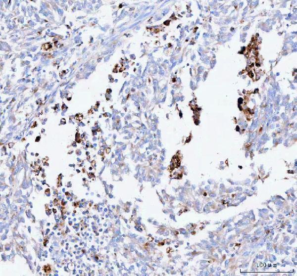
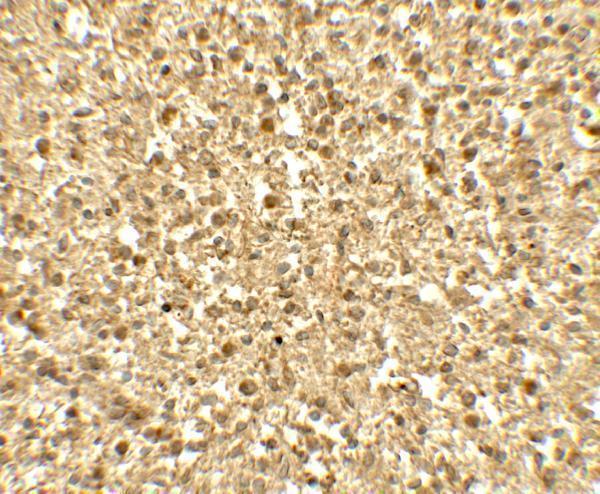
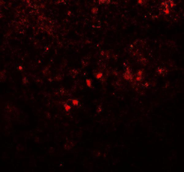
Anti-SREBF1 antibodies have IHC images from human spleen and paraffin-embedded human melanoma, plus an IF image from human spleen (catalog image captions: A00282, A00282-2).

Real IHC data IHC analysis of SREBF1 using anti-SREBF1 antibody (A00282-2). SREBF1 was detected in a paraffin-embedded section of human melanoma tissue. Heat mediated antigen retrieval was performed in EDTA buffer (pH 8.0, epitope retrieval solution). The tissue section was blocked with 10% goat serum. The tissue section was then incubated with 1:100 rabbit anti-SREBF1 Antibody (A00282-2) overnight at 4°C. Peroxidase Conjugated Goat Anti-rabbit IgG was used as secondary antibody and incubated for 30 minutes at 37°C. The tissue section was developed using HRP Conjugated Rabbit IgG Super Vision Assay Kit (Catalog # SV0002) with DAB as the chromogen.
Anti-SREBF1 Antibody
Cat # A00282-2
Real IHC data Immunohistochemistry of SREBF1 in human spleen tissue with SREBF1 antibody at 5 μg/mL.
Anti-SREBF1 Antibody
Cat # A00282

A00282 has human spleen IHC and IF images and lists human and mouse reactivity (catalog image captions and reactivity list: A00282). A00282-2 has a paraffin-embedded human melanoma IHC image and lists IHC, IF and ICC applications with human and rat reactivity (catalog IHC image caption and application/reactivity lists: A00282-2).

Which to pick: For tissue IHC, choose A00282 if its human spleen example fits your sample, or A00282-2 for a paraffin-section example in human melanoma; the A00282-2 caption does not report the fixative (catalog IHC image captions: A00282, A00282-2). For IF, A00282 has a human spleen image; for ICC or rat reactivity, A00282-2 lists ICC and IF, lists rat reactivity and is described as polyclonal (catalog IF image caption: A00282; catalog application/reactivity lists and dilution_raw: A00282-2). For mouse reactivity, A00282 lists mouse, while its supplied images show human spleen (catalog reactivity list and image captions: A00282).

Each figure is that product's own IHC / IF validation image from its datasheet.

References

  1. UniProt Consortium. UniProt entry P36956 (SRBP1_HUMAN, Sterol regulatory element-binding protein 1).
  2. Human Protein Atlas. SREBF1 tissue IHC expression (reliability: Uncertain).
  3. Human Protein Atlas. SREBF1 subcellular location (ICC-IF): Localized to the nucleoplasm..
  4. Human Protein Atlas. SREBF1 antibody validation summary (2 antibodies).
  5. Histone H2A Lys130 acetylation epigenetically regulates androgen production in prostate cancer. Nature communications 2023 — PMC10256812.
  6. Spatial transcriptomics reveals gene expression characteristics in invasive micropapillary carcinoma of the breast. Cell death & disease 2021 — PMC8605000.
  7. Reactive Oxygen Species Induces Lipid Droplet Accumulation in HepG2 Cells by Increasing Perilipin 2 Expression. International journal of molecular sciences 2018 — PMC6274801.
  8. Sesamin attenuates atherosclerosis by alleviating vascular endothelial ferroptosis-related injury via m(6)A-dependent regulation of SREBF1 expression. Frontiers in cell and developmental biology 2026 — PMC13337896.
  9. PubMed PMID:8402897 — UniProt-cited evidence.
  10. PubMed PMID:7759101 — UniProt-cited evidence.
  11. PubMed PMID:18267114 — UniProt-cited evidence.