Click image to see more details
Product Info Summary
| SKU: | A00282-2 |
|---|---|
| Size: | 100 μl/vial |
| Reactive Species: | Human, Rat |
| Host: | Rabbit |
| Application: | ELISA, IP, IF, IHC, ICC, WB |
Customers Who Bought This Also Bought

Product info
Product Name
Anti-SREBF1 Antibody
SKU/Catalog Number
A00282-2
Size
100 μl/vial
Form
Liquid
Description
Boster Bio Anti-SREBF1 Antibody catalog # A00282-2. Tested in WB, IHC, ICC, IF, IP, ELISA applications. This antibody reacts with Human, Rat.
Storage & Handling
12 months from date of receipt,-20℃ as supplied. 6 months 2 to 8℃ after reconstitution. Avoid repeated freezing and thawing.
Cite This Product
Anti-SREBF1 Antibody (Boster Biological Technology, Pleasanton CA, USA, Catalog # A00282-2)
Host
Rabbit
Contents
500 μg/ml antibody with PBS, 0.02% NaN3, 1 mg stabilizing protein and 50% glycerol
*This antibody is supplied in a stabilized formulation.
Compatibility with conjugation reactions depends on the chemistry of the conjugation method used.
For conjugation methods that are not compatible with the stabilizing components present in this formulation, a carrier-free antibody format is required.
Clonality
Polyclonal
Immunogen
E.coli-derived human SREBF1 recombinant protein (Position: E3-A330).
Reactive Species
A00282-2 is reactive to SREBF1 in Human, Rat
Calculated molecular weight
121.7 kDa
Background of SREBF1
This gene encodes a basic helix-loop-helix-leucine zipper (bHLH-Zip) transcription factor that binds to the sterol regulatory element-1 (SRE1), which is a motif that is found in the promoter of the low density lipoprotein receptor gene and other genes involved in sterol biosynthesis. The encoded protein is synthesized as a precursor that is initially attached to the nuclear membrane and endoplasmic reticulum. Following cleavage, the mature protein translocates to the nucleus and activates transcription. This cleaveage is inhibited by sterols. This gene is located within the Smith-Magenis syndrome region on chromosome 17. Alternative promoter usage and splicing result in multiple transcript variants, including SREBP-1a and SREBP-1c, which correspond to RefSeq transcript variants 2 and 3, respectively.
Antibody Validation
Boster validates all antibodies on WB, IHC, ICC, Immunofluorescence, and ELISA with known positive control and negative samples to ensure specificity and high affinity, including thorough antibody incubations.
Application & Images
Applications
A00282-2 is guaranteed for ELISA, IP, IF, IHC, ICC, WB Boster Guarantee
Recommend Dilution
| Application | Dilution | Species |
|---|---|---|
| Western blot | 1:500-2000 | |
| Immunohistochemistry | 1:50-400 | |
| Immunocytochemistry/Immunofluorescence | 1:50-400 | |
| Immunoprecipitation | 1:50 | |
| ELISA | 1:100-1000 |
Validation Images & Assay Conditions
Click image to see more details
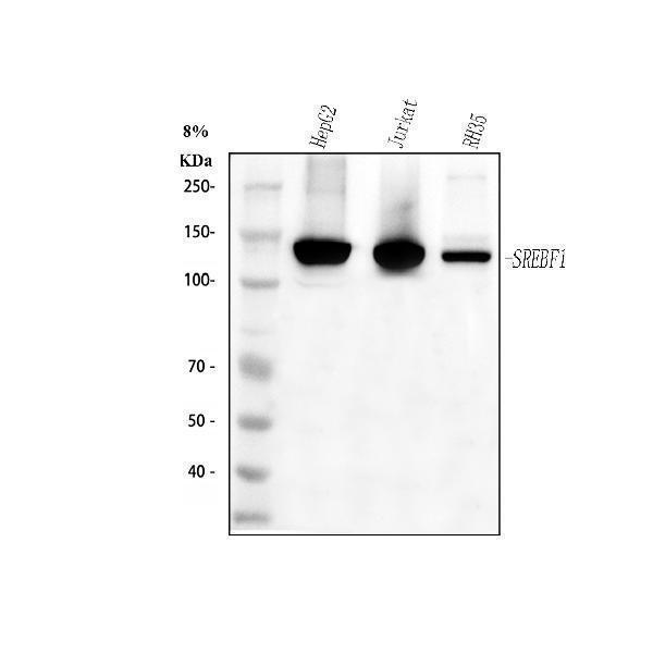
Western blot analysis of SREBF1 using anti-SREBF1 antibody (A00282-2).
Electrophoresis was performed on a 10% SDS-PAGE gel at 80V (Stacking gel) / 120V (Resolving gel) for 2 hours. The sample well of each lane was loaded with 30 ug of sample under reducing conditions.
Lane 1: human HepG2 whole cell lysates,
Lane 2: human Jurkat whole cell lysates,
Lane 3: rat RH-35 whole cell lysates.
After electrophoresis, proteins were transferred to a nitrocellulose membrane at 150 mA for 50-90 minutes. Blocked the membrane with 5% non-fat milk/TBS for 1.5 hour at RT. The membrane was incubated with rabbit anti-SREBF1 antigen affinity purified polyclonal antibody (A00282-2) at 1:1000 overnight at 4°C, then washed with TBS-0.1%Tween 3 times with 5 minutes each and probed with a goat anti-rabbit IgG-HRP secondary antibody at a dilution of 1:5000 for 1.5 hour at RT. The signal is developed using an ECL Plus Western Blotting Substrate (Catalog # AR1196-200) with Tanon 5200 system. A specific band was detected for SREBF1 at approximately 122 kDa. The expected band size for SREBF1 is at 122 kDa.

Click image to see more details
IHC analysis of SREBF1 using anti-SREBF1 antibody (A00282-2).
SREBF1 was detected in a paraffin-embedded section of human melanoma tissue. Heat mediated antigen retrieval was performed in EDTA buffer (pH 8.0, epitope retrieval solution). The tissue section was blocked with 10% goat serum. The tissue section was then incubated with 1:100 rabbit anti-SREBF1 Antibody (A00282-2) overnight at 4°C. Peroxidase Conjugated Goat Anti-rabbit IgG was used as secondary antibody and incubated for 30 minutes at 37°C. The tissue section was developed using HRP Conjugated Rabbit IgG Super Vision Assay Kit (Catalog # SV0002) with DAB as the chromogen.

Click image to see more details
IF analysis of SREBF1 using anti-SREBF1 antibody (A00282-2).
SREBF1 was detected in an immunocytochemical section of Hela cells. Enzyme antigen retrieval was performed using IHC enzyme antigen retrieval reagent (AR0022) for 15 mins. The cells were blocked with 10% goat serum. And then incubated with 1:100 rabbit anti-SREBF1 Antibody (A00282-2) overnight at 4°C. Cy3 Conjugated Goat Anti-Rabbit IgG (BA1032) was used as secondary antibody at 1:500 dilution and incubated for 30 minutes at 37°C. The section was counterstained with DAPI. Visualize using a fluorescence microscope and filter sets appropriate for the label used.
Specific Publications For Anti-SREBF1 Antibody (A00282-2)
Loading publications
Recommended Resources
Here are featured tools and databases that you might find useful.
- Boster's Pathways Library
- Protein Databases
- Bioscience Research Protocol Resources
- Data Processing & Analysis Software
- Photo Editing Software
- Scientific Literature Resources
- Research Paper Management Tools
- Molecular Biology Software
- Primer Design Tools
- Bioinformatics Tools
- Phylogenetic Tree Analysis
Customer Reviews
Have you used Anti-SREBF1 Antibody?
Share your experimental results or join a short interview to earn up to $1,000 in product credits or other rewards.
0 Reviews For Anti-SREBF1 Antibody
Customer Q&As
Have a question?
Find answers in Q&As, reviews.
Can't find your answer?
Submit your question
