This website uses cookies to ensure you get the best experience on our website.
- Table of Contents
Facts about SPARC-related modular calcium-binding protein 1.
.
Mouse | |
---|---|
Gene Name: | Smoc1 |
Uniprot: | Q8BLY1 |
Entrez: | 64075 |
Belongs to: |
---|
No superfamily |
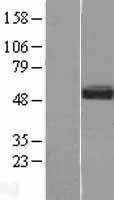
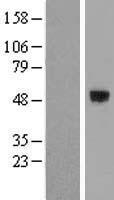
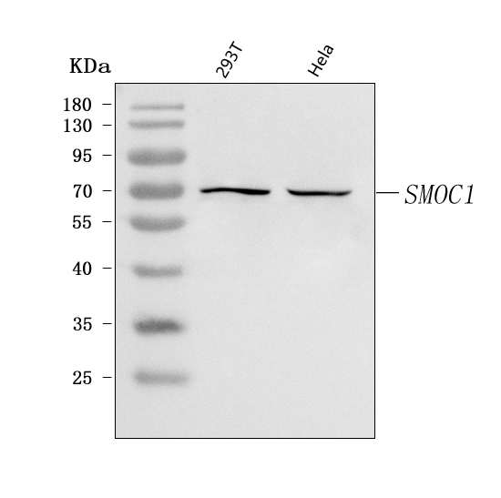
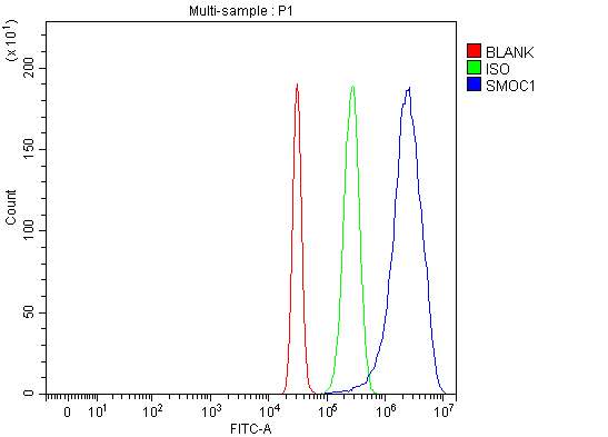
Secreted modular calcium-binding protein 1; SMOC1; SMOC-1; SPARC related modular calcium binding 1; SPARC-related modular calcium-binding protein 1
Mass (kDA):
51.076 kDA
Mouse | |
---|---|
Location: | 12|12 D1 |
Sequence: | 12; |
Widely expressed in many tissues with a strongest signal in ovary.
This article will discuss the SMOC1 Marker as well as its validation. We'll also discuss the applications for the SMOC1 Marker. This technique is available to scientists from all over the globe. It is a powerful tool for biomarker analysis. The SMOC1 can be used in a variety experiments and is a useful tool for researchers.
The SMOC1 Gene is highly expressed within the human body and is involved at various biological processes. It is involved with cell cycle progression, tumor development, attachment to the surface of cells, and cell cycle progression. Some cancers have a lower expression of SMOC1, while others have a higher level. The SMOC1 genes is also expressed in many normal cells including the lining and kidneys.
This protein regulates post-gastrulation development, and influences BMP signaling. Moreover, SMOC1 can act as an antagonist of BMP signaling and regulate MAPK-mediated Smad phosphorylation in the linker region. It has been shown to regulate cell fate specification. Its functions are many and well-documented. There are several research methods available to identify SMOC-1.
Digoxigenin glycan detect kits can be used to detect SMOC-1 if human serum-free supernatants are used. This kit uses 250 ng affinity-purified SMOC-1. The samples were separated on a 4--15% polyacrylamidegel and transferred to the nitrocellulose. Further treatment was done according to the manufacturer's instructions.
The SMOC1 genes has been shown to play a critical role for bone mineralization and wound healing. It also affects the expression of matrix turn-over proteins. Researchers have found that SMOC1 can play a vital role in tissue healing, cellular proliferation, as well as angiogenesis. Scientists can use the SMOC1 gene to identify damaged tissues and treat them accordingly. The gene is found in the basement membrane and is therefore a useful tool for cancer research.
Different types of tumors express the SMOC1 Gene and are correlated with different functional States. Different types have positive correlations between SMOC1 gene expression and SMOC1. It is interesting to note that SMOC1 has a chemoattractive effect that inhibits tenascin C-induced effects. Despite lack of concrete evidence supporting this, the findings offer new insights and avenues for further research.
A recent study has identified a novel genetic marker for glioma, the SMOC1 gene. This marker co-expresses with several genes that inhibit leukocyte migration, lymphocyte proliferation, adaptive immune response, and neutrophil-mediated immunity. It also inhibits T cell activation, Th27 cell differentiation. It was discovered that higher levels SMOC1 were associated to better outcomes in LGG patients. These results need to be confirmed by further research.
SMOC1 comes from the family of secreted module calcium-binding proteins. This protein can be found in basement membranes. These membranes anchor many tissues and cells during embryonic developmental. It binds to a variety protein and regulates activity of growth factors. Among these, it promotes osteoblast differentiation. It is important to validate SMOC1 markers in different tissues.
The expression level of SMOC1 was higher among asymptomatic AD patients compared to controls. This indicates that the SMOC1 marker has greater relevance than other markers in the detection or diagnosis of AD. Asymptomatic AD patients had higher levels than controls of osteopontin, UCHL1, and other markers. The study authors also found higher levels of SMOC1 in asymptomatic AD patients than in asymptomatic controls.
Research has shown that SCG2 as well as SMOC1 have been implicated in the regulation and maintenance of angiogenesis. SCG2 and SMOC1 are expressed in endothelial cell. Tube formation is inhibited if either gene is blocked. This inhibitory effect is dependent upon the levels of SCG2 and SMOC1. Inhibition of both genes partially prevents fasting/refeeding-induced angiogenesis.
SMOC1 is involved in the regulation of cell cycle progression as well as the process of calciumification. Further research is needed to determine how SMOC1 regulates the receptor-mediated signaling. This marker may prove useful for diagnosing cancer and other diseases. Further research is needed to determine the role SMOC plays in controlling cell adhesion as well as angiogenesis. In the long run, SMOC1 may prove to be an effective therapeutic target for many types of disease.
SMOC2 is a key component of cancer development and progression. Overexpression of SMOC2 in HCC tissues inhibits cell proliferation, migration, and growth. SMOC2 also has a positive association with survival in patients suffering from colorectal and lung carcinomas. Although SMOC2 is thought to play a role in the development of cancer, there are still questions. In one study, SMOC2 was shown to increase tumor progression in mice expressing SMOC2. Huang et.al. In a recent study, Huang et.al. reported that SMOC2 expression was reduced in HCC tissues when chemotherapy was administered. Another study revealed that HCC tumors expressing SMOC2 had a lower survival rate and a higher quality life.
SMOC1 has been successfully identified as a secreted modular glycoprotein in 2002. It is located in chromosome 14, 24.2 and contains 434 amino acids. It has a molecular weight of around 48,000. It also contains five structural domains including a follistatin domain, two thyroglobulin domains and an EC hand calcium binding domain. It is highly expressed in the brain, thymus and ovary.
Studies on the extracellular matrix-associated proteins SMOC1 have shown that SMOC1 is essential for bone development. It also regulates BMP2 signaling pathways, which is critical for anatomic patterns and cell or tissue fate specification. Chondrocalcinosis, which is a form of excessive calcification, can be caused by excessive limb and bone growth. In this study, SMOC2 wasn't detected in neonatal mouse calvaria cellular cells. This suggests that SMOC plays a less important regulatory role in MC3T3–E1 cells.
The SMOC1 gene marker is an oncogene that has a unique domain. It is correlated with genes that identify immune cells in various types of cancer. It has been proven to be a prognostic indicator for patients with glioma or LGG. SMOC1 expression also was found to be decreased in some types and increased in others. Its expression was increased in chromophobe and LUAD-like tumors, but not in LGG and GBM.
It has been reported that the SMOC1 marker can be used in cell culture. The SMOC1 Gene is expressed in basement membranes, as well in other tissues containing oocytes (bone and skin). However, SMOC-1 doesn't amplify RTPCR with normal blood RNA, so the gene may not be functionally important in these other tissues. However, the gene is still very important because it is involved for the development certain cancers like lung cancer, multiple sclerosis, and others.
The regulation of osteogenic differentiation in human bone marrow stem cells has been implicated by SMOC1. A 16-amino acid-rich peptide derived SMOC1's extracellular Calcium binding domain stimulates osteogenic differentiate of human BMSCs, in vitro and in vivo. Moreover, SMOC1 knockout mice as well as genetically engineered Smoc1 mutated mice exhibit similar skeletal phenotypes that OAS patients.
Ang II-induced myocardial fibrosis is inhibited by SMOC1 silencing in MFB cells. This gene silencing has a significant impact on myocardial fibrisis therapy. A SMOC-1 RNA siRNA can block the activity of the SMOC1 gene. This gene silencing can be applied in different clinical settings. More research is needed to understand the role of SMOC1 for fibrosis.
These kits can be used to detect Digoxigenin Glycans. SMOC-1 was affinity purified, and then treated with PNGaseF. Finally, the samples were separated on a 4- to 5-% SDS-polyacrylamide gel, and stained with Coomassie Brilliant Blue. The SMOC1 antibodies was not reactive with other BM-40 family members. This enzyme is highly specific for SMOC-1.
PMID: 21194678 by Okada I., et al. SMOC1 is essential for ocular and limb development in humans and mice.
PMID: 21750680 by Rainger J., et al. Loss of the BMP antagonist, SMOC-1, causes Ophthalmo-acromelic (Waardenburg Anophthalmia) syndrome in humans and mice.