This website uses cookies to ensure you get the best experience on our website.


- Table of Contents
Plan TERF2 chromogenic IHC on paraffin sections using the catalog antibody at 0.5–1 μg/ml (datasheet A00650-1). Assess nuclear staining against the HPA tissue profile, including high staining in tonsil germinal center cells (HPA tissue IHC).

Expected localisation, validated protocols, controls and antibodies — the at-a-glance facts below, then the full design guide.
| Expected localisation | Nuclear staining in tissue (HPA tissue IHC) | |
| Staining pattern | Widespread nuclear staining across tissue cell types (HPA tissue IHC) | |
| Antigen retrieval | EDTA pH 8.0 HIER, heat-mediated (datasheet A00650-1) | |
| Positive control | Appendix+4 more · see all | |
| Negative control | None in HPA (detected in all 45 tissues); use no-primary + isotype controls |
| Fixation | Keep fixation consistent across sections. (standard IHC practice; not target-specific) | |
| Caveat | Staining–RNA agreement has medium consistency (HPA tissue IHC) | |
| Regulation | Low tissue specificity (HPA tissue RNA) | |
| Isoform / epitope | 2 isoforms; verify antibody epitope coverage (UniProt) |
Compare the catalog antibody’s IHC-P protocol (datasheet A00650-1) with four published TERF2 IHC protocols (PMC5190092; PMC11425175; PMC10240986; PMC12881650).
| Sample | Paraffin-embedded human tonsil tissue; fixative not specified (datasheet A00650-1) |
| Fixation | Image fixative and duration unreported (datasheet A00650-1); verify before use. |
| Sectioning | 4–5 µm sections on charged slides (standard) |
| Deparaffinisation | Xylene, graded ethanol series to water (standard) |
| Antigen retrieval | Heat retrieval: EDTA pH 8.0 (datasheet A00650-1); 20 min, 95–100 °C (standard) |
| Peroxidase block | 3% H2O2, 10 min, room temperature (standard) |
| Blocking | 10% goat serum (datasheet A00650-1) |
| Primary antibody | Rabbit anti-TERF2, 0.5-1μg/ml (datasheet A00650-1) |
| Primary incubation | Overnight at 4 °C (datasheet A00650-1) |
| Detection | Streptavidin-biotin complex (SABC), DAB chromogen (datasheet A00650-1) |
| Counterstain | Hematoxylin, blue, dehydrate and mount (standard) |
| Expected result | TERF2-positive staining in lymphoid tissue of appendix (HPA tissue IHC: High). HPA tissue profile: Ubiquitous nuclear expression. No signal in the no-primary control. |
TERF2 is a nuclear, telomere-associated protein without a transmembrane segment (UniProt Q15554). In paraffin-section IHC, expect nuclear staining across many cell types (HPA: ubiquitous nuclear expression; IHC reliability Enhanced). Strong examples include lymph-node germinal center cells, fallopian-tube glandular cells and testicular preleptotene spermatocytes (HPA: High). Staining intensity varies by tissue and cell type (HPA: High, Medium and Low observations).
| Clear nuclear staining in germinal center cells, with weaker staining in some neighboring cells. | This fits the reported pattern: germinal center cells stain High in lymph node and tonsil (HPA: High), while the broader tissue profile is ubiquitous and nuclear (HPA: tissue IHC). Compare cells within the same section before interpreting weaker nuclei as negative. |
| Staining is predominantly cytoplasmic or membranous while nuclei remain pale. | That distribution conflicts with nuclear and telomeric localization (UniProt Q15554) and the ubiquitous nuclear IHC profile (HPA: tissue IHC). Review controls, detection chemistry and staining conditions; compartment alone cannot identify the source of an artefact (general IHC practice). |
| An isolated cell population stains strongly outside the expected nuclear pattern. | TERF2 expression is widespread (UniProt Q15554; HPA: tissue IHC), so an unexpected cell type alone does not prove cross-reactivity. Strong nonnuclear staining, especially in cells lacking matching nuclear signal, warrants checks for nonspecific binding or endogenous detection activity (general IHC practice). |
| Color is spread across stroma, extracellular spaces or most of the section without crisp nuclei. | Diffuse background cannot be scored as TERF2-positive nuclei against the reported nuclear profile (HPA: tissue IHC). Check the no-primary control and detection reagents, then optimize blocking, antibody concentration and wash conditions (general IHC practice); the payload supplies no TERF2-specific cause of background. |
| No convincing nuclear signal appears in a known High-staining cell population. | Absent staining in lymph-node germinal center cells or fallopian-tube glandular cells conflicts with their reported High signal (HPA: tissue IHC). First check section quality, assay controls and antibody use (general IHC practice). One negative section does not establish biological absence. |
| Tissue and cell selection | Use a reported High population, such as lymph-node germinal center cells, as a positive reference (HPA: High). Parathyroid glandular cells and cardiomyocytes are reported Low (HPA: Low), so weak staining there is not a validated negative control. |
| Evidence strength | The tissue profile carries Enhanced IHC reliability, with medium consistency between antibody staining and RNA data (HPA: tissue IHC). HPA001907 and HPA002735 have Enhanced IHC validation; CAB010451 and CAB080271 are Supported (HPA: antibodies). Interpret an unusual pattern alongside controls. |
| IF/ICC: what pattern is expected? | Expect nucleoplasm and nuclear bodies (HPA: supported ICC-IF locations). UniProt places TERF2 at telomeres in interphase and chromosome ends in metaphase (UniProt Q15554). These observations guide localization; this IHC section provides no IF/ICC protocol. |
| Molecular features and antibody scope | Two isoforms and several modified residues are recorded (UniProt Q15554), but the supplied evidence does not map an antibody epitope or link these features to IHC intensity. Do not infer an isoform-specific pattern or a retrieval requirement from them. |
| Situation | Likely cause | Next action |
|---|---|---|
| Expected positive tissue shows no nuclear stain. | The assay may have failed, or the examined section may lack the reported High-staining cells (general IHC practice; HPA: High in specified cell populations). | Confirm the relevant cells are present, review the positive and no-primary controls, then verify antibody dilution and detection steps against the validated IHC procedure (general IHC practice). |
| Only cytoplasm or membrane stains. | This conflicts with nuclear localization (UniProt Q15554; HPA: ubiquitous nuclear expression); nonspecific staining or detection artefact is possible (general IHC practice). | Compare with a no-primary control, inspect nuclear counterstain and cell boundaries, and optimize blocking and antibody concentration if background persists (general IHC practice). |
| Most of the section has diffuse color. | High background can obscure the nuclear pattern (general IHC practice; HPA: ubiquitous nuclear expression). The supplied sources do not establish a TERF2-specific background mechanism. | Check no-primary staining and reagent controls; review blocking, washes and chromogen development before scoring nuclei (general IHC practice). |
| A reported Low population looks nearly unstained. | Low HPA staining indicates a weaker observed signal, not confirmed protein absence (HPA: Low in parathyroid glandular cells, cardiomyocytes and other listed populations). | Compare with a reported High population processed in the same run and score the Low population cautiously (HPA: High and Low observations; general IHC practice). |
| Strong color appears in an unexpected cell population. | TERF2 is broadly expressed (UniProt Q15554; HPA: ubiquitous nuclear expression), so cell identity alone cannot establish cross-reactivity; endogenous detection activity is also possible (general IHC practice). | Check whether color is nuclear, examine no-primary and detection controls, and repeat with an independently validated antibody if the discrepancy remains (general IHC practice; HPA: antibody validation). |
| Nuclear signal differs between runs. | Run-to-run differences can reflect section processing, antibody concentration or detection conditions (general IHC practice). No TERF2-specific fixation sensitivity is reported in the supplied UniProt or HPA evidence. | Compare matched positive-control tissue and recorded assay conditions across runs before attributing the difference to TERF2 biology (general IHC practice; HPA: High populations). |


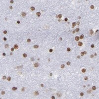
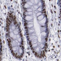


Comprehensive Human Protein Atlas IHC scoring per tissue (reliability: Enhanced — Medium consistency between antibody staining and RNA expression data.). Rows are taken directly from the HPA tissue chart — click any row's HPA link to view the source.
| Tissue | Cell type | Level | Evidence | Source |
|---|---|---|---|---|
| Appendix | Lymphoid tissue | High | Protein (IHC) | HPA → |
| Fallopian tube | Glandular cells | High | Protein (IHC) | HPA → |
| Lung | Alveolar cells type I | High | Protein (IHC) | HPA → |
| Lymph node | Germinal center cells | High | Protein (IHC) | HPA → |
| Testis | Preleptotene spermatocytes | High | Protein (IHC) | HPA → |
| Tissue | Cell type | Level | Evidence | Source |
|---|---|---|---|---|
| None in HPA: TERF2 is detected in all 45 scored tissues. Use a no-primary (secondary-only) and an isotype control instead. | ||||
Troubleshoot TERF2 staining in paraffin section IHC using nuclear localisation, the catalog antibody’s tissue result, and appropriate controls.
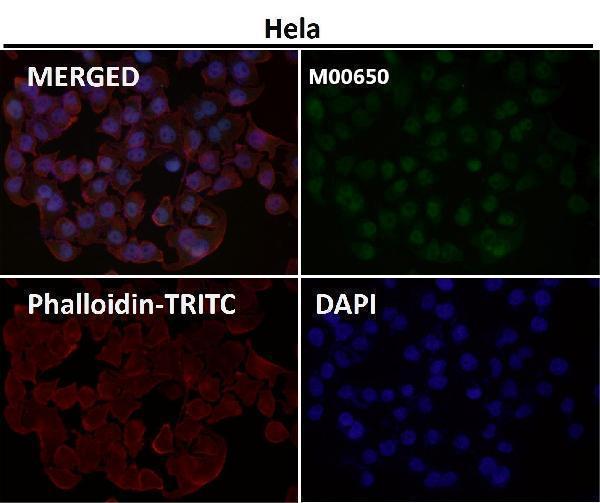
Anti-TERF2 antibodies have IHC images from human tonsil and lung cancer paraffin sections (A00650-1 IHC captions) and an IF image with no specimen identified (M00650 IF caption).
A00650-1 shows IHC staining in human tonsil and lung cancer paraffin sections (A00650-1 IHC captions). M00650 lists human IHC and IF/ICC applications (M00650 catalog), but its IF image does not identify the specimen (M00650 IF caption).
Which to pick: Choose A00650-1 for human paraffin-section chromogenic IHC: its images document 1 μg/ml primary antibody, EDTA retrieval at pH 8.0, and DAB detection (A00650-1 IHC captions); the fixative is unreported (A00650-1 IHC captions). For IF/ICC, choose M00650, a rabbit monoclonal with human reactivity and listed IF/ICC applications (M00650 catalog). For work involving rat samples, A00650-1 lists rat reactivity (A00650-1 catalog), although its displayed IHC evidence is from human tissue (A00650-1 IHC captions).