This website uses cookies to ensure you get the best experience on our website.
- Table of Contents
2 Citations 7 Q&As
1 Citations 1 Q&As
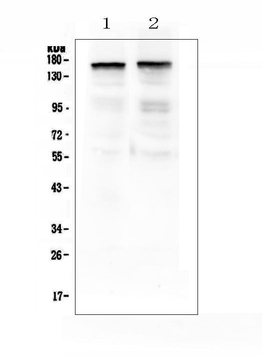
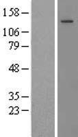
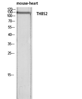
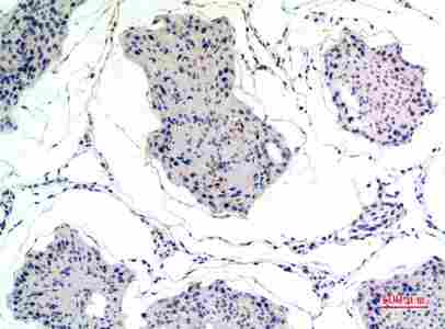
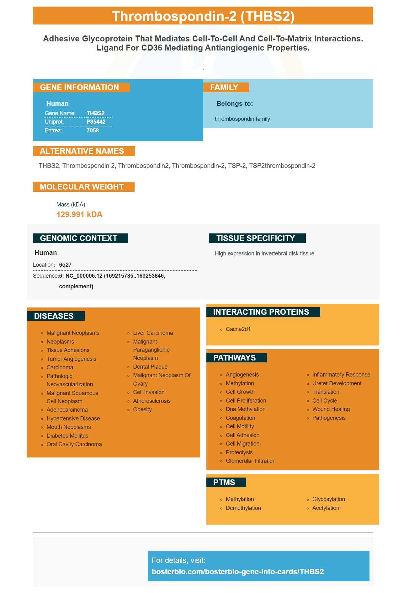
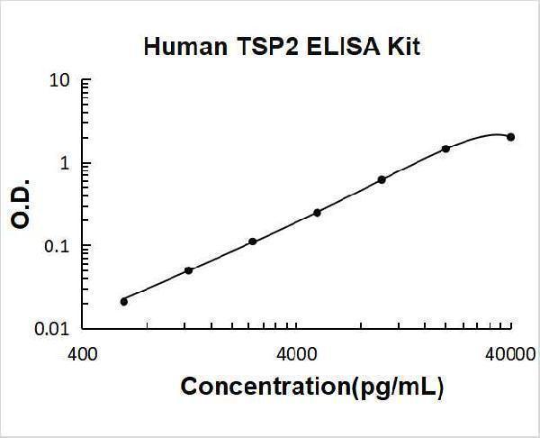
Facts about Thrombospondin-2.
.
Human | |
---|---|
Gene Name: | THBS2 |
Uniprot: | P35442 |
Entrez: | 7058 |
Belongs to: |
---|
thrombospondin family |
THBS2; thrombospondin 2; Thrombospondin2; Thrombospondin-2; TSP-2; TSP2thrombospondin-2
Mass (kDA):
129.991 kDA
Human | |
---|---|
Location: | 6q27 |
Sequence: | 6; NC_000006.12 (169215785..169253846, complement) |
High expression in invertebral disk tissue.
THBS2 is an inhibitor of angiogenesis that modulates tumor-associated macrophages (DCs) and could be used to predict the future. We will discuss the best uses of the THBS2 marker in cancer research. Here are some examples. Read on to find out more. * Kaplan-Meier survival curves: GSE17536 OS The GSE17536 OS, GSE17536 RFS, and GSE.
Thrombospondins (THBSs) are a disulfide-linked family of five homotrimeric glycoproteins that function in a variety of tissue remodeling environments. These proteins have been associated in various malignancies such as cancer of the gastrointestinal tract and breast cancer. They are also known for being controlled by the microRNA network.
Healthy subjects as well as CRC patients were examined for the presence of RBP4 (and the THBS2) and their respective effects on the body. Both RBP4 and THBS2 levels were significantly lower in patients suffering from CRC as compared with controls. However, they were not significantly different from healthy individuals. Therefore, serum RBP4 concentrations and THBS2 levels can be used to identify CRC risk.
In experiments in vivo, knockdown and overexpression of THBS2 increased the expression of glycolysis-related genes mRNA. THBS2 knockdown and overexpression also increased glucose consumption and production of lactate acid and ECAR. In addition, the knockdown of HIF-1a blocked the upregulation of several glycolysis-related genes. These genes help to ensure cell viability.
TLR4 interacts with THBS2 which boosts aerobic glycolysis and is a factor in the progression of CRC. Furthermore, the growth of tumors in orthotopic regions is slowed down by knockdown of THBS2, while THBS2 expression increases tumor growth. Additionally, orthotopic tumors that overexpressed THBS2 were found to have increased intensity of KI-67, a marker of the growth of tumor cells. These results confirm that THBS2 plays a significant role in CRC development.
To determine if THBS2 inhibits orthotopic tumor growth in mice, SW620 cells were injected with a compound that inhibits the expression of specific genes in the mouse. These cells caused tumors in mice. In this study, SW620 cells were given THBS2-OV and in vivo vector injections. The Student t-test was used to analyse the results.
These results suggest that THBS2 could be a therapeutic target in CRC. These findings must be confirmed in humans. The THBS2-TCGA dataset also analyzed mRNA expression and protein expression of THBS2 in patients with CRC. The overall survival results of this study suggest that THBS2 may inhibit aerobic glycolysis and increase angiogenesis.
TSP-1 is a multimodular calcium binding glycoprotein that is used to block angiogenesis. The TSP-1 can be activated by the latent TGFb1 complex, however TSP-2 does not have this property. TSPs hinder angiogenesis through interactions with CD36 in endothelial cell lines. This inhibition of endothelial cells expansion through a homeostatic process. It also inhibits the signaling of Nitric oxide.
TSP1 induces cell death in microvascular endothelial cell types, and interacts with the CD36 protein on the cell surface. CD-36 has been shown to induce anti-migratory actions in endothelial cells, while CD-36 and CD47 are involved in the production and release of nitric oxygen. These findings suggest that TSP2 might play a role in regulating the angiogenesis of the human body.
The cancer-associated protein THBS2 that is found in extracellular vesicles that are found in cancer cells. It is linked to the development of gastric, colon and pancreatic cancers. The THBS family contains members THBS1 to THBS4. They could all play a role in tumorigenesis together. THBS2 expression is associated with lower overall survival in COAD and STAD.
A crucial role for the microenvironment during cancer development and metastasis is played by tumor-associated macrophages or TAMs. Macrophages are an essential component of the tumor microenvironment and are classified into two subsets, the classical subset (M1) and the alternate subset (M2). While TAMs might have anti-tumor properties Their primary function is to promote the progression of the tumor by complex autocrine and paracrine pathways. Additionally cancer malignant proliferation as well as metastasis are closely related processes.
Researchers isolated murine macrophage-like RAW264.7 cells in the study, and then transformed them into subsets of M1 or M2 macrophages. They also examined the phenotype of these cells and their expression of SPP1 and VEGF. The results revealed that THP-1 derived M1 macrophages had the same expression patterns as RAW264.7-derived M2 macrophages.
Despite the numerous implications of this discovery researchers aren't quite sure what TSP1 affects tumor-associated macrophages and DCs. Macrophages associated with tumors can cause cancer by evading the innate immune surveillance. It is therefore crucial to determine if TSP1 is an anti-tumor agent or a tumor-inhibitor.
In the BRCA-basal class, knockdown of SDC1 decreased the risk of disease progression. Survival was related to the expression of MMP1 or CD24. Moreover, IL6 secretion was positively connected to survival. These findings suggest the possibility of role of SDC1 during immune treatment of the course of the disease. Additionally these findings suggest SDC1's role as a key factor in breast cancer.
Furthermore, tumor-associated fibrblasts may be located in the ECM. They have been linked to breast cancer growth and resistance to chemotherapy. For instance, Hoang et al. discovered that ERK5 is responsible for ECM and tumor proliferation and migration. The development of tumors is closely related to a dysfunctional lipid metabolism. FAC also functions as a cancerogene that is metabolic in nature. HBXIP modulates the metabolism of lipids and tumor growth by activation of FAS signaling.
TSP1 reduces adhesion to tumor cells in various ways. It reduced the adhesion of tumor cells through the upregulation of the plasmin activity and uPA-controlled. Therefore, TSP1 has a dual function in adhesion of tumor cells. Its proadhesive capacity is a key factor in cell-cell interactions as well cell-matrix interactions. Its proadhesive function, which is overriding, on the other hand, promotes tumor invasion and metastasis.
In breast cancer, expression of CD24 protein was found to be higher in breast cancer tissues than in para-cancerous tissue. Additionally, patients with breast cancer with overexpression of CD24 were less likely to survive. These findings suggest that SDC1 or CD24 could be targets for immunotherapy to combat breast cancer. Further research is required to determine their function as tumor-associated macrophages/DC Infiltrates.
The Boster Bio THBS2 protein plays an impact on the progression of pancreatic cancer. The expression of mRNA in this protein is associated with the development of gastric, pancreatic, and colon cancer. Its connections with THBS1 and THBS3 suggest that THBS2 may be a biomarker for cancer development.
The gene expression data of THBS2 were collected from seven datasets. A meta-analysis was conducted using THBS2 expression data and the data from disease-free survival. The results showed that the gene was strongly related to differentiation and TNM stage. The ROC method is a popular statistical tool to evaluate biomarkers and has been successfully used in clinical studies.
The study found that patients with CRC had higher HR values when the THBS2 gene was expressed. Furthermore it was observed to be more common in tumors that had budding, which is the hallmark of an EMT phenotype. THBS2 expression was also found to be linked with CDh2, CDH2, CDH2 and CDH2 which suggests a possible role for THBS2 in biomarkers for prognostic purposes.
More research is needed to determine the precise function of THBS2 in cancer progression. It also displayed high levels of RAS/MAPK signaling in cancer cells, and completely blocked cell cycle and DDR. This protein could play a role in the development of new treatments for pancreatic cancer, and thus reduce the risk of relapse.
The expression of the THBS2 protein was significantly higher in BEAS-2B A549 and h2299 cell lines. Both cells showed similar inhibition effects of miR-598. The inhibition of THBS2 caused by miR-598 was also observed in absence of miR-598 in NSCLC. However, the inhibitory effect of miR-598 was not evident in BEAS-2B cells, h2299 cells and H262 human breast cancer cell cells.
To test the antibodies in order to validate the antibodies, a lysate from THBS2 was isolated and mixed with anti b-actin and anti-gH2AX antibodies. After the incubation with these antibodies the membranes were analysed by immunoblotting for THBS2 and b-actin. The procedure was repeated a third group of patients.
The boster Bio THBS2 protein has demonstrated significant correlation with mPAP and TAPSE in patients with PAH. In addition, miR 29a-3p regulates THBS2 expression, making it a potential biomarker for this disease. These results are encouraging and suggest the possibility of THBS2 being used as a biomarker for prognosis.
THBS2 plays a role in the progression of fibrotic inflammation and also plays a role in cardiac tissues. Idiopathic PAH patients have higher levels of THBS2 than healthy subjects. The findings support the concept that THBS2 is a reliable biomarker of PAH. Its clinical utility will require further investigation. These findings are crucial in determining the course of the disease and also to identify the best treatment options for patients.
PMID: 8406456 by Labell T.L., et al. Sequence and characterization of the complete human thrombospondin 2 cDNA: potential regulatory role for the 3' untranslated region.
PMID: 1559694 by Labell T.L., et al. Thrombospondin II: partial cDNA sequence, chromosome location, and expression of a second member of the thrombospondin gene family in humans.
*More publications can be found for each product on its corresponding product page