TMC8 / Transmembrane channel-like protein 8 · IHC design guide

Design Immunohistochemistry for TMC8

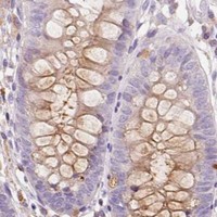
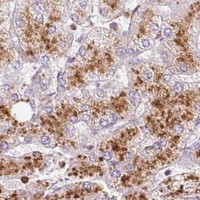
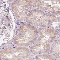
Plan paraffin-section TMC8 IHC using reported cytoplasmic staining in bone marrow hematopoietic cells and prostate glandular cells as reference patterns (HPA tissue IHC). The IHC-P catalog antibody starts at 5 μg/mL (datasheet: A06892-1); interpret staining cautiously because agreement with RNA data is low (HPA tissue IHC).

Evidence assembled Oct 2026 · For research use; verify linked source records and product datasheet before use
Immunohistochemistry protocol sheet for TMC8 (IHC for TMC8): expected localisation Cytoplasmic tissue staining; ER, Golgi and nuclear membranes are molecular expectations (HPA tissue IHC; UniProt), antibody A06892, validated IHC image, and IHC protocol steps
Printable TMC8 IHC protocol sheet — expected localisation Cytoplasmic tissue staining; ER, Golgi and nuclear membranes are molecular expectations (HPA tissue IHC; UniProt), antibody A06892, controls and protocol steps. Open the full TMC8 IHC guide →

TMC8 Immunohistochemistry Experimental Design Guide

Expected localisation, validated protocols, controls and antibodies — the at-a-glance facts below, then the full design guide.

Must know before staining
Expected localisation Cytoplasmic tissue staining; ER, Golgi and nuclear membranes are molecular expectations (HPA tissue IHC; UniProt)
Staining pattern Cytoplasmic staining in several tissues, including glandular cells (HPA tissue IHC)
Antigen retrieval Tris-EDTA pH 9.0 HIER, 95–98 °C, 20 min (rule: nuclear antigen)
Positive control ⓘ Bone marrow+4 more · see all
Negative control ⓘ Breast+4 more · see all
Important caveats
Reasons your staining may differ from the expected pattern.
Fixation Keep fixation consistent across sections. (standard IHC practice; not target-specific)
Caveat Staining has low consistency with RNA data (HPA tissue IHC)
Regulation Expression induction is unreported (UniProt)
Isoform / epitope 2 isoforms; epitope differences are unmapped, so check cytoplasmic versus lumenal location (UniProt)
Section 1

Recommended TMC8 IHC & IF Protocols

The catalog antibody protocol is paired with published TMC8 IHC methods from three articles (PMC8108259; PMC7138561; PMC13462283).

Recommended immunohistochemistry (IHC-P) protocol parameters
SampleParaffin-embedded human tonsil tissue; fixative not specified (datasheet A06892)
FixationImage fixative and duration unreported (datasheet A06892); verify before use.
Sectioning4–5 µm sections on charged slides (standard)
DeparaffinisationXylene, graded ethanol series to water (standard)
Antigen retrievalHeat-induced epitope retrieval in Tris-EDTA buffer, pH 9.0, 20 min at 95–98 °C (standard rule: nuclear antigen)
Peroxidase block3% H2O2, 10 min, room temperature (standard)
Blocking10% normal serum of the secondary host, 30 min, room temperature (standard)
Primary antibodyRabbit anti-TMC8, 1:100-1:300 (datasheet A06892)
Primary incubationOvernight at 4 °C (standard)
DetectionHRP-polymer secondary, DAB chromogen 5–10 min (standard)
CounterstainHematoxylin, blue, dehydrate and mount (standard)
Expected resultTMC8-positive staining in hematopoietic cells of bone marrow (HPA tissue IHC: High). HPA tissue profile: Cytoplasmic expression in several tissues. No signal in the no-primary control.
💡Decision noteStart with Tris-EDTA pH 9.0 HIER at 95–98 °C for 20 min (page retrieval); compare boiling citrate for 20 min (PMC8108259).
Section 2

What Is the Expected TMC8 Staining Pattern?

TMC8 is an 8-pass membrane protein at ER, Golgi and nuclear membranes in keratinocytes (UniProt Q8IU68 topology and subcellular location). In paraffin-section IHC, expect mainly cytoplasmic staining in selected cells, including prostate glandular cells, testis Leydig cells and bone marrow hematopoietic cells (HPA tissue IHC). Treat this as a provisional pattern: HPA rates tissue IHC reliability Uncertain because staining has low consistency with RNA expression (HPA tissue IHC).

What am I looking at on my slide?
Cytoplasmic stain in prostate glandular cells or testis Leydig cells, with visible cell boundaries.These cell types show High staining in HPA tissue IHC. Score the fraction and intensity of stained target cells; compare with adjacent cells and controls (HPA tissue IHC; general IHC practice).
Predominantly extracellular, or uniformly filled nuclear, signal with little cytoplasmic staining.This does not fit HPA’s cytoplasmic tissue profile (HPA tissue IHC). A nuclear-envelope rim can be plausible because UniProt places TMC8 at the nucleus membrane; verify compartment boundaries before rejecting the result (UniProt Q8IU68 subcellular location; general IHC practice).
Strong staining in parathyroid glandular cells while expected positive cells are weak.HPA reports parathyroid glandular cells as Not detected (HPA tissue IHC). Consider antibody cross-reactivity or endogenous detection activity; compare with an appropriate primary-antibody omission control (general IHC practice).
Diffuse color across cells and surrounding tissue, obscuring cellular detail.This pattern is difficult to score as TMC8 because HPA describes cellular cytoplasmic staining (HPA tissue IHC). Background can arise from nonspecific binding or detection chemistry; examine the negative control and tissue morphology (general IHC practice).
No staining in prostate glandular cells or testis Leydig cells.Both are High in HPA tissue IHC, so an absent signal calls for a control and workflow check (HPA tissue IHC; general IHC practice). HPA reliability is Uncertain, so one negative specimen alone does not settle target expression (HPA tissue IHC).
💡Expected TMC8 appearanceCall a result provisionally positive when selected glandular, Leydig or hematopoietic cells show interpretable cytoplasmic chromogen, potentially concentrated around membrane-bound organelles; broad extracellular or uniformly filled nuclear color warrants a control check (HPA tissue IHC; UniProt Q8IU68 subcellular location; general IHC practice).
How each factor affects the staining
Membrane topologyTMC8 has 8 transmembrane segments with cytoplasmic and lumenal regions (UniProt Q8IU68 topology). Epitope location is unspecified here, so topology alone cannot predict retrieval performance.
Reference tissue choiceProstate glandular cells and testis Leydig cells are High; parathyroid glandular cells are Not detected (HPA tissue IHC). Compare named cell types, not whole-section color.
IHC evidence strengthThe HPA tissue profile and antibody HPA054429 have Uncertain IHC assessments (HPA tissue IHC; HPA antibodies). Treat a matching pattern as supportive, not definitive.
RNA and protein agreementHPA reports group-enriched RNA in bone marrow, intestine and lymphoid tissue, but low consistency between RNA and antibody staining (HPA tissue IHC). Do not infer an individual cell’s stain intensity from tissue RNA.
IF/ICC Q&A: where should signal appear?HPA reports Golgi localization in HaCaT and MCF-7 images, rated Uncertain (HPA subcellular ICC-IF). UniProt also lists ER and nuclear membranes in keratinocytes (UniProt Q8IU68 subcellular location).
Isoforms and processingUniProt lists 2 isoforms, one chain spanning residues 1–726 and no annotated signal peptide or propeptide (UniProt Q8IU68). The supplied record does not establish which isoform an antibody detects or any shedding pattern.
Fixation and retrieval evidenceTarget-specific fixation effects are not established by the supplied assay evidence. Verify with a matched IHC source before attributing a result to fixation.
Why is my staining missing, weak or wrong?
SituationLikely causeNext action
Positive-control cells show no chromogen.The section, primary antibody, retrieval or detection step may have failed (general IHC practice); a TMC8-specific fixation effect is unreported in the supplied sources.Confirm that the control contains prostate glandular or testis Leydig cells reported High by HPA; check each IHC step with suitable process controls (HPA tissue IHC; general IHC practice).
Signal is strong across nearly every cell, including an expected negative population.Nonspecific antibody binding or endogenous detection activity is possible (general IHC practice). HPA reports parathyroid glandular cells as Not detected (HPA tissue IHC).Inspect primary-antibody omission and detection controls; assess whether color follows cell boundaries and whether the negative cell population remains negative (general IHC practice).
Only nuclei appear filled with chromogen.Filled nuclear staining differs from the HPA cytoplasmic tissue profile; UniProt’s nuclear-membrane location refers to a membrane, not the entire nucleus (HPA tissue IHC; UniProt Q8IU68 subcellular location).Review counterstain and focal plane, then compare nuclear interiors with nuclear rims and cytoplasm in control cells (general IHC practice).
Brown haze obscures glandular or marrow cell boundaries.Nonspecific background or detection activity can prevent cellular scoring (general IHC practice).Review blocking, washing and detection controls; score only signal that can be assigned to cells with intact morphology (general IHC practice).
One specimen disagrees with the HPA positive pattern.HPA rates the tissue IHC profile Uncertain owing to low staining–RNA consistency (HPA tissue IHC). A single mismatch cannot identify its cause.Compare another documented positive cell population and the specimen’s negative controls before interpreting the mismatch (HPA tissue IHC; general IHC practice).
A concentrated perinuclear signal seems different from broad cytoplasmic IHC.Golgi localization is reported by HPA ICC-IF with Uncertain confidence; UniProt also lists ER and Golgi membranes in keratinocytes (HPA subcellular ICC-IF; UniProt Q8IU68 subcellular location).Check that the signal remains cell-associated and compare it with the slide’s controls; do not require IF-like organelle resolution in chromogenic paraffin IHC (general IHC practice).

Sample controls for TMC8 IHC & IF

🧪Run prostate first: glandular cells should stain (HPA: High in prostate glandular cells). Use breast adipocytes as the negative tissue (HPA: Not detected in breast adipocytes); compare adjacent nonglandular areas on the prostate slide with background controls, without assuming those cells lack TMC8.
Positive control tissue: Bone marrow (Hematopoietic cells, HPA High)
Negative control tissue: Breast (HPA Not detected)
ICC-IF cell lines (HPA subcellular resource): HPA ICC-IF images show TMC8 in HaCaT, MCF-7, with annotated localisation: Golgi apparatus (uncertain) (HPA subcellular).
Technical controls: Include a no-primary (secondary-only) control, an isotype-matched control for a monoclonal primary or host-matched nonimmune IgG for a polyclonal primary, and a TMC8 knockout or cognate peptide-block control (selected-SKU tissue-IHC caption: peptide block shown for A06892 in tonsil). On prostate sections, quench endogenous peroxidase and distinguish glandular cell staining from luminal deposits before scoring (HPA: High in prostate glandular cells; standard IHC practice).
⚠️Feasibility: A target-specific fixation window or fixation effect is unreported in the supplied evidence; the A06892 paraffin-section tonsil caption does not report a fixative (selected-SKU tissue-IHC caption). TMC8-specific antigen retrieval requirements are unreported, so retrieval needs empirical optimization for the catalog antibody. The evidence does not establish whether frozen sections or IF/ICC are easier; for IF/ICC, interpret Golgi-like signal cautiously because that localization is uncertain (HPA subcellular: Golgi apparatus, uncertain).

HPA tissue IHC evidence for TMC8

Comprehensive Human Protein Atlas IHC scoring per tissue (reliability: Uncertain — Low consistency between antibody staining and RNA expression data. Pending external verification.). Rows are taken directly from the HPA tissue chart — click any row's HPA link to view the source.

Positive expression · recommended positive controls

TissueCell typeLevelEvidenceSource
Bone marrow Hematopoietic cells High Protein (IHC) HPA →
Cervix Glandular cells High Protein (IHC) HPA →
Epididymis Glandular cells High Protein (IHC) HPA →
Pancreas Exocrine glandular cells High Protein (IHC) HPA →
Prostate Glandular cells High Protein (IHC) HPA →

Undetected expression · recommended negative controls

TissueCell typeLevelEvidenceSource
Breast Adipocytes Not detected Protein (IHC) HPA →
Cerebellum Cells in granular layer Not detected Protein (IHC) HPA →
Cerebral cortex Glial cells Not detected Protein (IHC) HPA →
Endometrium Cells in endometrial stroma Not detected Protein (IHC) HPA →
Hippocampus Glial cells Not detected Protein (IHC) HPA →
Section 3

Advanced TMC8 IHC Tips

Troubleshoot TMC8 chromogenic IHC by checking retrieval, tissue handling, compartmental pattern and controls before assigning biological meaning to staining.

How should I troubleshoot weak TMC8 staining after antigen retrieval?
Start with Tris-EDTA at pH 9.0, heated to 95–98 °C for 20 min (page retrieval setting). Keep section thickness, heating and cooling consistent across test and control slides so retrieval differences do not masquerade as tissue differences (standard IHC practice). If staining remains weak, compare a shorter exposure or an alternative retrieval buffer on serial sections, recording tissue damage and background alongside signal (standard IHC practice). The page setting is a starting condition rather than evidence that TMC8 requires alkaline retrieval; its annotated locations are ER, Golgi and nuclear membranes (UniProt Q8IU68 localisation).
Could fixation explain inconsistent TMC8 staining between paraffin blocks?
Yes, fixation differences can change epitope access and tissue preservation in paraffin IHC (standard IHC practice). TMC8-specific fixation sensitivity is unknown here: the selected A06892 image shows paraffin-embedded human tonsil, but its caption does not state the fixative (A06892 tissue-IHC caption). Compare blocks with documented fixation histories, then stain matched sections in one run with the same retrieval and detection conditions (standard IHC practice). Record signal and morphology together, since weak staining in an overprocessed section cannot by itself establish low TMC8 abundance (standard IHC practice). Do not assign an optimal fixation duration without a matched comparison (standard IHC practice).
What staining pattern should prompt a closer localisation check?
Assess TMC8 primarily for cytoplasmic or membrane-associated staining, because tissue IHC reports cytoplasmic expression and UniProt places it at ER, Golgi and nuclear membranes (HPA tissue IHC profile; UniProt Q8IU68 localisation). A compact perinuclear pattern can be compatible with Golgi localisation, although that subcellular assignment is uncertain (HPA subcellular: Golgi apparatus, uncertain). Diffuse nuclear chromogen alone needs separate verification because nuclear membrane localisation does not establish nucleoplasmic staining (UniProt Q8IU68 localisation; standard IHC interpretation). Compare the pattern with tissue morphology and matched controls before calling a compartment positive, particularly because tissue staining has uncertain reliability (HPA tissue IHC reliability).
How do I assess whether retrieval or antibody binding misses a TMC8 isoform?
TMC8 has 2 annotated isoforms, but the supplied evidence does not map this antibody’s epitope to either one (UniProt Q8IU68 isoforms; A06892 tissue-IHC caption). Obtain the immunogen or epitope sequence and align it with both isoforms before claiming shared recognition (standard IHC validation practice). Topology places alternating cytoplasmic and lumenal regions around 8 transmembrane segments, so epitope position can inform an accessibility hypothesis without proving staining performance (UniProt Q8IU68 topology). Compare serial sections under the same retrieval conditions and use an independent antibody with a mapped epitope when isoform discrimination matters (standard IHC validation practice).
How can IF help check the cell type and compartment seen in TMC8 IHC?
On a separate IF/ICC workflow, multiplex TMC8 with a validated marker for the cell population being assessed in the chromogenic section (standard IF practice). For example, selecting a glandular-cell marker is pertinent when examining reported prostate glandular-cell staining (HPA tissue IHC: High in prostate glandular cells). Choose fluorophores after inspecting unstained tissue autofluorescence, and include single-label controls to assess spectral bleed-through (standard IF practice). Match permeabilisation to the mapped epitope: cytoplasmic stretches and lumenal loops have different membrane access, while this antibody’s epitope is unspecified (UniProt Q8IU68 topology; A06892 tissue-IHC caption). Interpret any Golgi overlap cautiously because the reported Golgi assignment is uncertain (HPA subcellular).
How do I separate TMC8 chromogen signal from nonspecific background?
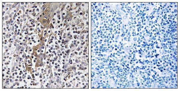
Inspect a no-primary control and the peptide-blocked comparison before treating brown deposits as antibody-dependent TMC8 staining (standard IHC practice; A06892 tissue-IHC caption). The selected tonsil image includes a synthesized-peptide block, which supports a competition check for that image but does not establish specificity across tissues (A06892 tissue-IHC caption; standard IHC interpretation). Apply an appropriate peroxidase block before DAB detection, and compare deposits with the no-primary section to identify endogenous enzyme contribution (standard chromogenic IHC practice). If diffuse background persists, titrate antibody and detection strength on serial sections while preserving a consistent counterstain (standard IHC practice).
What is a defensible way to quantify TMC8 staining across sections? ⚠ ANSWER MARKED FOR VERIFICATION
Define the relevant cell population and tissue area before scoring, then apply the same threshold and chromogen development across slides (standard IHC quantification practice). Report the percentage of positive cells and staining intensity as an H-score, or count positive cells per mm² when spatial density answers the question (standard IHC quantification practice). Normalise positive counts to the total eligible cells or viable tissue area, and exclude folds, edges and necrosis using prespecified rules (standard IHC quantification practice). Stratify results by cell type because reported staining differs across tissues and cells, while the tissue IHC evidence has uncertain reliability (HPA tissue IHC profile; HPA tissue IHC reliability).
When should a TMC8-positive IHC result be treated as questionable?
Treat a signal as more credible when it follows identifiable cells, shows a plausible cytoplasmic or membrane-associated pattern, and exceeds matched control staining (HPA tissue IHC profile; UniProt Q8IU68 localisation; standard IHC interpretation). Recheck isolated nucleoplasmic deposits or staining in an unexpected cell population against morphology and independent evidence rather than assigning them to TMC8 (UniProt Q8IU68 localisation; standard IHC interpretation). Edge staining, necrotic deposits and signal reproduced in a no-primary control suggest section or detection artefact, including endogenous enzyme activity (standard chromogenic IHC practice). A peptide-blocked tonsil image is available, but broader tissue conclusions remain limited by uncertain HPA staining reliability (A06892 tissue-IHC caption; HPA tissue IHC reliability).
Boster reagents

Best TMC8 / Transmembrane channel-like protein 8 IHC Antibodies

Both anti-TMC8 antibodies have human IHC images; one also has a human-cell IF image (catalog image captions). Human, mouse and rat reactivity is listed for both (catalog reactivity).

Real IHC data Immunohistochemistry analysis of paraffin-embedded human tonsil, using TMC8 Antibody. The picture on the right is blocked with the synthesized peptide.
Anti-TMC8/Ever2 Antibody
Cat # A06892
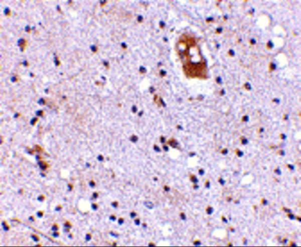
Real IHC data Immunohistochemistry of EVER2 in human brain with EVER2 antibody at 5 μg/mL.
Anti-EVER2 TMC8 Antibody
Cat # A06892-1

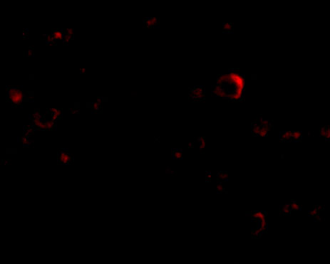
A06892 has an IHC image of paraffin-embedded human tonsil with peptide blocking, and lists IF as an application (A06892 image caption; A06892 applications). A06892-1 has an IHC image of human brain and an IF image of human brain cells (A06892-1 image captions).

Which to pick: For paraffin-section IHC, choose A06892 when the human tonsil example is most relevant; its caption identifies paraffin embedding but does not report the fixative (A06892 IHC image caption). For IF/ICC planning, choose A06892-1 for its human brain-cell IF image; ICC performance is unreported (A06892-1 IF image caption; A06892-1 applications). Both list human, mouse and rat reactivity, but their supplied images show human samples only; A06892-1 lists IHC-P, while the processing and fixative of its brain image are unreported (catalog reactivity; A06892-1 applications; catalog image captions).

Each figure is that product's own IHC / IF validation image from its datasheet.

References

  1. UniProt Consortium. UniProt entry Q8IU68 (TMC8_HUMAN, Transmembrane channel-like protein 8).
  2. Human Protein Atlas. TMC8 tissue IHC expression (reliability: Uncertain).
  3. Human Protein Atlas. TMC8 subcellular location (ICC-IF): Localized to the Golgi apparatus..
  4. Human Protein Atlas. TMC8 antibody validation summary (2 antibodies).
  5. Comprehensive co-expression analysis reveals TMC8 as a prognostic immune-associated gene in head and neck squamous cancer. Oncology letters 2021 — PMC8108259.
  6. Pan-Cancer Analysis Reveals the Signature of TMC Family of Genes as a Promising Biomarker for Prognosis and Immunotherapeutic Response. Frontiers in immunology 2021 — PMC8660091.
  7. Prognostic values of immune scores and immune microenvironment-related genes for hepatocellular carcinoma. Aging 2020 — PMC7138561.
  8. Elevated TMC8 expression correlates with CD8(+) T cell infiltration and favorable survival in oral squamous cell carcinoma. Translational cancer research 2026 — PMC13462283.
  9. PubMed PMID:12426567 — UniProt-cited evidence.
  10. PubMed PMID:12906855 — UniProt-cited evidence.
  11. PubMed PMID:16625196 — UniProt-cited evidence.