TMIGD2 / Transmembrane and immunoglobulin domain-containing protein 2 · IHC design guide

Design Immunohistochemistry for TMIGD2

Plan TMIGD2 chromogenic IHC in paraffin sections using the membranous glandular and epithelial pattern reported by HPA tissue IHC. Compare positive gallbladder glandular cells with an unstained tissue control, and check the antibody epitope against the 2 UniProt isoforms.

Evidence assembled Oct 2026 · For research use; verify linked source records and product datasheet before use
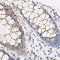
Immunohistochemistry protocol sheet for TMIGD2 (IHC for TMIGD2): expected localisation Glandular and epithelial cell membranes (HPA tissue IHC), antibody A11415, validated IHC image, and IHC protocol steps
Printable TMIGD2 IHC protocol sheet — expected localisation Glandular and epithelial cell membranes (HPA tissue IHC), antibody A11415, controls and protocol steps. Open the full TMIGD2 IHC guide →

TMIGD2 Immunohistochemistry Experimental Design Guide

Expected localisation, validated protocols, controls and antibodies — the at-a-glance facts below, then the full design guide.

Must know before staining
Expected localisation Glandular and epithelial cell membranes (HPA tissue IHC)
Staining pattern Membranous staining in most glandular and epithelial cells (HPA tissue IHC)
Antigen retrieval Citrate pH 6 HIER, heat-mediated (datasheet A11415)
Positive control ⓘ Gallbladder+4 more · see all
Negative control ⓘ Adipose tissue+4 more · see all
Important caveats
Reasons your staining may differ from the expected pattern.
Fixation Formaldehyde-fixed paraffin sections were used (datasheet A11415, IHC-P) (selected-SKU IHC image A11415)
Caveat Some glandular epithelia show no staining (HPA tissue IHC)
Regulation No staining-related regulation specified (UniProt)
Isoform / epitope 2 isoforms; check whether the epitope is extracellular or cytoplasmic (UniProt)
Section 1

Recommended TMIGD2 IHC & IF Protocols

The catalog antibody has a datasheet IHC-P protocol (datasheet: A11415). Published TMIGD2 IHC protocols cover oral squamous cell carcinoma (PMC6476002) and glioma (PMC10227580).

Recommended immunohistochemistry (IHC-P) protocol parameters
SampleFormaldehyde-fixed, paraffin-embedded human small intestine tissue (datasheet A11415)
FixationImage formalin-fixed; duration unreported (datasheet A11415); verify before use.
Sectioning4–5 µm sections on charged slides (standard)
DeparaffinisationXylene, graded ethanol series to water (standard)
Antigen retrievalHeat retrieval: Citrate pH 6 (datasheet A11415); 20 min, 95–100 °C (standard)
Peroxidase block3% H2O2, 10 min, room temperature (standard)
Blocking10% serum (datasheet A11415)
Primary antibodyRabbit anti-TMIGD2, 5 μg/mL (datasheet A11415)
Primary incubationOvernight at 4 °C (standard)
DetectionHRP-polymer secondary, DAB chromogen 5–10 min (standard)
CounterstainHematoxylin, blue, dehydrate and mount (standard)
Expected resultTMIGD2-positive staining in glandular cells of gallbladder (HPA tissue IHC: High). HPA tissue profile: Membranous expression in most glandular and epithelial cells. No signal in the no-primary control.
💡Decision noteStart with citrate pH 6 heat retrieval for the catalog antibody (datasheet: A11415). The glioma protocol used retrieval at 98°C for 20 min (PMC10227580).
Section 2

What Is the Expected TMIGD2 Staining Pattern?

TMIGD2 is a cell membrane protein with an extracellular region at residues 23–150 and one transmembrane segment at 151–171 (UniProt Q96BF3 topology). In paraffin section IHC, expect membranous staining in many glandular and epithelial cells (HPA tissue IHC: membranous expression). HPA rates its tissue staining evidence Approved, meaning external characterization supports the antibody staining (HPA tissue IHC: reliability description).

What am I looking at on my slide?
Membrane outlines in gallbladder glandular cells, with strong chromogen (HPA tissue IHC: High).This is a well supported positive pattern: the compartment matches TMIGD2's cell membrane location (UniProt Q96BF3: subcellular location), and HPA scores these cells High (HPA tissue IHC). Compare staining with neighboring structures on the same section before judging its extent.
Membrane outlines in colon or duodenal glandular cells, or kidney tubular cells (HPA tissue IHC: Medium).Moderate staining in these specified cells is consistent with HPA observations (HPA tissue IHC: Medium). A positive slide need not resemble the stronger gallbladder example; assess the named cell population and its membrane pattern together.
Strong nuclear staining, or broad cytoplasmic color without a discernible membrane pattern.Treat this as questionable for TMIGD2 because UniProt places it at the cell membrane (UniProt Q96BF3: subcellular location), and HPA describes membranous epithelial staining (HPA tissue IHC: profile). Check controls and detection background before assigning a biological meaning.
Signal in adipocytes or bone marrow hematopoietic cells (HPA tissue IHC: Not detected).These are unexpected cell populations in the cited HPA images (HPA tissue IHC: Not detected). Consider nonspecific antibody binding or endogenous chromogenic detection activity; verify cell identity and compare a control lacking primary antibody. A negative HPA observation is not proof that every specimen must be negative.
Uniform haze across tissue, or no signal in gallbladder glandular cells (HPA tissue IHC: High).Uniform haze limits interpretation and calls for a detection background check (general IHC practice). A blank expected positive area calls for review of tissue preservation, staining steps, and antibody performance (general IHC practice); HPA's High score is a reference pattern, not a guarantee for every section.
💡Expected TMIGD2 appearanceCall a result positive when chromogen outlines glandular or epithelial cell membranes, strongest in the HPA gallbladder example (High; HPA tissue IHC), while prominent nuclear color or widespread cell independent haze should trigger a false positive check (UniProt Q96BF3: cell membrane; general IHC practice).
How each factor affects the staining
Compartment and epitopeTMIGD2 has extracellular residues 23–150, a transmembrane segment at 151–171, and a cytoplasmic region at 172–282 (UniProt Q96BF3 topology). The supplied evidence does not locate this antibody's epitope, so these boundaries cannot specify a retrieval condition.
Cell population and tissueHPA scores gallbladder glandular cells High; colon and duodenal glandular cells, kidney tubular cells, and placental decidual cells Medium (HPA tissue IHC). Compare like cell populations when interpreting intensity; a whole tissue label alone can hide different cell types.
Source disagreementHPA reports Medium staining in cerebellar granular layer cells (HPA tissue IHC), whereas UniProt says cerebellum is not detected at protein level (UniProt Q96BF3: tissue specificity). Treat cerebellar staining as unresolved and avoid using it as the sole positive control.
Antibody evidenceHPA lists rabbit polyclonal HPA011081 as IHC Approved (HPA: antibodies; HPA tissue IHC: reliability). That supports use of its reported pattern as a reference, but the supplied record does not report an IHC Enhanced validation result.
IF/ICC Q: What location should be assessed?A: Assess membrane associated signal: HPA summarizes the ICC/IF location as Membrane, but lists no main location or cell line images (HPA subcellular ICC/IF). UniProt also places TMIGD2 at the cell membrane (UniProt Q96BF3: subcellular location).
Why is my staining missing, weak or wrong?
SituationLikely causeNext action
Gallbladder glandular cells are blank despite an intended positive control (HPA tissue IHC: High).A failed IHC step, unsuitable antibody dilution, or a specimen issue is possible (general IHC practice); the HPA score alone cannot identify which.Review section quality, reagent sequence, retrieval, antibody incubation, and chromogen development against the laboratory's established IHC workflow (general IHC practice).
Color covers nuclei more strongly than cell borders.This conflicts with the reported membrane location (UniProt Q96BF3: subcellular location; HPA tissue IHC: profile). Nonspecific staining is possible.Check a control lacking primary antibody and reassess whether glandular cell membranes are separately outlined (general IHC practice; HPA tissue IHC: membranous profile).
Adipocytes stain alongside glandular cells.HPA records adipocytes as Not detected in adipose tissue and breast (HPA tissue IHC). The adipocyte signal may reflect background or nonspecific binding.Score the two cell populations separately and compare a control lacking primary antibody; do not count adipocyte color as supporting TMIGD2 staining (general IHC practice; HPA tissue IHC).
Chromogen is diffuse across cells and empty areas of the section.Widespread color can arise from detection background or incomplete washing (general IHC practice), so membrane localization cannot be judged reliably.Inspect the control lacking primary antibody, washing, blocking, and chromogen development; then reassess the membrane pattern (general IHC practice; HPA tissue IHC: profile).
Cerebellar granular layer cells stain, but the result seems inconsistent.The sources differ: HPA scores these cells Medium, while UniProt reports cerebellum not detected at protein level (HPA tissue IHC; UniProt Q96BF3: tissue specificity).Record the observed cell type and pattern, and compare a better supported positive such as gallbladder glandular cells (HPA tissue IHC: High). Do not resolve the source disagreement by intensity alone.
A weak section is called negative because its signal is below the gallbladder example.HPA reports Medium staining in several specified cell populations, including colon glandular and kidney tubular cells (HPA tissue IHC); gallbladder is High.Judge membrane localization and the relevant cell population before applying an intensity threshold; document the tissue and cells assessed (HPA tissue IHC: profile and tissue levels).

Sample controls for TMIGD2 IHC & IF

🧪Run gallbladder first and score its glandular cells for staining (HPA: High in gallbladder glandular cells). Use adipose tissue as the negative comparator and score its adipocytes for background staining (HPA: Not detected in adipocytes); on the gallbladder slide, treat only demonstrably unstained cells as internal negatives, without assuming vascular cells are negative (UniProt Q96BF3 tissue specificity: vascular expression).
Positive control tissue: Gallbladder (Glandular cells, HPA High)
Negative control tissue: Adipose tissue (HPA Not detected)
ICC-IF cell lines (HPA subcellular resource): HPA carries no ICC-IF cell line for TMIGD2; derive a cell-line control from the positive tissue's cell type (Glandular cells) and confirm it by RNA or western blot first.
Technical controls: Include a no-primary, secondary-only control and a concentration-matched isotype control of the primary antibody’s host species and immunoglobulin class; use TMIGD2 knockout material or peptide competition, if a suitable immunizing peptide is available, as a biological specificity control (standard IHC practice). Block endogenous peroxidase and assess background on the gallbladder section before interpreting chromogenic signal (standard chromogenic IHC practice).
⚠️Feasibility: A target-specific fixation window or fixation effect is unreported, and the selected A11415 tissue-IHC caption’s fixative field is unreported (selected A11415 caption metadata). The paraffin-section example uses heat-mediated citrate retrieval at pH 6, but does not establish that retrieval is required (selected A11415 tissue-IHC caption). There is no matched frozen-section or IF/ICC evidence here to show either is easier; when scoring gallbladder, distinguish glandular signal from vascular staining because TMIGD2 is reported in vessels (UniProt Q96BF3 tissue specificity).

HPA tissue IHC evidence for TMIGD2

Comprehensive Human Protein Atlas IHC scoring per tissue (reliability: Approved — External characterization data supports antibody staining.). Rows are taken directly from the HPA tissue chart — click any row's HPA link to view the source.

Positive expression · recommended positive controls

TissueCell typeLevelEvidenceSource
Gallbladder Glandular cells High Protein (IHC) HPA →
Cerebellum Cells in granular layer Medium Protein (IHC) HPA →
Colon Glandular cells Medium Protein (IHC) HPA →
Duodenum Glandular cells Medium Protein (IHC) HPA →
Kidney Cells in tubules Medium Protein (IHC) HPA →

Undetected expression · recommended negative controls

TissueCell typeLevelEvidenceSource
Adipose tissue Adipocytes Not detected Protein (IHC) HPA →
Bone marrow Hematopoietic cells Not detected Protein (IHC) HPA →
Breast Adipocytes Not detected Protein (IHC) HPA →
Caudate Glial cells Not detected Protein (IHC) HPA →
Cervix Glandular cells Not detected Protein (IHC) HPA →
Section 3

Advanced TMIGD2 IHC Tips

Troubleshoot TMIGD2 staining by checking retrieval, cellular compartment, and tissue context before changing detection or scoring.

What should I change when TMIGD2 staining is weak after antigen retrieval?
Start with heat-mediated citrate retrieval at pH 6 for paraffin-section TMIGD2 IHC (datasheet A11415). The selected small-intestine image used this retrieval method, so compare your run with a matched positive control before changing conditions (selected-SKU caption). Check that sections received consistent heating and cooling, then titrate retrieval duration while keeping the buffer fixed (standard IHC practice). If staining remains weak, test an alternative retrieval buffer on adjacent sections as a fallback and assess tissue integrity alongside signal (standard IHC practice). Score membrane staining separately from diffuse cytoplasmic color because TMIGD2 is assigned to the cell membrane (UniProt Q96BF3).
Could fixation explain inconsistent TMIGD2 staining between paraffin blocks?
Target-specific TMIGD2 sensitivity to fixation is unknown; the selected image describes formaldehyde-fixed paraffin tissue without comparing fixatives or fixation times (selected-SKU caption). Record each block’s fixative and processing history before attributing differences in staining to biological expression (standard IHC practice). Compare blocks in the same staining run using the same pH 6 citrate retrieval and detection settings (datasheet A11415; standard IHC practice). Include a previously staining control section to reveal run-level variation, and inspect morphology for processing damage (standard IHC practice). Do not infer fixation sensitivity from membrane topology or reported tissue staining patterns (UniProt Q96BF3; HPA tissue IHC).
Where should convincing TMIGD2 signal appear in tissue sections?
Prioritize staining along cell boundaries because TMIGD2 is a cell-membrane protein and HPA describes membranous staining in most glandular and epithelial cells (UniProt Q96BF3; HPA tissue IHC). Its extracellular segment spans residues 23–150, followed by a transmembrane segment at 151–171 and cytoplasmic residues 172–282 (UniProt Q96BF3 topology). Examine intact glandular profiles and compare the signal with neighboring cells rather than judging isolated brown deposits (standard IHC practice). HPA reports high staining in gallbladder glandular cells and medium staining in colon glandular cells, useful comparison contexts (HPA tissue IHC). Treat predominantly nuclear staining as a localisation mismatch requiring control review (UniProt Q96BF3).
How could epitope location affect TMIGD2 IHC results?
TMIGD2 has 2 reported isoforms, but the supplied record does not map their sequence differences or this antibody’s epitope (UniProt Q96BF3; datasheet A11415). The extracellular region includes an Ig-like domain at residues 23–129 and glycosylation sites at 73, 105, and 127 (UniProt Q96BF3). The cytoplasmic region includes reported phosphorylation sites at 192, 220, and 222 (UniProt Q96BF3). Check the antibody’s documented immunogen or epitope before interpreting absent staining as isoform loss (standard IHC practice). Compare retrieval conditions on adjacent sections and retain morphology and membrane localisation as acceptance criteria (datasheet A11415; UniProt Q96BF3; standard IHC practice).
How should I check a TMIGD2 pattern by multiplex immunofluorescence?
Pair TMIGD2 with a validated marker for the expected epithelial, endothelial, or lymphocyte population, chosen for the tissue being examined (UniProt Q96BF3 tissue specificity; standard IF practice). Place the weaker signal in a bright, spectrally separated channel and inspect unstained tissue for autofluorescence before assigning fluorophores (standard IF practice). TMIGD2 spans extracellular residues 23–150 and cytoplasmic residues 172–282, so select permeabilisation according to the antibody’s documented epitope side (UniProt Q96BF3 topology). Minimize permeabilisation when testing an accessible extracellular epitope, while assessing intracellular access for a cytoplasmic epitope (standard IF practice). Confirm boundary signal with single-stain controls; no HPA ICC/IF images are supplied (HPA subcellular record; standard IF practice).
How can I reduce diffuse brown staining without losing TMIGD2 signal?
First compare the stained section with a no-primary control to distinguish primary-antibody signal from detection-system background (standard IHC practice). The selected A11415 image used 10% serum blocking for 1 hour at room temperature and an HRP secondary at 1:250 (selected-SKU caption). For chromogenic IHC, check the peroxidase block, washing, and DAB development time while keeping a positive control in each run (standard IHC practice). Titrate primary concentration around the reported 5 µg/ml if background persists, and assess membrane contrast rather than total brown area (selected-SKU caption; UniProt Q96BF3). Record whether staining also appears in the no-primary control (standard IHC practice).
How should TMIGD2 staining be scored across tissue samples? ⚠ ANSWER MARKED FOR VERIFICATION
Define the cell population and compartment before scoring, because expected TMIGD2 staining is membranous and varies by tissue cell type (UniProt Q96BF3; HPA tissue IHC). For epithelial regions, report the percentage of membrane-positive cells and an H-score from predefined intensity categories (standard IHC practice). For scattered immune cells, report positive-cell density per mm² within a consistently defined tissue area (standard IHC practice). Normalize cell counts to the number of eligible cells or analyzed tissue area, and exclude folds, necrosis, and section edges (standard IHC practice). Keep retrieval, exposure to DAB, and scoring thresholds consistent across the comparison set (datasheet A11415; standard IHC practice).
How do I distinguish true TMIGD2 staining from artefact?
A plausible positive follows cell boundaries in an appropriate population, consistent with membrane localisation and HPA’s epithelial staining profile (UniProt Q96BF3; HPA tissue IHC). Compare glandular cells with adjacent tissue and controls; HPA reports high gallbladder glandular staining but no detectable signal in adipocytes (HPA tissue IHC). Question staining confined to nuclei, cut edges, folds, or necrotic areas because these patterns do not establish membrane localisation (UniProt Q96BF3; standard IHC practice). A no-primary control helps identify endogenous enzyme activity or detection background in chromogenic sections (standard IHC practice). Interpret discrepancies cautiously because HPA reports medium cerebellar granular-layer staining while UniProt reports no cerebellar protein detection (HPA tissue IHC; UniProt Q96BF3).
Boster reagents

Best TMIGD2 / Transmembrane and immunoglobulin domain-containing protein 2 IHC Antibodies

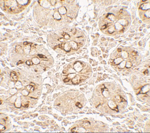
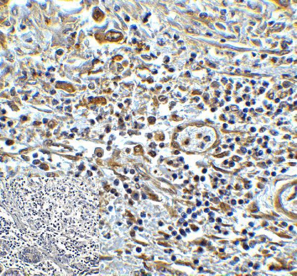
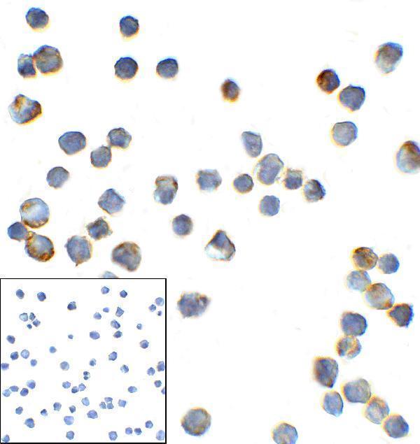
Human tissue IHC images show TMIGD2 staining in paraffin-embedded small intestine and colon carcinoma; IF tissue and ICC cell images are also available (catalog image captions).

Real IHC data Immunohistochemistry Validation of TMIGD2 in Human Small Intestine Tissue Immunohistochemical analysis of paraffin-embedded human small intestine tissue using anti-TMIGD2 antibody (A11415) at 5 μg /ml. Tissue was fixed with formaldehyde and blocked with 10% serum for 1 h at RT; antigen retrieval was by heat mediation with a citrate buffer (pH6). Samples were incubated with primary antibody overnight at 4˚ C. A goat anti-rabbit IgG H&L (HRP) at 1/250 was used as secondary. Counter stained with Hematoxylin.
Anti-TMIGD2 Antibody
Cat # A11415
Real IHC data Immunohistochemistry of TMIGD2 in human colon carcinoma tissue with TMIGD2 antibody at 5 μg/mL.
Anti-TMIGD2 Monoclonal Antibody [8A1]
Cat # M11415

A11415 has an IHC image from human paraffin-embedded small intestine fixed with formaldehyde (A11415 image caption). M11415 has an IHC image from human colon carcinoma, plus IF images from colon carcinoma tissue and ICC images from transfected HEK293 cells (M11415 image captions).

Which to pick: For paraffin-section IHC, choose rabbit antibody A11415 when its documented formaldehyde fixation and citrate-buffer retrieval conditions fit your sample (A11415 catalog entry and IHC image caption). Choose mouse monoclonal M11415 for IF or ICC; its IHC caption reports human colon carcinoma tissue but does not report the fixative (M11415 catalog entry and image captions). Neither is listed for cross-species work: both list Human reactivity only (catalog entries).

Each figure is that product's own IHC / IF validation image from its datasheet.

References

  1. UniProt Consortium. UniProt entry Q96BF3 (TMIG2_HUMAN, Transmembrane and immunoglobulin domain-containing protein 2).
  2. Human Protein Atlas. TMIGD2 tissue IHC expression (reliability: Approved).
  3. Human Protein Atlas. TMIGD2 subcellular location (ICC-IF): Membrane.
  4. Human Protein Atlas. TMIGD2 antibody validation summary (1 antibodies).
  5. The Expression Patterns and Associated Clinical Parameters of Human Endogenous Retrovirus-H Long Terminal Repeat-Associating Protein 2 and Transmembrane and Immunoglobulin Domain Containing 2 in Oral Squamous Cell Carcinoma. Disease markers 2019 — PMC6476002.
  6. TMIGD2 as a potential therapeutic target in glioma patients. Frontiers in immunology 2023 — PMC10227580.
  7. PubMed PMID:12975309 — UniProt-cited evidence.
  8. PubMed PMID:15489334 — UniProt-cited evidence.
  9. PubMed PMID:15340161 — UniProt-cited evidence.