This website uses cookies to ensure you get the best experience on our website.
- Table of Contents
16 Citations 8 Q&As
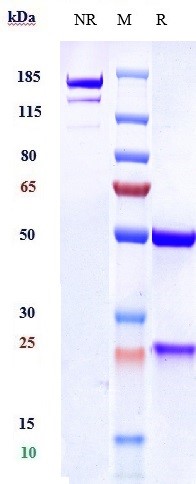
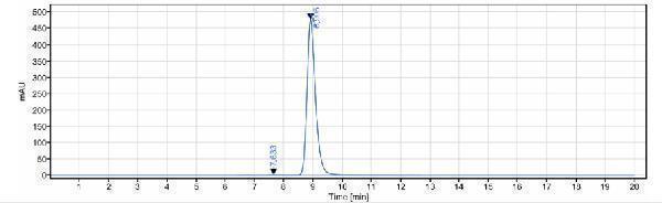
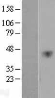
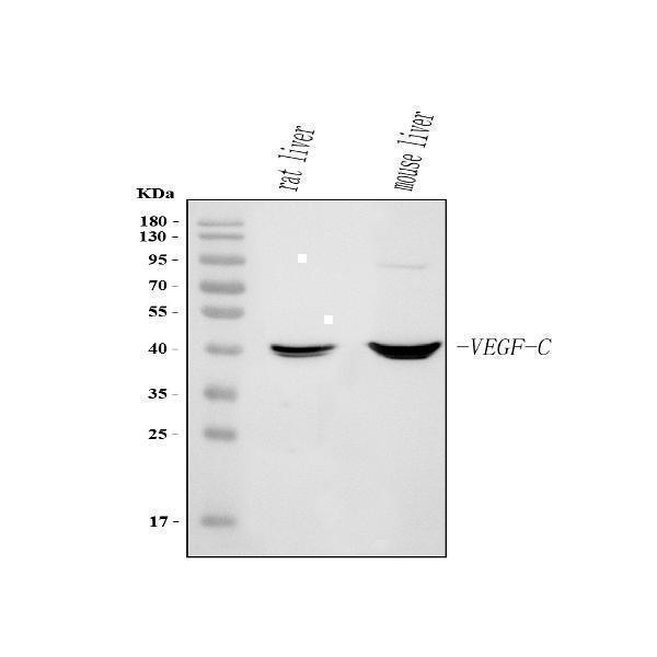
Facts about Vascular endothelial growth factor C.
Binds and activates KDR/VEGFR2 and FLT4/VEGFR3 receptors. .
Human | |
---|---|
Gene Name: | VEGFC |
Uniprot: | P49767 |
Entrez: | 7424 |
Belongs to: |
---|
PDGF/VEGF growth factor family |
Flt4 ligand; Flt4-L; vascular endothelial growth factor C; Vascular endothelial growth factor-related protein; VEGFC; VEGF-C; VRPFLT4 ligand DHM
Mass (kDA):
46.883 kDA
Human | |
---|---|
Location: | 4q34.3 |
Sequence: | 4; NC_000004.12 (176683538..176792922, complement) |
Spleen, lymph node, thymus, appendix, bone marrow, heart, placenta, ovary, skeletal muscle, prostate, testis, colon and small intestine and fetal liver, lung and kidney, but not in peripheral blood lymphocyte.
Secreted.
The body contains VEGF-C mRNA. It plays an important role in maintaining the development and growth of tissues. The VEGF-C mRNA can be detected in tumors by immunohistochemistry. This marker can also be found in breast and ovarian carcinomas. We will discuss the best methods for detecting VEGF-C mRNA.
The VEGFC gene encodes a protein that is part of the platelet-derived growth factor family. This protein is active in the angiogenesis and endothelial cell growth processes. It can also alter the blood vessel permeability. Complex proteolytic maturation is required to release the protein in multiple forms. The fully processed form can bind VEGFR-2 receptors. Other forms, such as fibroblasts, are synthesized within the cytoplasm.
The Boster Bio Anti -VEGFC marker reacts with Rabbits, Mouse, Rats, and Humans. It is supplied in PBS containing 0.09% sodium azide and purified through a protein G column. Antibodies were formed from immunized mice who were injected intramuscularly with recombinant VEGFC. A blocking peptide was also added (sequence ID no. 8), which recognizes amino acids 104-120 of SEQ ID NO. 8 can be bought.
VEGF-C gene promoter is located 2.4 kb upstream of the translation initiation site. This region includes a 5' cDNA sequence that is non-coding and a putative promoter area. The position 2632 is the start of the sequence for gene promoter. This sequence contains G- and C residues, but it does not include the consensus TATA/CCAAT sequences.
VEGF-C has biological activities that include stimulation and inhibition, respectively, of angiogenesis. These polypeptides are useful for tissue transplantation, eye diseases, and as an inducer of angiogenesis. They also can increase the number of granulocytes and neutrophils in the body. They could also be useful in the treatment of granulocytopenia. VEGF C can also be a useful biomarker in a variety of applications.
VEGF C gene sequences derived from nonhuman animals are processed in the same manner as human VEGF C. The amino acids sequences in avian (quail), murine VEGF–C precursors share high conservation. This shows that VEGFC is a universal biomarker. This biomarker is useful in monitoring the growth and progression of tumor cells.
Three cysteines compose the VEGFC gene in a single tRNA. Each cysteine is a unique protein with unique biological activities. Because they differ from other mammals, it is possible to identify cancer using the VEGFC genome. The mutation occurs in the same amino acids as the human VEGF165 precursor. The mutant peptide thus is VEGFL58C.
VEGFC has the ability to activate receptors intracellularly. Using the Miles method, the VEGFC DNDCHisC156S also has the ability to alter vascular permeability. However, the short VEGF-C is more difficult to activate receptors than its long counterpart. The dissociation coefficients of mature VEGF–C indicate the optimal concentration for biological effects.
The VEGFC gene promotes tumor cell growth, invasion, and angiogenesis. It also promotes metastasis. VEGF-C is expressed in a large proportion of breast cancer cells. While most VEGFC studies focus on lymphatic metastasis of the gene, some others have focused more on the tumor’s ability to grow within the body.
The immunohistochemical method used in previous studies to detect the presence VEGFC in tumor tissues was used. The tumor tissue samples were first paraffinized using 100% of xylene. After three washes with PBS, antigen retrieval in citrate buffer (pH 6.8) was done. The ELISA plates were read on an RT-2100C Microplate Reader. The results were expressed as pg/ml.
VEGFC-C is abundantly expressed in the heart, skeletal and placenta. Other tissues such as the prostate and breast express the VEGFC gene. The VEGFC gene family may form heterodimers and interact to exert their physiological functions. It is believed that the gene VEGFC acts on both vascular and non-vascular targets. It is also expressed in the ovarian follicles and endometrium.
The RNAi treatment of VEGF-1C promotes apoptosis among 4T1 breast-cancer cells. The RNAi treatment stops the AKT and ERK signals, which are vital for the growth breast cancer cells. It may also directly hinder the growth of breast-cancer cells. This study is representative of three independent experiments.
KLK3 was a prostatic polypeptide used in the creation of VEGFC. The protein is a protease and plays a role in seminal clot liquidification. Human seminal plasma KLK3 cleaves VEGF–C between the N end propeptide VEGF–C homology domain.
VEGF-C is a homodimeric ligand of the VEGF R3/Flt-4 receptor. It is synthesized by adding N-terminal propeptides. Fully processed VEGF C contains a VEGF homology region. VEGFC binds to VEGF R2/KDR/Flk-1 and activates it. VEGF -C interacts with VEGF –R3 in lymphangiogenesis. This is vital for the formation of lymphatic vessel. They are generally co-expressed during embryonic development, and are also expressed in several pathological conditions.
The cells were then infected for 10 days with 50 ng/mL dextran. The cells were then separated into four groups. The cells were then treated using the nanocomplexes and collected as previously described. The Matrigel basement layer matrix was diluted with serum-free DMEM (1/1), and added to a 24-well cultivation plate. The cells were allowed to gel for 10 minutes at 37°C.
VEGF-C has been associated with many cancers and is often expressed in cancer tissues. It is involved in the development and maintenance of lymphangiogenesis. In fact, oxLDL is known to increase the expression of VEGF-C in gastric cancer cells. The oxLDL activates the NFKB signaling pathway via the LOX protein. LOX-1 has also been shown to play a role in the growth and development many types of cancers.
Moreover, ADSCs are associated with VEGF-C and express METTL3 and LYVE-1. They also regulate VEGFC-mediated Lymphangiogenesis in DFU mice. This study confirms that ADSCs can be used as therapeutic targets to repair wounds in DFUs. Qualified researchers have access to raw data supporting the conclusions made in this manuscript.
Although the KLK3 Study provides biochemical evidence concerning the regulation VEGFC, it does not provide any biological and tissue-level data. In addition, the cleavage of KLK3 would be more intriguing if it provided evidence of the mechanism of KLK3 activation. Additionally, research on KLK3 cleavage will increase the interest in this study. For analyzing the role VEGF-C plays in sperm biology, additional biochemical evidence and in vivo information would be helpful.
BosterBio: Best Uses Of VeGF-C Marker - Numerous biochemical studies have been performed
Semi-quantitative, RTPCR analysis was used to assess the expression VEGFR-3 levels in human CD34+PEGFR-3+EPCs. Then, PEI alginate nanoparticles with VEGFR-3-siRNA were applied to cells, inhibiting viability, proliferation, tube formation, and cell viability.
PMID: 8617204 by Joukov V., et al. A novel vascular endothelial growth factor, VEGF-C, is a ligand for the Flt4 (VEGFR-3) and KDR (VEGFR-2) receptor tyrosine kinases.
PMID: 8700872 by Lee J., et al. Vascular endothelial growth factor-related protein: a ligand and specific activator of the tyrosine kinase receptor Flt4.
*Showing only the more recent 20. More publications can be found for each product on its corresponding product page