| SKU | A01424-2 |
|---|---|
| Application | Western Blot |
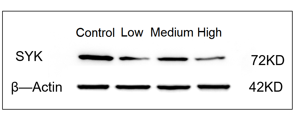
| Sample | Mouse HT-22 cells |
| Sample Processing Description | After digestion, cells were collected by centrifugation and lysed in 1 mL of RIPA lysis buffer supplemented with a protease inhibitor cocktail on ice for 1 h. The lysates were then centrifuged, and the supernatants were collected. After protein quantification, 5× loading buffer was added, and the samples were denatured in a 100 °C water bath for 10 min before loading for electrophoresis. |
| Other Reagents | Blocking buffer |
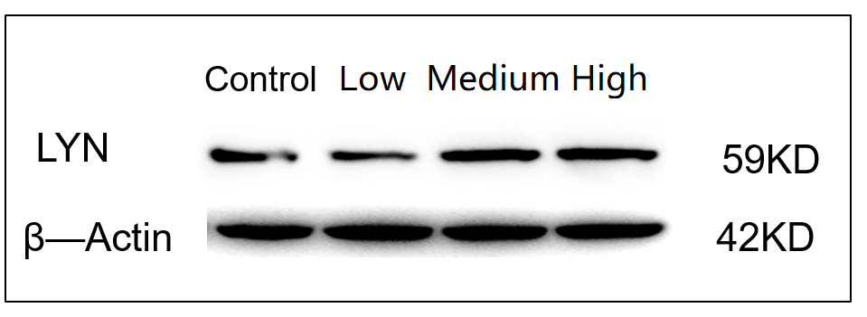
| Primary Antibody | Lyn Antibody Picoband® |
| Primary Incubation | 1:1000, overnight at 4 ℃ |
| Secondary Antibody | HRP Conjugated AffiniPure Goat Anti-Rabbit IgG (H+L) |
| Secondary Incubation | 1:2000, 1 hour in room temperature |
| Detection | Substrate: ECL, Imaging system:ChemiDoc MP |
| Results Summary | The control represents HT22 cells without drug treatment, while Low, Medium, and High correspond to experimental groups treated with three different drug concentrations for 24 h. The Western blot results obtained with this antibody are clear and well-defined, allowing distinct comparison of expression levels between the groups. It can be observed that the expression of the target protein slightly increases with increasing drug concentration. |