| SKU | A08719-2 |
|---|---|
| Application | Western Blot |
| Sample | rat colon tissue |
| Sample Processing Description | RIPA lysis buffer with protease inhibitor PMSF (100:1) was used to lyse the sample for 10 minutes, followed by centrifugation at 12,000 rpm for 15 minutes. The supernatant was mixed with 5× loading buffer, denatured at 100°C for 10 minutes, and then loaded onto SDS-PAGE. |
| Other Reagents | Blocking buffer |
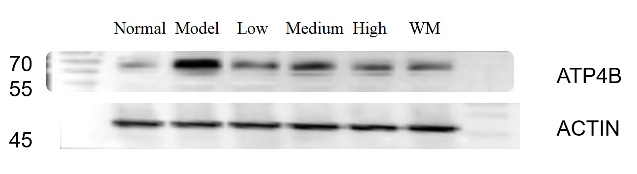
| Primary Antibody | ATP4B Antibody Picoband® |
| Primary Incubation | 1:1000, overnight at 4 ℃ |
| Secondary Antibody | HRP Conjugated AffiniPure Goat Anti-Rabbit IgG (H+L) |
| Secondary Incubation | 1:5000, 1 hour in room temperature |
| Detection | Substrate: ECL, Imaging system:ChemiDoc MP |
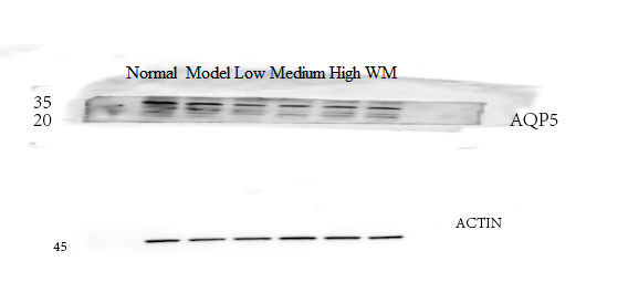
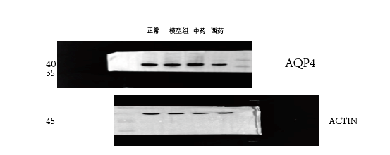
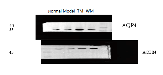
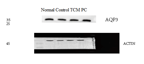
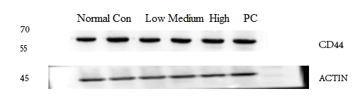
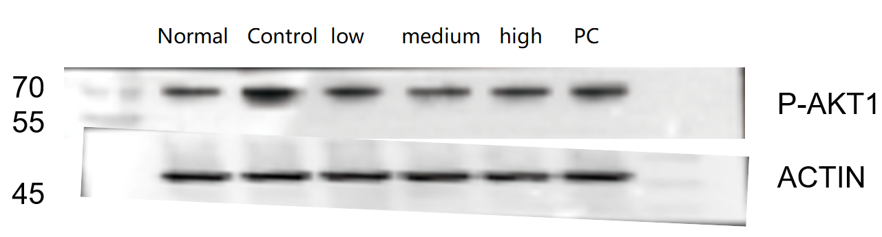
| Results Summary | The figure shows a schematic representation of Western blot results for the target protein ATP4B and the loading control Actin in rat colon across the normal group, model group, low-, medium-, and high-dose traditional Chinese medicine groups, and the western medicine group. Expression was increased in the model group, and among the traditional Chinese medicine groups, the high-dose group showed the best efficacy. The target bands are clear and distinct, and the experimental results are satisfactory. |