| SKU | DZ41720 |
|---|---|
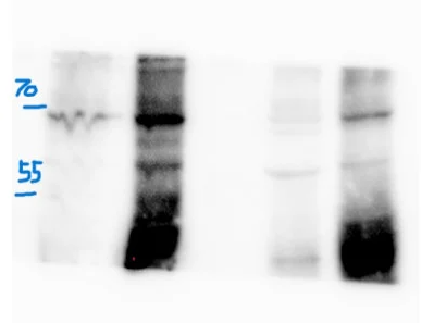
| Application | Western Blot |
| Sample | HEK293T whole cell lysates |
| Sample Processing Description | HEK293T cells were transfected with a plasmid expressing SP only (and not Env or Rem) and lysed using RIPA buffer. A total of 80 µg of protein was resolved on a 4–12% gradient SDS-PAGE gel and transferred to a membrane. The membrane was blocked for 1 hour at room temperature with 5% non-fat milk in PBS-Tween (PBS-T). After washing three times with 1× PBS-T (10 minutes each), the membrane was incubated overnight at 4 °C with varying dilutions ofthe SP antibody prepared in 2% non-fat milk (1:500; 1:1000; 1:2000). Following three additional washes with PBS-T (10 minutes each), the membrane was incubated with anti-rabbit secondary antibody, washed again three times, and developed using ECL Plus. |
| Primary Incubation | The membrane was incubated with the SP primary antibody (1:500; 1:1000; 1:2000) overnight at 4 °C. |
| Secondary Antibody | Anti-rabbit-HRP-conjugated secondary antibody |
| Secondary Incubation | Dilution: 1:25,000 in PBS-T/2% milk |
| Other Reagents used | 1× RIPA lysis buffer 1× PBS-T washing buffer (T = 1% Tween 20) Non-fat milk (for blocking and antibody dilution) Anti-rabbit HRP-conjugated secondary antibody |
| Detection | Signal was developed using ECL Plus chemiluminescent substrate |
| Results Summary | The SP antibody demonstrated strong performance across all tested dilutions, producing clear and specific signal detection. Dilutions of 1:1000 and 1:2000 yielded the most optimal results, showing minimal background compared to 1:500, which highlights the antibody’s high specificity and sensitivity at these concentrations. To ensure equal protein loading across lanes, the membrane was also probed with a β-actin antibody, confirming equal sample loading and validating the observed signal intensity for SP. Overall, these results demonstrate that the SP antibody is reliable for detecting SP in RIPA-lysed HEK293T samples. |