| SKU | M00007 |
|---|---|
| Application | Western Blot |
| Sample | rat colon tissue |
| Sample Processing Description | RIPA lysis buffer with protease inhibitor PMSF (100:1) was used to lyse the sample for 10 minutes, followed by centrifugation at 12,000 rpm for 15 minutes. The supernatant was mixed with 5× loading buffer, denatured at 100°C for 10 minutes, and then loaded onto SDS-PAGE. |
| Other Reagents | Blocking buffer |
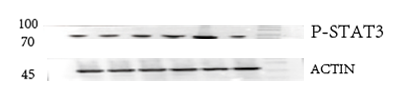
| Primary Antibody | Phospho-STAT3 (Y705) Rabbit Monoclonal Antibody |
| Primary Incubation | 1:1000, overnight at 4 ℃ |
| Secondary Antibody | HRP Conjugated AffiniPure Goat Anti-Rabbit IgG (H+L) |
| Secondary Incubation | 1:5000, 1 hour in room temperature |
| Detection | Substrate: ECL, Imaging system:ChemiDoc MP |
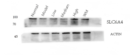
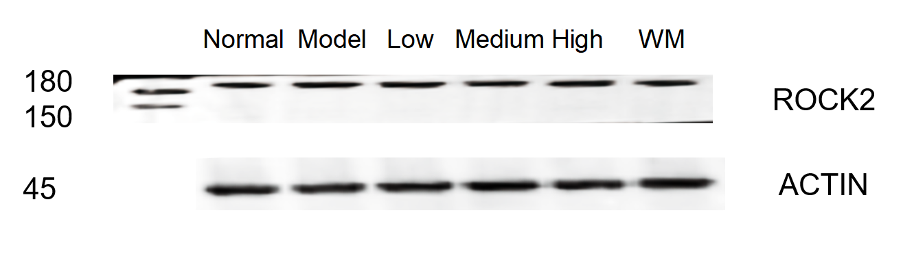
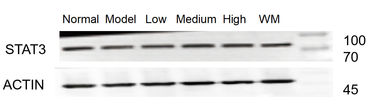
| Results Summary | The figure shows Western blot results of the target protein STAT3 and the loading control Actin in the colon of normal rats, model rats, low/medium/high dose Chinese medicine treatment groups, and Western medicine treatment group. No differences were observed between the groups. The target bands are clear and distinct, and the experimental results are satisfactory. |