Click image to see more details
Product Info Summary
| SKU: | A01434-1 |
|---|---|
| Size: | 100 μg/vial |
| Reactive Species: | Human |
| Host: | Rabbit |
| Application: | WB |
Customers Who Bought This Also Bought

Product info
Product Name
Anti-Bestrophin/BEST1 Antibody Picoband®
SKU/Catalog Number
A01434-1
Size
100 μg/vial
Form
Lyophilized
Description
Boster Bio Anti-Bestrophin/BEST1 Antibody Picoband® catalog # A01434-1. Tested in WB applications. This antibody reacts with Human. The brand Picoband indicates this is a premium antibody that guarantees superior quality, high affinity, and strong signals with minimal background in Western blot applications. Only our best-performing antibodies are designated as Picoband, ensuring unmatched performance.
Storage & Handling
Store at -20˚C for one year from date of receipt. After reconstitution, at 4˚C for one month. It can also be aliquotted and stored frozen at -20˚C for six months. Avoid repeated freeze-thaw cycles.
Cite This Product
Anti-Bestrophin/BEST1 Antibody Picoband® (Boster Biological Technology, Pleasanton CA, USA, Catalog # A01434-1)
Host
Rabbit
Contents
Each vial contains 4 mg Trehalose, 0.9 mg NaCl and 0.2 mg Na2HPO4.
Clonality
Polyclonal
Isotype
Rabbit IgG
Immunogen
A synthetic peptide corresponding to a sequence at the N-terminus of human Bestrophin, which shares 62.5% amino acid (aa) sequence identity with mouse Bestrophin.
Cross-reactivity
No cross-reactivity with other proteins.
Reactive Species
A01434-1 is reactive to BEST1 in Human
Observed Molecular Weight
68 kDa
Calculated molecular weight
67.7 kDa
Background of BEST1
Bestrophin-1 (Best1) is a protein that, in humans, is encoded by the BEST1 gene. This gene encodes a member of the bestrophin gene family. This small gene family is characterized by proteins with a highly conserved N-terminus with four to six transmembrane domains. Bestrophins may form chloride ion channels or may regulate voltage-gated L-type calcium-ion channels. Bestrophins are generally believed to form calcium-activated chloride-ion channels in epithelial cells but they have also been shown to be highly permeable to bicarbonate ion transport in retinal tissue. Mutations in this gene are responsible for juvenile-onset vitelliform macular dystrophy (VMD2), also known as Best macular dystrophy, in addition to adult-onset vitelliform macular dystrophy (AVMD) and other retinopathies. Alternative splicing results in multiple variants encoding distinct isoforms.
Antibody Validation
Boster validates all antibodies on WB, IHC, ICC, Immunofluorescence, and ELISA with known positive control and negative samples to ensure specificity and high affinity, including thorough antibody incubations.
Application & Images
Applications
A01434-1 is guaranteed for WB Boster Guarantee
Recommend Dilution
| Application | Dilution | Species |
|---|---|---|
| Western blot | 0.1-0.5μg/ml | Human |
Tested application
Suggested blocking solution with 5% non-fat milk or BSA; (*)Recommended protein loading: 20-40 µg per lane
Validation Images & Assay Conditions
Click image to see more details
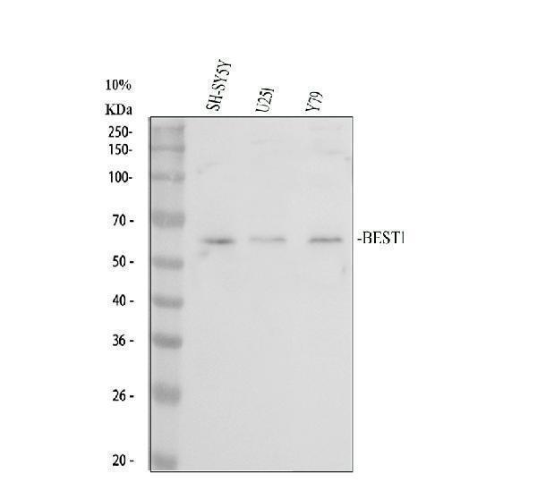
Western blot analysis of BEST1 using anti-BEST1 antibody (A01407-2).
Electrophoresis was performed on a 10% SDS-PAGE gel at 80V (Stacking gel) / 120V (Resolving gel) for 2 hours. The sample well of each lane was loaded with 30 ug of sample under reducing conditions.
Lane 1: human SH-SY5Y whole cell lysates,
Lane 2: human U251 whole cell lysates,
Lane 3: human Y79 whole cell lysates.
After electrophoresis, proteins were transferred to a nitrocellulose membrane at 150 mA for 50-90 minutes. Blocked the membrane with 5% non-fat milk/TBS for 1.5 hour at RT. The membrane was incubated with rabbit anti-BEST1 antigen affinity purified polyclonal antibody (A01407-2) at 0.5 μg/mL overnight at 4°C, then washed with TBS-0.1%Tween 3 times with 5 minutes each and probed with a goat anti-rabbit IgG-HRP secondary antibody (Catalog # BA1054) at a dilution of 1:5000 for 1.5 hour at RT. The signal is developed using an ECL Plus Western Blotting Substrate (Catalog # AR1196-200) with Tanon 5200 system. A specific band was detected for BEST1 at approximately 68 kDa. The expected band size for BEST1 is at 68 kDa.
Specific Publications For Anti-Bestrophin/BEST1 Antibody Picoband® (A01434-1)
Loading publications
Recommended Resources
Here are featured tools and databases that you might find useful.
- Boster's Pathways Library
- Protein Databases
- Bioscience Research Protocol Resources
- Data Processing & Analysis Software
- Photo Editing Software
- Scientific Literature Resources
- Research Paper Management Tools
- Molecular Biology Software
- Primer Design Tools
- Bioinformatics Tools
- Phylogenetic Tree Analysis
Customer Reviews
Have you used Anti-Bestrophin/BEST1 Antibody Picoband®?
Share your experimental results or join a short interview to earn up to $1,000 in product credits or other rewards.
0 Reviews For Anti-Bestrophin/BEST1 Antibody Picoband®
Customer Q&As
Have a question?
Find answers in Q&As, reviews.
Can't find your answer?
Submit your question
