Click image to see more details
Product Info Summary
| SKU: | A01289-1 |
|---|---|
| Size: | 100 μg/vial |
| Reactive Species: | Human, Mouse, Rat |
| Host: | Rabbit |
| Application: | ELISA, IHC, WB |
Customers Who Bought This Also Bought

Product info
Product Name
Anti-BRD7 Antibody Picoband®
SKU/Catalog Number
A01289-1
Size
100 μg/vial
Form
Lyophilized
Description
Boster Bio Anti-BRD7 Antibody Picoband® catalog # A01289-1. Tested in ELISA, IHC, WB applications. This antibody reacts with Human, Mouse, Rat. The brand Picoband indicates this is a premium antibody that guarantees superior quality, high affinity, and strong signals with minimal background in Western blot applications. Only our best-performing antibodies are designated as Picoband, ensuring unmatched performance.
Storage & Handling
Store at -20˚C for one year from date of receipt. After reconstitution, at 4˚C for one month. It can also be aliquotted and stored frozen at -20˚C for six months. Avoid repeated freeze-thaw cycles.
Cite This Product
Anti-BRD7 Antibody Picoband® (Boster Biological Technology, Pleasanton CA, USA, Catalog # A01289-1)
Host
Rabbit
Contents
Each vial contains 4mg Trehalose, 0.9mg NaCl, 0.2mg Na2HPO4, 0.05mg NaN3.
Clonality
Polyclonal
Isotype
Rabbit IgG
Immunogen
E. coli-derived human BRD7 recombinant protein (Position: T514-R567).
Cross-reactivity
No cross-reactivity with other proteins.
Reactive Species
A01289-1 is reactive to BRD7 in Human, Mouse, Rat
Observed Molecular Weight
85 kDa
Calculated molecular weight
74.1 kDa
Background of BRD7
Bromodomain-containing protein 7 is a protein that in humans is encoded by the BRD7 gene. This gene encodes a protein which is a member of the bromodomain-containing protein family. The product of this gene has been identified as a component of one form of the SWI/SNF chromatin remodeling complex, and as a protein which interacts with p53 and is required for p53-dependent oncogene-induced senescence which prevents tumor growth. Pseudogenes have been described on chromosomes 2, 3, 6, 13 and 14. Alternative splicing results in multiple transcript variants.
Antibody Validation
Boster validates all antibodies on WB, IHC, ICC, Immunofluorescence, and ELISA with known positive control and negative samples to ensure specificity and high affinity, including thorough antibody incubations.
Application & Images
Applications
A01289-1 is guaranteed for ELISA, IHC, WB Boster Guarantee
Recommend Dilution
| Application | Dilution | Species |
|---|---|---|
| Western blot | 0.1-0.5μg/ml | |
| Immunohistochemistry (Paraffin-embedded Section) | 0.5-1μg/ml | |
| ELISA | 0.1-0.5μg/ml |
Tested application
Suggested blocking solution with 5% non-fat milk or BSA; (*)Recommended protein loading: 20-40 µg per lane
Use TE buffer pH 9.0 for antigen retrieval; (*) citrate buffer pH 6.0 is an alternative.
Validation Images & Assay Conditions
Click image to see more details
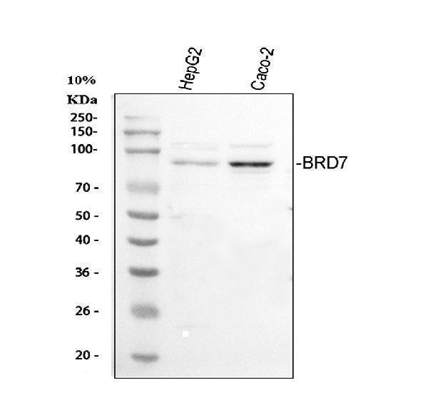
Western blot analysis of BRD7 using anti-BRD7 antibody (A01289-1).
Electrophoresis was performed on a 5-20% SDS-PAGE gel at 70V (Stacking gel) / 90V (Resolving gel) for 2-3 hours. The sample well of each lane was loaded with 30 ug of sample under reducing conditions.
Lane 1: human HepG2 whole cell lysates,
Lane 2: human CACO-2 whole cell lysates.
After electrophoresis, proteins were transferred to a nitrocellulose membrane at 150 mA for 50-90 minutes. Blocked the membrane with 5% non-fat milk/TBS for 1.5 hour at RT. The membrane was incubated with rabbit anti-BRD7 antigen affinity purified polyclonal antibody (A01289-1) at 0.5 μg/mL overnight at 4°C, then washed with TBS-0.1%Tween 3 times with 5 minutes each and probed with a goat anti-rabbit IgG-HRP secondary antibody at a dilution of 1:5000 for 1.5 hour at RT. The signal is developed using an Enhanced Chemiluminescent detection (ECL) kit (Catalog # EK1002) with Tanon 5200 system. A specific band was detected for BRD7 at approximately 85 kDa. The expected band size for BRD7 is at 74 kDa.

Click image to see more details
IHC analysis of BRD7 using anti-BRD7 antibody (A01289-1).
BRD7 was detected in paraffin-embedded section of human colon cancer tissue. Heat mediated antigen retrieval was performed in citrate buffer (pH6, epitope retrieval solution) for 20 mins. The tissue section was blocked with 10% goat serum. The tissue section was then incubated with 1μg/ml rabbit anti-BRD7 Antibody (A01289-1) overnight at 4°C. Biotinylated goat anti-rabbit IgG was used as secondary antibody and incubated for 30 minutes at 37°C. The tissue section was developed using Strepavidin-Biotin-Complex (SABC)(Catalog # SA1022) with DAB as the chromogen.

Click image to see more details
IHC analysis of BRD7 using anti-BRD7 antibody (A01289-1).
BRD7 was detected in paraffin-embedded section of human lung cancer tissue. Heat mediated antigen retrieval was performed in citrate buffer (pH6, epitope retrieval solution) for 20 mins. The tissue section was blocked with 10% goat serum. The tissue section was then incubated with 1μg/ml rabbit anti-BRD7 Antibody (A01289-1) overnight at 4°C. Biotinylated goat anti-rabbit IgG was used as secondary antibody and incubated for 30 minutes at 37°C. The tissue section was developed using Strepavidin-Biotin-Complex (SABC)(Catalog # SA1022) with DAB as the chromogen.
Specific Publications For Anti-BRD7 Antibody Picoband® (A01289-1)
Loading publications
Recommended Resources
Here are featured tools and databases that you might find useful.
- Boster's Pathways Library
- Protein Databases
- Bioscience Research Protocol Resources
- Data Processing & Analysis Software
- Photo Editing Software
- Scientific Literature Resources
- Research Paper Management Tools
- Molecular Biology Software
- Primer Design Tools
- Bioinformatics Tools
- Phylogenetic Tree Analysis
Customer Reviews
Have you used Anti-BRD7 Antibody Picoband®?
Share your experimental results or join a short interview to earn up to $1,000 in product credits or other rewards.
0 Reviews For Anti-BRD7 Antibody Picoband®
Customer Q&As
Have a question?
Find answers in Q&As, reviews.
Can't find your answer?
Submit your question
4 Customer Q&As for Anti-BRD7 Antibody Picoband®
Question
I see that the anti-BRD7 antibody A01289-1 works with IHC, what is the protocol used to produce the result images on the product page?
Verified Customer
Verified customer
Asked: 2019-11-04
Answer
You can find protocols for IHC on the "support/technical resources" section of our navigation menu. If you have any further questions, please send an email to support@bosterbio.com
Boster Scientific Support
Answered: 2019-11-04
Question
Thank you for helping with my inquiry over the phone. Here are the WB image, lot number and protocol we used for brain skin using anti-BRD7 antibody A01289-1. Let me know if you need anything else.
Verified Customer
Verified customer
Asked: 2019-03-06
Answer
We appreciate the data. You have provided everything we needed. Our lab team are working to resolve your inquiry as quickly as possible, and we appreciate your patience and understanding! Please let me know if there is anything you need in the meantime.
Boster Scientific Support
Answered: 2019-03-06
Question
Would anti-BRD7 antibody A01289-1 work on primate IHC with fetal brain?
Verified Customer
Verified customer
Asked: 2018-11-16
Answer
Our lab technicians have not tested anti-BRD7 antibody A01289-1 on primate. You can run a BLAST between primate and the immunogen sequence of anti-BRD7 antibody A01289-1 to see if they may cross-react. If the sequence homology is close, then you can perform a pilot test. Keep in mind that since we have not validated primate samples, this use of the antibody is not covered by our guarantee. However we have an innovator award program that if you test this antibody and show it works in primate fetal brain in IHC, you can get your next antibody for free.
Boster Scientific Support
Answered: 2018-11-16
Question
We are currently using anti-BRD7 antibody A01289-1 for human tissue, and we are well pleased with the ELISA results. The species of reactivity given in the datasheet says human, mouse, rat. Is it likely that the antibody can work on dog tissues as well?
Verified Customer
Verified customer
Asked: 2017-08-25
Answer
The anti-BRD7 antibody (A01289-1) has not been tested for cross reactivity specifically with dog tissues, but there is a good chance of cross reactivity. We have an innovator award program that if you test this antibody and show it works in dog you can get your next antibody for free. Please contact me if I can help you with anything.
Boster Scientific Support
Answered: 2017-08-25
