Click image to see more details
-

-
-
-
-
+2
Product Info Summary
| SKU: | M00223-1 |
|---|---|
| Size: | 100 μg/vial |
| Reactive Species: | Human, Mouse, Rat |
| Host: | Mouse |
| Application: | Flow Cytometry, IHC, WB |
Customers Who Bought This Also Bought

Product info
Product Name
Anti-BRG1 SMARCA4 Antibody Picoband® (monoclonal, 3F4)
SKU/Catalog Number
M00223-1
Size
100 μg/vial
Form
Lyophilized
Description
Boster Bio Anti-BRG1 SMARCA4 Antibody Picoband® (monoclonal, 3F4) catalog # M00223-1. Tested in Flow Cytometry, IHC, WB applications. This antibody reacts with Human, Mouse, Rat. The brand Picoband indicates this is a premium antibody that guarantees superior quality, high affinity, and strong signals with minimal background in Western blot applications. Only our best-performing antibodies are designated as Picoband, ensuring unmatched performance.
Storage & Handling
Store at -20˚C for one year from date of receipt. After reconstitution, at 4˚C for one month. It can also be aliquotted and stored frozen at -20˚C for six months. Avoid repeated freeze-thaw cycles.
Cite This Product
Anti-BRG1 SMARCA4 Antibody Picoband® (monoclonal, 3F4) (Boster Biological Technology, Pleasanton CA, USA, Catalog # M00223-1)
Host
Mouse
Contents
Each vial contains 4mg Trehalose, 0.9mg NaCl, 0.2mg Na2HPO4, 0.05mg NaN3.
Clonality
Monoclonal
Clone Number
3F4
Isotype
Mouse IgG1
Immunogen
E. coli-derived human BRG1 recombinant protein (Position: Q555-E763).
Cross-reactivity
No cross-reactivity with other proteins.
Reactive Species
M00223-1 is reactive to SMARCA4 in Human, Mouse, Rat
Observed Molecular Weight
181 kDa
Calculated molecular weight
184.6 kDa
Background of SMARCA4
Transcription activator BRG1 also known as ATP-dependent helicase SMARCA4 is a protein that in humans is encoded by the SMARCA4 gene. The protein encoded by this gene is a member of the SWI/SNF family of proteins and is similar to the brahma protein of Drosophila. Members of this family have helicase and ATPase activities and are thought to regulate transcription of certain genes by altering the chromatin structure around those genes. The encoded protein is part of the large ATP-dependent chromatin remodeling complex SNF/SWI, which is required for transcriptional activation of genes normally repressed by chromatin. In addition, this protein can bind BRCA1, as well as regulate the expression of the tumorigenic protein CD44. Mutations in this gene cause rhabdoid tumor predisposition syndrome type 2. Multiple transcript variants encoding different isoforms have been found for this gene.
Antibody Validation
Boster validates all antibodies on WB, IHC, ICC, Immunofluorescence, and ELISA with known positive control and negative samples to ensure specificity and high affinity, including thorough antibody incubations.
Application & Images
Applications
M00223-1 is guaranteed for Flow Cytometry, IHC, WB Boster Guarantee
Recommend Dilution
| Application | Dilution | Species |
|---|---|---|
| Western blot | 0.1-0.5μg/ml | |
| Immunohistochemistry (Paraffin-embedded Section) | 0.5-1μg/ml | |
| Flow Cytometry (Fixed) | 1-3μg/1x106 cells |
Tested application
Suggested blocking solution with 5% non-fat milk or BSA; (*)Recommended protein loading: 20-40 µg per lane
Use TE buffer pH 9.0 for antigen retrieval; (*) citrate buffer pH 6.0 is an alternative.
Validation Images & Assay Conditions
Click image to see more details
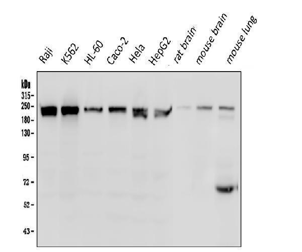
Western blot analysis of BRG1 using anti-BRG1 antibody (M00223-1).
Electrophoresis was performed on a 5-20% SDS-PAGE gel at 70V (Stacking gel) / 90V (Resolving gel) for 2-3 hours. The sample well of each lane was loaded with 50ug of sample under reducing conditions.
Lane 1: human Raji whole cell lysates;
Lane 2: human K562 whole cell lysates;
Lane 3: human HL-60 whole cell lysates;
Lane 4: human Caco-2 whole cell lysates;
Lane 5: human Hela whole cell lysates;
Lane 6: human HepG2 whole cell lysates;
Lane 7: rat brain tissue lysates;
Lane 8: mouse brain tissue lysates;
Lane 9: mouse lung tissue lysates.
After Electrophoresis, proteins were transferred to a Nitrocellulose membrane at 150mA for 50-90 minutes. Blocked the membrane with 5% Non-fat Milk/ TBS for 1.5 hour at RT. The membrane was incubated with mouse anti-BRG1 antigen affinity purified monoclonal antibody (Catalog # M00223-1) at 0.5 μg/mL overnight at 4°C, then washed with TBS-0.1%Tween 3 times with 5 minutes each and probed with a goat anti-mouse IgG-HRP secondary antibody at a dilution of 1:10000 for 1.5 hour at RT. The signal is developed using an Enhanced Chemiluminescent detection (ECL) kit (Catalog # EK1001) with Tanon 5200 system. A specific band was detected for BRG1 at approximately 181KD. The expected band size for BRG1 is at 181KD.

Click image to see more details
IHC analysis of BRG1 using anti-BRG1 antibody (M00223-1).
BRG1 was detected in paraffin-embedded section of human lung cancer tissue. Heat mediated antigen retrieval was performed in EDTA buffer (pH8.0, epitope retrieval solution). The tissue section was blocked with 10% goat serum. The tissue section was then incubated with 1μg/ml mouse anti-BRG1 Antibody (M00223-1) overnight at 4°C. Biotinylated goat anti-mouse IgG was used as secondary antibody and incubated for 30 minutes at 37°C. The tissue section was developed using Strepavidin-Biotin-Complex (SABC) (Catalog # SA1021) with DAB as the chromogen.

Click image to see more details
IHC analysis of BRG1 using anti-BRG1 antibody (M00223-1).
BRG1 was detected in paraffin-embedded section of human rectal cancer tissue. Heat mediated antigen retrieval was performed in EDTA buffer (pH8.0, epitope retrieval solution). The tissue section was blocked with 10% goat serum. The tissue section was then incubated with 1μg/ml mouse anti-BRG1 Antibody (M00223-1) overnight at 4°C. Biotinylated goat anti-mouse IgG was used as secondary antibody and incubated for 30 minutes at 37°C. The tissue section was developed using Strepavidin-Biotin-Complex (SABC) (Catalog # SA1021) with DAB as the chromogen.

Click image to see more details
IHC analysis of BRG1 using anti-BRG1 antibody (M00223-1).
BRG1 was detected in paraffin-embedded section of rat intestine tissue. Heat mediated antigen retrieval was performed in EDTA buffer (pH8.0, epitope retrieval solution). The tissue section was blocked with 10% goat serum. The tissue section was then incubated with 1μg/ml mouse anti-BRG1 Antibody (M00223-1) overnight at 4°C. Biotinylated goat anti-mouse IgG was used as secondary antibody and incubated for 30 minutes at 37°C. The tissue section was developed using Strepavidin-Biotin-Complex (SABC) (Catalog # SA1021) with DAB as the chromogen.

Click image to see more details
Flow Cytometry analysis of U20S cells using anti- BRG1 antibody (M00223-1).
Overlay histogram showing U20S cells stained with M00223-1 (Blue line). To facilitate intracellular staining, cells were fixed with 4% paraformaldehyde and permeabilized with permeabilization buffer. The cells were blocked with 10% normal goat serum. And then incubated with mouse anti- BRG1 Antibody (M00223-1, 1μg/1x106 cells) for 30 min at 20°C. DyLight®488 conjugated goat anti-mouse IgG (BA1126, 5-10μg/1x106 cells) was used as secondary antibody for 30 minutes at 20°C. Isotype control antibody (Green line) was mouse IgG (1μg/1x106) used under the same conditions. Unlabelled sample without incubation with primary antibody and secondary antibody (Red line) was used as a blank control.
Specific Publications For Anti-BRG1 SMARCA4 Antibody Picoband® (monoclonal, 3F4) (M00223-1)
Loading publications
Recommended Resources
Here are featured tools and databases that you might find useful.
- Boster's Pathways Library
- Protein Databases
- Bioscience Research Protocol Resources
- Data Processing & Analysis Software
- Photo Editing Software
- Scientific Literature Resources
- Research Paper Management Tools
- Molecular Biology Software
- Primer Design Tools
- Bioinformatics Tools
- Phylogenetic Tree Analysis
Customer Reviews
Have you used Anti-BRG1 SMARCA4 Antibody Picoband® (monoclonal, 3F4)?
Share your experimental results or join a short interview to earn up to $1,000 in product credits or other rewards.
0 Reviews For Anti-BRG1 SMARCA4 Antibody Picoband® (monoclonal, 3F4)
Customer Q&As
Have a question?
Find answers in Q&As, reviews.
Can't find your answer?
Submit your question
2 Customer Q&As for Anti-BRG1 SMARCA4 Antibody Picoband® (monoclonal, 3F4)
Question
My boss were happy with the WB result of your anti-BRG1 antibody (monoclonal, 3F4). However we have been able to see positive staining in tendon of biceps brachii nucleus using this antibody. Is that expected? Could you tell me where is SMARCA4 supposed to be expressed?
Verified Customer
Verified customer
Asked: 2019-10-17
Answer
From what I have seen in literature, tendon of biceps brachii does express SMARCA4. Generally SMARCA4 expresses in nucleus. Regarding which tissues have SMARCA4 expression, here are a few articles citing expression in various tissues:
Cervix carcinoma, Pubmed ID: 16964243, 17081983, 18669648, 20068231
Cervix carcinoma, and Erythroleukemia, Pubmed ID: 23186163
Embryonic kidney, Pubmed ID: 17525332
Fetal brain, Pubmed ID: 8208605
Leukemic T-cell, Pubmed ID: 19690332
Liver, Pubmed ID: 24275569
Lung, Pubmed ID: 18386774
Boster Scientific Support
Answered: 2019-10-17
Question
We have seen staining in mouse cervix carcinoma. What should we do? Is anti-BRG1 antibody (monoclonal, 3F4) supposed to stain cervix carcinoma positively?
D. Parker
Verified customer
Asked: 2017-02-21
Answer
According to literature cervix carcinoma does express SMARCA4. According to Uniprot.org, SMARCA4 is expressed in tendon of biceps brachii, fetal brain, lung, cervix carcinoma, embryonic kidney, leukemic t-cell, cervix carcinoma erythroleukemia, liver, among other tissues. Regarding which tissues have SMARCA4 expression, here are a few articles citing expression in various tissues:
Cervix carcinoma, Pubmed ID: 16964243, 17081983, 18669648, 20068231
Cervix carcinoma, and Erythroleukemia, Pubmed ID: 23186163
Embryonic kidney, Pubmed ID: 17525332
Fetal brain, Pubmed ID: 8208605
Leukemic T-cell, Pubmed ID: 19690332
Liver, Pubmed ID: 24275569
Lung, Pubmed ID: 18386774
Boster Scientific Support
Answered: 2017-02-21
