Click image to see more details
-

-
-
-
-
+2
Product Info Summary
| SKU: | PA1874-1 |
|---|---|
| Size: | 100 μg/vial |
| Reactive Species: | Human |
| Host: | Rabbit |
| Application: | IHC, WB |
Customers Who Bought This Also Bought

Product info
Product Name
Anti-CD163 Antibody Picoband®
SKU/Catalog Number
PA1874-1
BA13856 is an alternative SKU for this antibody, used in previous lots.
Size
100 μg/vial
Form
Lyophilized
Description
Boster Bio Anti-CD163 Antibody catalog # PA1874-1. Tested in IHC, WB applications. This antibody reacts with Human. The brand Picoband indicates this is a premium antibody that guarantees superior quality, high affinity, and strong signals with minimal background in Western blot applications. Only our best-performing antibodies are designated as Picoband, ensuring unmatched performance.
Storage & Handling
Store at -20˚C for one year from date of receipt. After reconstitution, at 4˚C for one month. It can also be aliquotted and stored frozen at -20˚C for six months. Avoid repeated freeze-thaw cycles.
Cite This Product
Anti-CD163 Antibody Picoband® (Boster Biological Technology, Pleasanton CA, USA, Catalog # PA1874-1)
Host
Rabbit
Contents
Each vial contains 4 mg Trehalose, 0.9 mg NaCl and 0.2 mg Na2HPO4.
Clonality
Polyclonal
Isotype
Rabbit IgG
Immunogen
E. coli-derived human CD163 recombinant protein (Position: I1056-L1156).
Cross-reactivity
No cross-reactivity with other proteins
Reactive Species
PA1874-1 is reactive to CD163 in Human
Observed Molecular Weight
150 kDa
Calculated molecular weight
125.5 kDa
Background of CD163
CD163 (Cluster of Differentiation 163) is a protein that in humans is encoded by the CD163 gene. The protein encoded by this gene is a member of the scavenger receptor cysteine-rich (SRCR) superfamily, and is exclusively expressed in monocytes and macrophages. It functions as an acute phase-regulated receptor involved in the clearance and endocytosis of hemoglobin/haptoglobin complexes by macrophages, and may thereby protect tissues from free hemoglobin-mediated oxidative damage. This protein may also function as an innate immune sensor for bacteria and inducer of local inflammation. Alternatively spliced transcript variants encoding different isoforms have been described for this gene.
Antibody Validation
Boster validates all antibodies on WB, IHC, ICC, Immunofluorescence, and ELISA with known positive control and negative samples to ensure specificity and high affinity, including thorough antibody incubations.
Application & Images
Applications
PA1874-1 is guaranteed for IHC, WB Boster Guarantee
Recommend Dilution
| Application | Dilution | Species |
|---|---|---|
| Western blot | 0.1-0.5μg/ml | Human |
| Immunohistochemistry (Paraffin-embedded Section) | 2-5μg/ml | Human |
Tested application
Suggested blocking solution with 5% non-fat milk or BSA; (*)Recommended protein loading: 20-40 µg per lane
Use TE buffer pH 9.0 for antigen retrieval; (*) citrate buffer pH 6.0 is an alternative.
Validation Images & Assay Conditions
Click image to see more details
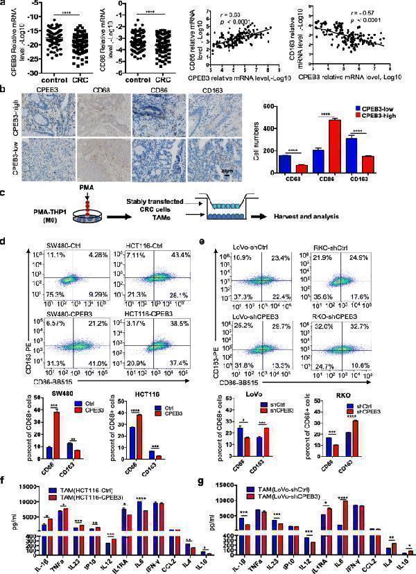
Decreased CPEB3 in human CRC correlates with low CD86 + TAM content and high CD163 + TAM content (a) The expression of CPEB3 and CD86 in 82 pairs of CRC tissues and adjacent non-tumor tissues was detected using qRT-PCR. Correlation between CPEB3 and CD86 or CD163 expression levels in 82 colorectal cancer tissues; error bars, SEM. (b) The protein expression of CPEB3, CD68, CD86, and CD163 in a human colorectal cancer tissue array was detected by IHC staining. Representative photos are shown (400× magnification). The number of CD68 + , CD86 + and CD163 + cells per high-power field in tissues from colorectal cancer patients with different levels of CPEB3 expression; error bars, SEM. (c) Schema for an in vitro model of stably transfected CRC cells co-cultured with TAMs. (d) Flow cytometry was used to explore the surface expression of CD86 and CD163 in SW480-Ctrl/CPEB3 and HCT116-Ctrl/CPEB3 cells; error bars, SEM. (e) Flow cytometry was used to explore the surface expression of CD86 and CD163 in LoVo-shCtrl/shCPEB3 and RKO-shCtrl/shCPEB3 cells; error bars, SEM. (f) We measured the expression of the respective inflammatory cytokines in cell culture supernatants of TAMs co-cultured HCT116-Ctrl/CPEB3 cells using ProcartaPlex combinable panels; error bars, SEM. (g) We measured the expression of the respective inflammatory cytokines in cell culture supernatants of TAM-co-cultured LoVo-shCtrl/shCPEB3 cells using ProcartaPlex combinable panels; error bars, SEM; ns, not significant; * P < 0.05; ** P < 0.01; *** P < 0.001; **** P < 0.0001
Index in PubMed under a CC BY license. PMID: 32653013

Click image to see more details
CPEB3 modulates CCL2 secretion in CRC cell supernatants to regulate TAM polarization (a) We measured the expression of the respective inflammatory cytokines in cell culture supernatants of HCT116-Ctrl/CPEB3 cells by ProcartaPlex combinable panels; error bars, SEM. (b) We measured the expression of the respective inflammatory cytokines in the supernatants of LoVo-shCtrl/shCPEB3 cells by ProcartaPlex combinable panels; error bars, SEM. (c) THP-1 macrophages were co-cultured with LoVo-shCtrl/shCPEB3 with or without CCL2-neutralizing antibody (1 μg/mL) for 24 h. Flow cytometry was used to explore the surface expression of CD86 and CD163 in the differentiated macrophages; error bars, SEM. (d) THP-1 macrophages were co-cultured with RKO-shCtrl/shCPEB3 cells with or without a CCL2-neutralizing antibody (1 μg/mL) for 24 h. Flow cytometry was used to explore the surface expression of CD86 and CD163 in the differentiated macrophages. Error bars, SEM. * P < 0.05; ** P < 0.01; *** P < 0.001; **** P < 0.0001
Index in PubMed under a CC BY license. PMID: 32653013

Click image to see more details
CPEB3 attenuates tumorigenesis and TAM polarization in vivo (a) Schematic of the procedure for separating tumor cells and TAMs. (b) HCT116 cells were stably infected with Ctrl and CPEB3 lentivirus, and LoVo cells were stably infected with shCtrl and shCPEB3 sequences. Tumorigenesis assay of Balb/c nude mice subcutaneously injected with HCT116-Ctrl/CPEB3 cells and LoVo-shCtrl/shCPEB3 cells ( n = 20). Representative photos of tumors from mice in various groups. ( c) IHC staining of Ki67 positive cells was counted per high-power field (PHF), while E-cadherin and vimentin expression scores were counted in tumor tissues in a mouse xenograft model; error bars, SEM. (d) The mice with intra-spleen injection of LoVo-shCtrl/shCPEB3 cells were treated with tocilizumab (5 mg/kg) weekly via intraperitoneally injection. The number of liver metastatic sites (indicated by arrows) was counted under the microscope; error bars, SEM. (e) Macrophages were separated from murine tumor tissues using Percoll-layered liquid. Surface expression of CD86 and CD163 was detected in macrophages using flow cytometry. The percentage of CD86 + or CD163 + cells in macrophages was reported using error bars and SEM. (f) Expression of JAK1, pJAK1, STAT3, and pSTAT3 in the tumor tissues of the two groups were analyzed by western blot analysis. (g) Schematic overview of the mechanisms by which CPEB3 modulate TAM polarization and inhibit colorectal cancer EMT. ** P < 0.01; *** P < 0.001; **** P < 0.0001
Index in PubMed under a CC BY license. PMID: 32653013

Click image to see more details
Western blot analysis of CD163 using anti-CD163 antibody (PA1874-1).
Electrophoresis was performed on a 5-20% SDS-PAGE gel at 70V (Stacking gel) / 90V (Resolving gel) for 2-3 hours. The sample well of each lane was loaded with 30 ug of sample under reducing conditions.
Lane 1: human hepatocellular carcinoma tumor tissue (HCCT) lysates.
After electrophoresis, proteins were transferred to a nitrocellulose membrane at 150 mA for 50-90 minutes. Blocked the membrane with 5% non-fat milk/TBS for 1.5 hour at RT. The membrane was incubated with rabbit anti-CD163 antigen affinity purified polyclonal antibody (Catalog # PA1874-1) at 0.5 μg/mL overnight at 4°C, then washed with TBS-0.1%Tween 3 times with 5 minutes each and probed with a goat anti-rabbit IgG-HRP secondary antibody at a dilution of 1:5000 for 1.5 hour at RT. The signal is developed using an Enhanced Chemiluminescent detection (ECL) kit (Catalog # EK1002) with Tanon 5200 system. A specific band was detected for CD163 at approximately 150 kDa. The expected band size for CD163 is at 125 kDa.

Click image to see more details
IHC analysis of CD163 using anti-CD163 antibody (PA1874-1).
CD163 was detected in a paraffin-embedded section of human liver cancer tissue. Heat mediated antigen retrieval was performed in EDTA buffer (pH 8.0, epitope retrieval solution). The tissue section was blocked with 10% goat serum. The tissue section was then incubated with 2 μg/ml rabbit anti-CD163 Antibody (PA1874-1) overnight at 4°C. Peroxidase Conjugated Goat Anti-rabbit IgG was used as secondary antibody and incubated for 30 minutes at 37°C. The tissue section was developed using HRP Conjugated Rabbit IgG Super Vision Assay Kit (Catalog # SV0002) with DAB as the chromogen.

Click image to see more details
IHC analysis of CD163 using anti-CD163 antibody (PA1874-1).
CD163 was detected in a paraffin-embedded section of human lung cancer tissue. Heat mediated antigen retrieval was performed in EDTA buffer (pH 8.0, epitope retrieval solution). The tissue section was blocked with 10% goat serum. The tissue section was then incubated with 2 μg/ml rabbit anti-CD163 Antibody (PA1874-1) overnight at 4°C. Peroxidase Conjugated Goat Anti-rabbit IgG was used as secondary antibody and incubated for 30 minutes at 37°C. The tissue section was developed using HRP Conjugated Rabbit IgG Super Vision Assay Kit (Catalog # SV0002) with DAB as the chromogen.
Specific Publications For Anti-CD163 Antibody Picoband® (PA1874-1)
Loading publications
Recommended Resources
Here are featured tools and databases that you might find useful.
- Boster's Pathways Library
- Protein Databases
- Bioscience Research Protocol Resources
- Data Processing & Analysis Software
- Photo Editing Software
- Scientific Literature Resources
- Research Paper Management Tools
- Molecular Biology Software
- Primer Design Tools
- Bioinformatics Tools
- Phylogenetic Tree Analysis
Customer Reviews
Have you used Anti-CD163 Antibody Picoband®?
Share your experimental results or join a short interview to earn up to $1,000 in product credits or other rewards.
0 Reviews For Anti-CD163 Antibody Picoband®
Customer Q&As
Have a question?
Find answers in Q&As, reviews.
Can't find your answer?
Submit your question
16 Customer Q&As for Anti-CD163 Antibody Picoband®
Question
Does PA1874-1 anti-CD163 antibody work on parafin embedded sections? If so, which fixation method do you recommend we use (PFA, paraformaldehyde, other)?
Verified Customer
Verified customer
Asked: 2020-04-10
Answer
You can see on the product datasheet, PA1874-1 anti-CD163 antibody as been validated on WB. It is best to use PFA for fixation because it has better tissue penetration ability. PFA needs to be prepared fresh before use. Long term stored PFA turns into formalin, as the PFA molecules congregate and become formalin.
Boster Scientific Support
Answered: 2020-04-10
Question
Is there a BSA free version of anti-CD163 antibody PA1874-1 available?
Verified Customer
Verified customer
Asked: 2020-03-16
Answer
I appreciate your recent telephone inquiry. I can confirm that some lots of this anti-CD163 antibody PA1874-1 are BSA free. For now, these lots are available and we can make a BSA free formula for you free of charge. It will take 3 extra days to prepare. If you require this antibody BSA free again in future, please do not hesitate to contact me and I will be pleased to check which lots we have in stock that are BSA free.
Boster Scientific Support
Answered: 2020-03-16
Question
Our team were satisfied with the WB result of your anti-CD163 antibody. However we have seen positive staining in plasma cell membrane using this antibody. Is that expected? Could you tell me where is CD163 supposed to be expressed?
Verified Customer
Verified customer
Asked: 2020-02-05
Answer
From literature, plasma does express CD163. Generally CD163 expresses in soluble cd163: secreted, cell membrane. Regarding which tissues have CD163 expression, here are a few articles citing expression in various tissues:
Liver, Pubmed ID: 19159218, 24275569
Plasma, Pubmed ID: 16335952
Spleen, Pubmed ID: 15489334
Boster Scientific Support
Answered: 2020-02-05
Question
We have observed staining in human plasma. What should we do? Is anti-CD163 antibody supposed to stain plasma positively?
Verified Customer
Verified customer
Asked: 2019-12-17
Answer
Based on literature plasma does express CD163. Based on Uniprot.org, CD163 is expressed in esophagus, spleen, plasma, liver, among other tissues. Regarding which tissues have CD163 expression, here are a few articles citing expression in various tissues:
Liver, Pubmed ID: 19159218, 24275569
Plasma, Pubmed ID: 16335952
Spleen, Pubmed ID: 15489334
Boster Scientific Support
Answered: 2019-12-17
Question
Is this PA1874-1 anti-CD163 antibody reactive to the isotypes of CD163?
Verified Customer
Verified customer
Asked: 2019-12-06
Answer
The immunogen of PA1874-1 anti-CD163 antibody is E. coli-derived human CD163 recombinant protein(Position: F1056-L1165). Could you tell me which isotype you are interested in so I can help see if the immunogen is part of this isotype?
Boster Scientific Support
Answered: 2019-12-06
Question
We appreciate helping with my inquiry over the phone. Here are the WB image, lot number and protocol we used for liver using anti-CD163 antibody PA1874-1. Let me know if you need anything else.
Verified Customer
Verified customer
Asked: 2019-11-01
Answer
Thank you for the data. You have provided everything we needed. Our lab team are working to resolve your inquiry as quickly as possible, and we appreciate your patience and understanding! Please let me know if there is anything you need in the meantime.
Boster Scientific Support
Answered: 2019-11-01
Question
I was wanting to use your anti-CD163 antibody for WB for human liver on frozen tissues, but I want to know if it has been tested for this particular application. Has this antibody been tested and is this antibody a good choice for human liver identification?
Verified Customer
Verified customer
Asked: 2019-09-10
Answer
As indicated on the product datasheet, PA1874-1 anti-CD163 antibody has been validated for IHC, WB on human tissues. We have an innovator award program that if you test this antibody and show it works in human liver in IHC-frozen, you can get your next antibody for free.
Boster Scientific Support
Answered: 2019-09-10
Question
See attached the WB image, lot number and protocol we used for liver using anti-CD163 antibody PA1874-1. Please let me know if you require anything else.
Verified Customer
Verified customer
Asked: 2019-08-16
Answer
Thank you very much for the data. Our lab team are working to resolve this as quickly as possible, and we appreciate your patience and understanding! You have provided everything we needed. Please let me know if there is anything you need in the meantime.
Boster Scientific Support
Answered: 2019-08-16
Question
Will anti-CD163 antibody PA1874-1 work for WB with liver?
Verified Customer
Verified customer
Asked: 2018-07-12
Answer
According to the expression profile of liver, CD163 is highly expressed in liver. So, it is likely that anti-CD163 antibody PA1874-1 will work for WB with liver.
Boster Scientific Support
Answered: 2018-07-12
Question
My question regarding product PA1874-1, anti-CD163 antibody. I was wondering if it would be possible to conjugate this antibody with biotin. I would need it to be without BSA or sodium azide. I am planning on using a buffer exchange of sodium azide with PBS only. Would there be problems for me to conjugate the antibody and store it in -20 degrees in small aliquots?
G. Mangal
Verified customer
Asked: 2018-06-07
Answer
It is not recommended storing this antibody with PBS buffer only in -20 degrees. If you want to store it in -20 degrees it is best to add some cryoprotectant like glycerol. If you want carrier free PA1874-1 anti-CD163 antibody, we can provide it to you in a special formula with trehalose and/or glycerol. These molecules will not interfere with conjugation chemistry and provide a good level of protection for the antibody from degradation. Please be sure to specify this in your purchase order.
Boster Scientific Support
Answered: 2018-06-07
Question
Our lab want to know about using your anti-CD163 antibody for receptor-mediated endocytosis studies. Has this antibody been tested with western blotting on intestinal cancer tissue? We would like to see some validation images before ordering.
Verified Customer
Verified customer
Asked: 2017-07-10
Answer
Thanks for your inquiry. This PA1874-1 anti-CD163 antibody is tested on mammary cancer tissue, intestinal cancer tissue. It is guaranteed to work for IHC, WB in human. Our Boster guarantee will cover your intended experiment even if the sample type has not been be directly tested.
Boster Scientific Support
Answered: 2017-07-10
Question
We are currently using anti-CD163 antibody PA1874-1 for human tissue, and we are happy with the IHC results. The species of reactivity given in the datasheet says human. Is it true that the antibody can work on primate tissues as well?
D. Krishna
Verified customer
Asked: 2016-09-21
Answer
The anti-CD163 antibody (PA1874-1) has not been validated for cross reactivity specifically with primate tissues, though there is a good chance of cross reactivity. We have an innovator award program that if you test this antibody and show it works in primate you can get your next antibody for free. Please contact me if I can help you with anything.
Boster Scientific Support
Answered: 2016-09-21
Question
I see that the anti-CD163 antibody PA1874-1 works with WB, what is the protocol used to produce the result images on the product page?
A. Banerjee
Verified customer
Asked: 2016-04-13
Answer
You can find protocols for WB on the "support/technical resources" section of our navigation menu. If you have any further questions, please send an email to support@bosterbio.com
Boster Scientific Support
Answered: 2016-04-13
Question
We bought anti-CD163 antibody for WB on liver in a previous project. I am using human, and We intend to use the antibody for IHC next. I would like examining liver as well as spleen in our next experiment. Could you please give me some suggestion on which antibody would work the best for IHC?
P. Banerjee
Verified customer
Asked: 2016-01-21
Answer
I took a look at the website and datasheets of our anti-CD163 antibody and I see that PA1874-1 has been validated on human in both WB and IHC. Thus PA1874-1 should work for your application. Our Boster satisfaction guarantee will cover this product for IHC in human even if the specific tissue type has not been validated. We do have a comprehensive range of products for IHC detection and you can check out our website bosterbio.com to find out more information about them.
Boster Scientific Support
Answered: 2016-01-21
Question
Is a blocking peptide available for product anti-CD163 antibody (PA1874-1)?
Z. Collins
Verified customer
Asked: 2015-04-06
Answer
We do provide the blocking peptide for product anti-CD163 antibody (PA1874-1). If you would like to place an order for it please contact support@bosterbio.com and make a special request.
Boster Scientific Support
Answered: 2015-04-06
Question
I am interested in to test anti-CD163 antibody PA1874-1 on human liver for research purposes, then I may be interested in using anti-CD163 antibody PA1874-1 for diagnostic purposes as well. Is the antibody suitable for diagnostic purposes?
N. Banerjee
Verified customer
Asked: 2013-08-15
Answer
The products we sell, including anti-CD163 antibody PA1874-1, are only intended for research use. They would not be suitable for use in diagnostic work. If you have the means to develop a product into diagnostic use, and are interested in collaborating with us and develop our product into an IVD product, please contact us for more discussions.
Boster Scientific Support
Answered: 2013-08-15
