Click image to see more details
-

-
-
-
-
+1
Product Info Summary
| SKU: | M02285-3 |
|---|---|
| Size: | 100 μl/vial |
| Reactive Species: | Human, Mouse, Rat |
| Host: | Rabbit |
| Application: | IP, IF, IHC, ICC, WB |
Customers Who Bought This Also Bought

Product info
Product Name
Anti-CD206/MRC1 Antibody (Monoclonal, 31M03)
SKU/Catalog Number
M02285-3
Size
100 μl/vial
Form
Liquid
Description
Boster Bio Anti-CD206/MRC1 Antibody (Monoclonal, 31M03) catalog # M02285-3. Tested in WB, IHC, IF, ICC/IF, IP applications. This antibody reacts with Human, Mouse, Rat.
Storage & Handling
Store at -20°C for one year. For short term storage and frequent use, store at 4°C for up to one month. Avoid repeated freeze-thaw cycles.
Cite This Product
Anti-CD206/MRC1 Antibody (Monoclonal, 31M03) (Boster Biological Technology, Pleasanton CA, USA, Catalog # M02285-3)
Host
Rabbit
Contents
Rabbit IgG in stabilizing components, phosphate buffered saline, pH 7.4, 150mM NaCl, 0.02% sodium azide and 50% glycerol.
This antibody is supplied in a stabilized formulation.
Compatibility with conjugation reactions depends on the chemistry of the conjugation method used.
For conjugation methods that are not compatible with the stabilizing components present in this formulation, a carrier-free antibody format is required.
Clonality
Monoclonal
Clone Number
31M03
Immunogen
E.coli-derived mouse Mannose Receptor/MRC1 recombinant protein (Position: L3-F407).
Reactive Species
M02285-3 is reactive to MRC1 in Human, Mouse, Rat
Observed Molecular Weight
180-200 kDa
Calculated molecular weight
165.0 kDa
Background of MRC1
The mannose receptor (Cluster of Differentiation 206,CD206) is a C-type lectin primarily present on the surface of macrophages,immature dendritic cells and liver sinusoidal endothelial cells,but is also expressed on the surface of skin cells such as human dermal fibroblasts and keratinocytes. It is mapped to 10p12.33. The recognition of complex carbohydrate structures on glycoproteins is an important part of several biological processes,including cell-cell recognition,serum glycoprotein turnover,and neutralization of pathogens. The protein encoded by this gene is a type I membrane receptor that mediates the endocytosis of glycoproteins by macrophages. The protein has been shown to bind high-mannose structures on the surface of potentially pathogenic viruses,bacteria,and fungi so that they can be neutralized by phagocytic engulfment.
Antibody Validation
Boster validates all antibodies on WB, IHC, ICC, Immunofluorescence, and ELISA with known positive control and negative samples to ensure specificity and high affinity, including thorough antibody incubations.
Application & Images
Applications
M02285-3 is guaranteed for IP, IF, IHC, ICC, WB Boster Guarantee
Recommend Dilution
| Application | Dilution | Species |
|---|---|---|
| Western blot | 1:500-2000 | |
| Immunohistochemistry | 1:50-200 | |
| Immunofluorescence | 1:50-200 | |
| Immunocytochemistry/Immunofluorescence | 1:50-200 | |
| ImmunoPrecipitation | 1:50 |
Tested application
Suggested blocking solution with 5% non-fat milk or BSA; (*)Recommended protein loading: 20-40 µg per lane
Use TE buffer pH 9.0 for antigen retrieval; (*) citrate buffer pH 6.0 is an alternative.
Validation Images & Assay Conditions
Click image to see more details
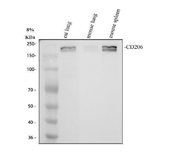
Western blot analysis of CD206/MRC1 using anti-CD206/MRC1 antibody (M02285-3).
Electrophoresis was performed on a 8% SDS-PAGE gel at 80V (Stacking gel) / 120V (Resolving gel) for 2 hours. The sample well of each lane was loaded with 30 ug of sample under reducing conditions.
Lane 1: rat lung tissue lysates,
Lane 2: mouse lung tissue lysates,
Lane 3: mouse spleen tissue lysates.
After electrophoresis, proteins were transferred to a nitrocellulose membrane at 150 mA for 50-90 minutes. Blocked the membrane with 5% non-fat milk/TBS for 1.5 hour at RT. The membrane was incubated with rabbit anti-CD206/MRC1 antigen affinity purified monoclonal antibody (M02285-3) at 1:1000 overnight at 4°C, then washed with TBS-0.1%Tween 3 times with 5 minutes each and probed with a goat anti-rabbit IgG-HRP secondary antibody at a dilution of 1:5000 for 1.5 hour at RT. The signal is developed using an ECL Plus Western Blotting Substrate (Catalog # AR1196-200) with Tanon 5200 system. A specific band was detected for CD206/MRC1 at approximately 180-200 kDa. The expected band size for CD206/MRC1 is at 165 kDa.

Click image to see more details
IHC analysis of CD206/MRC1 using anti-CD206/MRC1 antibody (M02285-3).
CD206/MRC1 was detected in a paraffin-embedded section of human liver cancer tissue. Heat mediated antigen retrieval was performed in EDTA buffer (pH 8.0, epitope retrieval solution). The tissue section was blocked with 10% goat serum. The tissue section was then incubated with 1:200 rabbit anti-CD206/MRC1 Antibody (M02285-3) overnight at 4°C. Peroxidase Conjugated Goat Anti-rabbit IgG was used as secondary antibody and incubated for 30 minutes at 37°C. The tissue section was developed using HRP Conjugated Rabbit IgG Super Vision Assay Kit (Catalog # SV0002) with DAB as the chromogen.

Click image to see more details
IHC analysis of CD206/MRC1 using anti-CD206/MRC1 antibody (M02285-3).
CD206/MRC1 was detected in a paraffin-embedded section of human placenta tissue. Heat mediated antigen retrieval was performed in EDTA buffer (pH 8.0, epitope retrieval solution). The tissue section was blocked with 10% goat serum. The tissue section was then incubated with 1:200 rabbit anti-CD206/MRC1 Antibody (M02285-3) overnight at 4°C. Peroxidase Conjugated Goat Anti-rabbit IgG was used as secondary antibody and incubated for 30 minutes at 37°C. The tissue section was developed using HRP Conjugated Rabbit IgG Super Vision Assay Kit (Catalog # SV0002) with DAB as the chromogen.

Click image to see more details
IHC analysis of CD206/MRC1 using anti-CD206/MRC1 antibody (M02285-3).
CD206/MRC1 was detected in a paraffin-embedded section of mouse liver tissue. Heat mediated antigen retrieval was performed in EDTA buffer (pH 8.0, epitope retrieval solution). The tissue section was blocked with 10% goat serum. The tissue section was then incubated with 1:200 rabbit anti-CD206/MRC1 Antibody (M02285-3) overnight at 4°C. Peroxidase Conjugated Goat Anti-rabbit IgG was used as secondary antibody and incubated for 30 minutes at 37°C. The tissue section was developed using HRP Conjugated Rabbit IgG Super Vision Assay Kit (Catalog # SV0002) with DAB as the chromogen.

Click image to see more details
IHC analysis of CD206/MRC1 using anti-CD206/MRC1 antibody (M02285-3).
CD206/MRC1 was detected in a paraffin-embedded section of rat liver tissue. Heat mediated antigen retrieval was performed in EDTA buffer (pH 8.0, epitope retrieval solution). The tissue section was blocked with 10% goat serum. The tissue section was then incubated with 1:200 rabbit anti-CD206/MRC1 Antibody (M02285-3) overnight at 4°C. Peroxidase Conjugated Goat Anti-rabbit IgG was used as secondary antibody and incubated for 30 minutes at 37°C. The tissue section was developed using HRP Conjugated Rabbit IgG Super Vision Assay Kit (Catalog # SV0002) with DAB as the chromogen.
Specific Publications For Anti-CD206/MRC1 Antibody (Monoclonal, 31M03) (M02285-3)
Loading publications
Recommended Resources
Here are featured tools and databases that you might find useful.
- Boster's Pathways Library
- Protein Databases
- Bioscience Research Protocol Resources
- Data Processing & Analysis Software
- Photo Editing Software
- Scientific Literature Resources
- Research Paper Management Tools
- Molecular Biology Software
- Primer Design Tools
- Bioinformatics Tools
- Phylogenetic Tree Analysis
Customer Reviews
Have you used Anti-CD206/MRC1 Antibody (Monoclonal, 31M03)?
Share your experimental results or join a short interview to earn up to $1,000 in product credits or other rewards.
0 Reviews For Anti-CD206/MRC1 Antibody (Monoclonal, 31M03)
Customer Q&As
Have a question?
Find answers in Q&As, reviews.
Can't find your answer?
Submit your question
