Click image to see more details
-

-
-
-
-
+10
Product Info Summary
| SKU: | A00602-1 |
|---|---|
| Size: | 100 μg/vial |
| Reactive Species: | Human |
| Host: | Rabbit |
| Application: | ELISA, IF, IHC |
Customers Who Bought This Also Bought

Product info
Product Name
Anti-CD68 Antibody
SKU/Catalog Number
A00602-1
Size
100 μg/vial
Form
Lyophilized
Description
Boster Bio Anti-CD68 Antibody catalog # A00602-1. Tested in ELISA, IF, IHC applications. This antibody reacts with Human.
Storage & Handling
At -20°C for one year from date of receipt. After reconstitution, at 4°C for one month. It can also be aliquotted and stored frozen at -20°C for six months. Avoid repeated freezing and thawing.
Cite This Product
Anti-CD68 Antibody (Boster Biological Technology, Pleasanton CA, USA, Catalog # A00602-1)
Host
Rabbit
Contents
Each vial contains 4 mg Trehalose, 0.9 mg NaCl, 0.2 mg Na2HPO4.
Clonality
Polyclonal
Isotype
Rabbit IgG
Immunogen
E.coli-derived human CD68 recombinant protein (Position: K154-L354).
Cross-reactivity
No cross-reactivity with other proteins.
Reactive Species
A00602-1 is reactive to CD68 in Human
Calculated molecular weight
37.4 kDa
Background of CD68
CD68, cluster of differentiation, is a 110-kD transmembrane glycoprotein that is highly expressed by human monocytes and tissue macrophages. CD68 is a member of a family of hematopoietic mucin-like molecules that includes leukosialin/CD43 and stem cell antigen CD34. The CD68 gene is mapped to 17p13.1. Immunohistochemistry can be used to identify the presence of CD68, which is found in the cytoplasmic granules of a range of different blood cells. It is particularly useful as a marker for the various cells of the macrophage lineage, including monocytes, histiocytes, giant cells, Kupffer cells, and osteoclasts. This allows it to be used to distinguish diseases of otherwise similar appearance, such as the monocyte/macrophage and lymphoid forms of leukaemia (the latter being CD68 negative). Its presence in macrophages also makes it useful in diagnosing conditions related to proliferation or abnormality of these cells, such as malignant histiocytosis, histiocytic lymphoma, and Gaucher's disease.
Antibody Validation
Boster validates all antibodies on WB, IHC, ICC, Immunofluorescence, and ELISA with known positive control and negative samples to ensure specificity and high affinity, including thorough antibody incubations.
Application & Images
Applications
A00602-1 is guaranteed for ELISA, IF, IHC Boster Guarantee
Recommend Dilution
| Application | Dilution | Species |
|---|---|---|
| Immunohistochemistry(Paraffin-embedded Section) | 2-5 μg/ml | Human |
| Immunofluorescence | 5 μg/ml | Human |
| ELISA | 0.1-0.5 μg/ml | - |
Tested application
Use TE buffer pH 9.0 for antigen retrieval; (*) citrate buffer pH 6.0 is an alternative.
Validation Images & Assay Conditions
Click image to see more details
IHC analysis of CD68 using anti-CD68 antibody (A00602-1).
CD68 was detected in a paraffin-embedded section of human tonsil tissue. Heat mediated antigen retrieval was performed in EDTA buffer (pH 8.0, epitope retrieval solution). The tissue section was blocked with 10% goat serum. The tissue section was then incubated with 2 μg/ml rabbit anti-CD68 Antibody (A00602-1) overnight at 4°C. Peroxidase Conjugated Goat Anti-rabbit IgG was used as secondary antibody and incubated for 30 minutes at 37°C. The tissue section was developed using HRP Conjugated Rabbit IgG Super Vision Assay Kit (Catalog # SV0002) with DAB as the chromogen.

Click image to see more details
IHC analysis of CD68 using anti-CD68 antibody (A00602-1).
CD68 was detected in a paraffin-embedded section of human spleen tissue. Heat mediated antigen retrieval was performed in EDTA buffer (pH 8.0, epitope retrieval solution). The tissue section was blocked with 10% goat serum. The tissue section was then incubated with 2.5 μg/ml rabbit anti-CD68 Antibody (A00602-1) overnight at 4°C. HRP-AffiniPure Goat Anti-Rabbit IgG was used as secondary antibody and incubated for 30 minutes at 37°C. The tissue section was developed using HRP Conjugated Rabbit IgG Super Vision Assay Kit (Catalog # SV0002) with DAB as the chromogen.

Click image to see more details
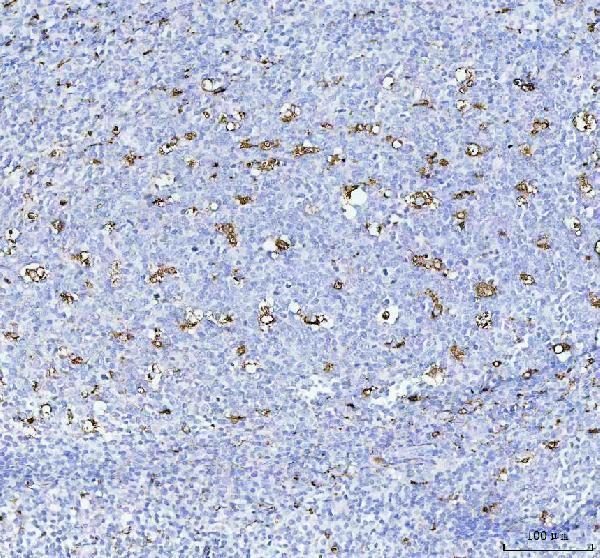
IHC analysis of CD68 using anti-CD68 antibody (A00602-1).
CD68 was detected in a paraffin-embedded section of human liver cancer tissue. Heat mediated antigen retrieval was performed in EDTA buffer (pH 8.0, epitope retrieval solution). The tissue section was blocked with 10% goat serum. The tissue section was then incubated with 2.5 μg/ml rabbit anti-CD68 Antibody (A00602-1) overnight at 4°C. HRP-AffiniPure Goat Anti-Rabbit IgG was used as secondary antibody and incubated for 30 minutes at 37°C. The tissue section was developed using HRP Conjugated Rabbit IgG Super Vision Assay Kit (Catalog # SV0002) with DAB as the chromogen.

Click image to see more details
IHC analysis of CD68 using anti-CD68 antibody (A00602-1).
CD68 was detected in a paraffin-embedded section of human prostate cancer tissue. Heat mediated antigen retrieval was performed in EDTA buffer (pH 8.0, epitope retrieval solution). The tissue section was blocked with 10% goat serum. The tissue section was then incubated with 2.5 μg/ml rabbit anti-CD68 Antibody (A00602-1) overnight at 4°C. HRP-AffiniPure Goat Anti-Rabbit IgG was used as secondary antibody and incubated for 30 minutes at 37°C. The tissue section was developed using HRP Conjugated Rabbit IgG Super Vision Assay Kit (Catalog # SV0002) with DAB as the chromogen.

Click image to see more details
IHC analysis of CD68 using anti-CD68 antibody (A00602-1).
CD68 was detected in a paraffin-embedded section of human breast cancer tissue. Heat mediated antigen retrieval was performed in EDTA buffer (pH 8.0, epitope retrieval solution). The tissue section was blocked with 10% goat serum. The tissue section was then incubated with 2.5 μg/ml rabbit anti-CD68 Antibody (A00602-1) overnight at 4°C. HRP-AffiniPure Goat Anti-Rabbit IgG was used as secondary antibody and incubated for 30 minutes at 37°C. The tissue section was developed using HRP Conjugated Rabbit IgG Super Vision Assay Kit (Catalog # SV0002) with DAB as the chromogen.

Click image to see more details
IF analysis of CD68 using anti-CD68 antibody (A00602-1).
CD68 was detected in a paraffin-embedded section of human tonsil tissue. Heat mediated antigen retrieval was performed in EDTA buffer (pH 8.0, epitope retrieval solution). The tissue section was blocked with 10% goat serum. The tissue section was then incubated with 5 μg/mL rabbit anti-CD68 Antibody (A00602-1) overnight at 4°C. Cy3 Conjugated Goat Anti-Rabbit IgG (BA1032) was used as secondary antibody at 1:500 dilution and incubated for 30 minutes at 37°C. The section was counterstained with DAPI. Visualize using a fluorescence microscope and filter sets appropriate for the label used.

Click image to see more details
IF analysis of CD68 using anti-CD68 antibody (A00602-1).
CD68 was detected in a paraffin-embedded section of human tonsil tissue. Heat mediated antigen retrieval was performed in EDTA buffer (pH 8.0, epitope retrieval solution). The tissue section was blocked with 10% goat serum. The tissue section was then incubated with 5 μg/mL rabbit anti-CD68 Antibody (A00602-1) overnight at 4°C. DyLight488 Conjugated Goat Anti-Rabbit IgG (BA1127) was used as secondary antibody at 1:500 dilution and incubated for 30 minutes at 37°C. The section was counterstained with DAPI. Visualize using a fluorescence microscope and filter sets appropriate for the label used.

Click image to see more details
IF analysis of CD68 using anti-CD68 antibody (A00602-1).
CD68 was detected in a paraffin-embedded section of human tonsil tissue. Heat mediated antigen retrieval was performed in EDTA buffer (pH 8.0, epitope retrieval solution). The tissue section was blocked with 10% goat serum. The tissue section was then incubated with 2.5 μg/mL rabbit anti-CD68 Antibody (A00602-1) overnight at 4°C. DyLight 488 Conjugated AffiniPure Goat Anti-rabbit IgG (H+L) (BA1127) was used as secondary antibody at 1:100 dilution and incubated for 30 minutes at 37°C. The section was counterstained with DAPI. Visualize using a fluorescence microscope and filter sets appropriate for the label used.

Click image to see more details
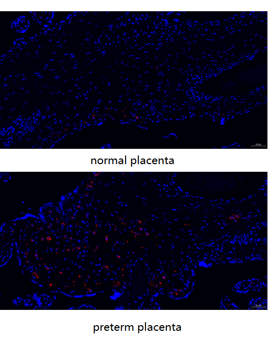
IF analysis of CD68 using anti-CD68 antibody (A00602-1).
CD68 was detected in a paraffin-embedded section of human normal placenta and preterm placentar tissue. Heat mediated antigen retrieval was performed in EDTA buffer (pH 8.0, epitope retrieval solution). The tissue section was blocked with 10% goat serum. The tissue section was then incubated with 1:500 rabbit anti-CD68 Antibody (A00602-1) overnight at 4°C. DyLight 594-conjugated Donkey Anti-Mouse IgG (H+L)(BA1148) was used as secondary antibody at 1:500 dilution and incubated for 30 minutes at 37°C. The section was counterstained with DAPI. Visualize using a fluorescence microscope and filter sets appropriate for the label used.

Click image to see more details
IF analysis of CD68 using anti-CD68 antibody (A00602-1).
CD68 was detected in a paraffin-embedded section of human prostate cancer tissue. Heat mediated antigen retrieval was performed in EDTA buffer (pH 8.0, epitope retrieval solution). The tissue section was blocked with 10% goat serum. The tissue section was then incubated with 2.5 μg/mL rabbit anti-CD68 Antibody (A00602-1) overnight at 4°C. DyLight 488 Conjugated AffiniPure Goat Anti-rabbit IgG (H+L) (BA1127) was used as secondary antibody at 1:100 dilution and incubated for 30 minutes at 37°C. The section was counterstained with DAPI. Visualize using a fluorescence microscope and filter sets appropriate for the label used.

Click image to see more details
Coordination of the hepatocyte IL-1β transcription-translation-mature tRNAome axis by monocytes . A Representative images of monocytes (light microscopy and hematoxylin staining). The identification of monocytes was confirmed by a rabbit anti-CD68 polyclonal antibody (green), and the results were compared with those of DEFs. DAPI was used for nuclear staining. B The impact of monocytes on the transcription of proinflammatory cytokines in infected hepatocytes. The hepatocytes were cocultured with a total of 5.0 × 10 6 PMBCs per well or with the corresponding monocytes derived from the same number of PBMCs. A total of 2.0 × 10 7 copies per well of DHAV were subsequently used. Samples were collected at 24 hpc. RT‒qPCR was used to quantify the expression of IL-1β, IL-6, and TNF-α compared with that in uninfected DPHs ( n = 3). Statistical significance was determined via unpaired t tests; *, p < 0.05; **, p < 0.01; ***, p < 0.001. C The impact of PBMCs or monocytes on the 23 selected tRNAs in infected DPHs. The tRNA data were normalized to those of the uninfected DPHs ( n = 3). D Western blotting of the IL-1β protein in infected DPHs cocultured with PBMCs or monocytes at 24 hpc, with the same number of cells used as in panel B. The gray value of the IL-1β protein was then quantitatively evaluated via ImageJ software. E Dynamic changes in the expression of IL-1β mRNA in DHAV-infected DPHs cocultured with monocytes from 12 to 48 hpc ( n = 3). DHAV-infected DPHs lacking monocyte coculture were used as a control, and uninfected DPHs were used as a negative control for gene normalization. F Dynamic changes in the 23 selected tRNAs in DPHs cocultured with monocytes. The tRNA data were normalized to those of the uninfected DPHs ( n = 3). G Western blotting of IL-1β in infected DPHs during different durations of coculture with monocytes (12 hpc, 24 hpc, and 48 hpc). The amount of IL-1β protein was determined by analysing the gray value via ImageJ software.
Index in PubMed under a CC BY license. PMID: 41108011

Click image to see more details
TUDCA promoted macrophages shift to M2-like phenotype and impacted endogenous NSCs morphology. (A) Immunofluorescent staining of M1-like (iNOS, red) and M2-like macrophages (CD163, green) at the margin of the lesion site at day 7 after SCI. (B, C) Quantification the number of iNOS + or CD163 cells. (D) Macrophages (CD68, red) and endogenous NSCs (Nestin, green) at the margin of the lesion site at day 7 after SCI. All experiments were performed in triplicated and data were presented means ± SEM, n = 3 per group. **P < 0.01, ***P < 0.001 .
Index in PubMed under a CC BY license. PMID: 40276612

Click image to see more details
TUDCA regulated macrophages and reactive astrocytes distribution. Co-immunofluorescence images showed macrophages (CD68, red) and reactive astrocytes (GFAP, green) at day 3 and day 7 after SCI.
Index in PubMed under a CC BY license. PMID: 40276612

Click image to see more details
TUDCA regulated macrophages and endogenous NSCs distribution. Co-immunofluorescence images showed macrophages (CD68, red) and endogenous NSCs (Nestin, green) at day 3 and day 7 after SCI.
Index in PubMed under a CC BY license. PMID: 40276612
Specific Publications For Anti-CD68 Antibody (A00602-1)
Loading publications
Recommended Resources
Here are featured tools and databases that you might find useful.
- Boster's Pathways Library
- Protein Databases
- Bioscience Research Protocol Resources
- Data Processing & Analysis Software
- Photo Editing Software
- Scientific Literature Resources
- Research Paper Management Tools
- Molecular Biology Software
- Primer Design Tools
- Bioinformatics Tools
- Phylogenetic Tree Analysis
Customer Reviews
Have you used Anti-CD68 Antibody?
Share your experimental results or join a short interview to earn up to $1,000 in product credits or other rewards.
1 Reviews For Anti-CD68 Antibody
Immunofluorescence analysis using the Anti-CD68 antibody (A00602-1) on human normal and preterm placenta showed minimal CD68 expression in normal samples and abundant expression in preterm inflamed samples, consistent with expected results.
Excellent
| SKU | A00602-1 |
|---|---|
| Application | Immunofluorescence |
| Sample | human normal and preterm placenta |
| Sample Processing Description | Human normal-term and preterm placentas were collected clinically and processed for longitudinal paraffin embedding, allowing visualization of both the amnion and chorion membranes. |
| Other Reagents | Goat serum, DAPI, anti-fade mounting medium |
| Primary Antibody | Anti-CD68 Antibody |
| Primary Incubation | 1:1000, overnight at 4 ℃ |
| Secondary Antibody | DyLight 594-conjugated Donkey Anti-Mouse IgG (H+L)(BA1148) |
| Secondary Incubation | 1:500, 45 min in 37℃ |
| Detection | Image system:Leica DM2500 |
| Results Summary | In normal placentas, macrophages are scarce or nearly absent, showing only sporadic CD68 expression, whereas in preterm placentas with inflammation, macrophages are abundant and CD68 expression is markedly increased, consistent with expected theoretical data. |
Yuan Wang, Shandong First Medical University
Verified customer
Submitted 2026-02-27
Customer Q&As
Have a question?
Find answers in Q&As, reviews.
Can't find your answer?
Submit your question
