Click image to see more details
Product Info Summary
| SKU: | A02300-1 |
|---|---|
| Size: | 100 μg/vial |
| Reactive Species: | Mouse, Rat |
| Host: | Rabbit |
| Application: | ELISA, Flow Cytometry, IHC |
Customers Who Bought This Also Bought

Product info
Product Name
Anti-CD82 Antibody
SKU/Catalog Number
A02300-1
Size
100 μg/vial
Form
Lyophilized
Description
Boster Bio Anti-CD82 Antibody catalog # A02300-1. Tested in ELISA, Flow Cytometry, IHC applications. This antibody reacts with Mouse, Rat.
Storage & Handling
Store at -20˚C for one year from date of receipt. After reconstitution, at 4˚C for one month. It can also be aliquotted and stored frozen at -20˚C for six months. Avoid repeated freeze-thaw cycles.
Cite This Product
Anti-CD82 Antibody (Boster Biological Technology, Pleasanton CA, USA, Catalog # A02300-1)
Host
Rabbit
Contents
Each vial contains 4mg Trehalose, 0.9mg NaCl, 0.2mg Na2HPO4, 0.01mg NaN3.
Clonality
Polyclonal
Isotype
Rabbit IgG
Immunogen
E.coli-derived mouse CD82 recombinant protein (Position: V32-Y266).
Cross-reactivity
No cross-reactivity with other proteins.
Reactive Species
A02300-1 is reactive to Cd82 in Mouse, Rat
Calculated molecular weight
29.6 kDa
Background of Cd82
CD82 (Cluster of Differentiation 82), also named KAI1, is a human protein encoded by the CD82 gene. The gene is mapped to 11p11.2. This metastasis suppressor gene product is a membrane glycoprotein that is a member of the transmembrane 4 superfamily. Expression of this gene has been shown to be downregulated in tumor progression of human cancers and can be activated by p53 through a consensus binding sequence in the promoter. The expression of CD82 protein appears to be correlated with lymph node metastasis in esophageal squamous cell carcinoma (ESCC). And the CD82 overexpression can suppress tumor invasiveness and metastatic potential by inducing MMP9 inactivation via upregulation of TIMP1.
Antibody Validation
Boster validates all antibodies on WB, IHC, ICC, Immunofluorescence, and ELISA with known positive control and negative samples to ensure specificity and high affinity, including thorough antibody incubations.
Application & Images
Applications
A02300-1 is guaranteed for ELISA, Flow Cytometry, IHC Boster Guarantee
Recommend Dilution
| Application | Dilution | Species |
|---|---|---|
| Immunohistochemistry (Paraffin-embedded Section) | 1-2μg/ml | Mouse, Rat |
| Flow Cytometry (Fixed) | 1-3μg/1x106 cells | Mouse |
| ELISA | 0.1-0.5μg/ml | - |
Tested application
Use TE buffer pH 9.0 for antigen retrieval; (*) citrate buffer pH 6.0 is an alternative.
Validation Images & Assay Conditions
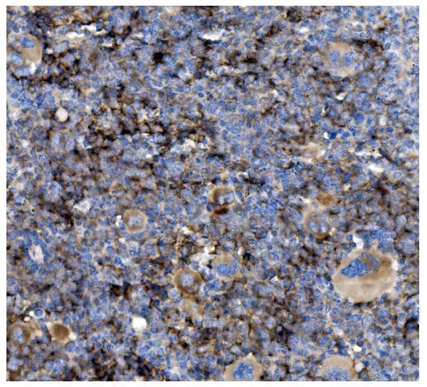
Click image to see more details
IHC analysis of CD82 using anti-CD82 antibody (A02300-1).
CD82 was detected in paraffin-embedded section of mouse spleen tissue. Heat mediated antigen retrieval was performed in EDTA buffer (pH8.0, epitope retrieval solution). The tissue section was blocked with 10% goat serum. The tissue section was then incubated with 2μg/ml rabbit anti-CD82 Antibody (A02300-1) overnight at 4°C. Biotinylated goat anti-rabbit IgG was used as secondary antibody and incubated for 30 minutes at 37°C. The tissue section was developed using Strepavidin-Biotin-Complex (SABC) (Catalog # SA1022) with DAB as the chromogen.

Click image to see more details
IHC analysis of CD82 using anti-CD82 antibody (A02300-1).
CD82 was detected in paraffin-embedded section of rat spleen tissue. Heat mediated antigen retrieval was performed in EDTA buffer (pH8.0, epitope retrieval solution). The tissue section was blocked with 10% goat serum. The tissue section was then incubated with 2μg/ml rabbit anti-CD82 Antibody (A02300-1) overnight at 4°C. Biotinylated goat anti-rabbit IgG was used as secondary antibody and incubated for 30 minutes at 37°C. The tissue section was developed using Strepavidin-Biotin-Complex (SABC) (Catalog # SA1022) with DAB as the chromogen.

Click image to see more details
Flow Cytometry analysis of mouse PBMC cells using anti-CD82 antibody (A02300-1).
Overlay histogram showing mouse PBMC cells stained with A02300-1 (Blue line). The cells were fixed with 4% paraformaldehyde and blocked with 10% normal goat serum. And then incubated with rabbit anti-CD82 Antibody (A02300-1,1μg/1x106 cells) for 30 min at 20°C. DyLight®488 conjugated goat anti-rabbit IgG (BA1127, 5-10μg/1x106 cells) was used as secondary antibody for 30 minutes at 20°C. Isotype control antibody (Green line) was rabbit IgG (1μg/1x106) used under the same conditions. Unlabelled sample without incubation with primary antibody and secondary antibody (Red line) was used as a blank control.

Click image to see more details
Flow Cytometry analysis of mouse spleen tissues using anti-CD82 antibody (A02300-1).
Overlay histogram showing mouse spleen tissues stained with A02300-1 (Blue line). The tissues were fixed with 4% paraformaldehyde and blocked with 10% normal goat serum. And then incubated with rabbit anti-CD82 Antibody (A02300-1,1μg/1x106 cells) for 30 min at 20°C. DyLight®488 conjugated goat anti-rabbit IgG (BA1127, 5-10μg/1x106 cells) was used as secondary antibody for 30 minutes at 20°C. Isotype control antibody (Green line) was rabbit IgG (1μg/1x106) used under the same conditions. Unlabelled sample without incubation with primary antibody and secondary antibody (Red line) was used as a blank control.
Specific Publications For Anti-CD82 Antibody (A02300-1)
Loading publications
Recommended Resources
Here are featured tools and databases that you might find useful.
- Boster's Pathways Library
- Protein Databases
- Bioscience Research Protocol Resources
- Data Processing & Analysis Software
- Photo Editing Software
- Scientific Literature Resources
- Research Paper Management Tools
- Molecular Biology Software
- Primer Design Tools
- Bioinformatics Tools
- Phylogenetic Tree Analysis
Customer Reviews
Have you used Anti-CD82 Antibody?
Share your experimental results or join a short interview to earn up to $1,000 in product credits or other rewards.
0 Reviews For Anti-CD82 Antibody
Customer Q&As
Have a question?
Find answers in Q&As, reviews.
Can't find your answer?
Submit your question
