Click image to see more details
-

-
-
-
-
+3
Product Info Summary
| SKU: | M00166-4 |
|---|---|
| Size: | 100 μg/vial |
| Reactive Species: | Human, Mouse, Rat |
| Host: | Mouse |
| Application: | IHC, WB |
Customers Who Bought This Also Bought

Product info
Product Name
Anti-Cdk2 Antibody Picoband® (monoclonal, 6D5B5)
SKU/Catalog Number
M00166-4
Size
100 μg/vial
Form
Lyophilized
Description
Boster Bio Anti-Cdk2 Antibody Picoband® (monoclonal, 6D5B5) catalog # M00166-4. Tested in IHC, WB applications. This antibody reacts with Human, Mouse, Rat. The brand Picoband indicates this is a premium antibody that guarantees superior quality, high affinity, and strong signals with minimal background in Western blot applications. Only our best-performing antibodies are designated as Picoband, ensuring unmatched performance.
Storage & Handling
At -20°C for one year from date of receipt. After reconstitution, at 4°C for one month. It can also be aliquotted and stored frozen at -20°C for six months. Avoid repeated freezing and thawing.
Cite This Product
Anti-Cdk2 Antibody Picoband® (monoclonal, 6D5B5) (Boster Biological Technology, Pleasanton CA, USA, Catalog # M00166-4)
Host
Mouse
Contents
Each vial contains 4 mg Trehalose, 0.9 mg NaCl and 0.2 mg Na2HPO4.
Clonality
Monoclonal
Clone Number
6D5B5
Isotype
Mouse IgG2b
Immunogen
E.coli-derived human Cdk2 recombinant protein (Position: E81-L298). Human Cdk2 shares 98.6% amino acid (aa) sequence identity with rat Cdk2.
Cross-reactivity
No cross-reactivity with other proteins.
Reactive Species
M00166-4 is reactive to CDK2 in Human, Mouse, Rat
Observed Molecular Weight
30 kDa
Calculated molecular weight
33.9 kDa
Background of CDK2
CDK2, Cyclin-Dependent Kinase2, is also known as P33. The CDK2 protein was highly homologous to p34(CDC2) kinase and more significantly homologous to Xenopus Eg1 kinase, suggesting that CDK2 is the human homolog of Eg1. The CDK2 gene is mapped to 12q13, the same region to which the CDK4 gene maps. Human cyclin A binds independently to 2 kinases, p34(cdc2) or p33. In adenovirus-transformed cells, the viral E1A oncoprotein seems to associate with p33/cyclin A but not with p34(cdc2)/cyclin A. The gene for p33 shares 65% sequence identity with p34(cdc2). P33(cdk2) plays a unique role in cell cycle regulation of vertebrate cells.
Antibody Validation
Boster validates all antibodies on WB, IHC, ICC, Immunofluorescence, and ELISA with known positive control and negative samples to ensure specificity and high affinity, including thorough antibody incubations.
Application & Images
Applications
M00166-4 is guaranteed for IHC, WB Boster Guarantee
Assay Dilutions Recommendation
The recommendations below provide a starting point for assay optimization. The actual working concentration varies and should be decided by the user.
Western blot, 0.25-0.5 μg/ml, Human, Mouse, Rat
Immunohistochemistry(Paraffin-embedded Section), 2-5 μg/ml, Human, Mouse, Rat
Positive Control
WB: human Jurkat whole cell, human HepG2 whole cell, human U20S whole cell, rat heart tissue, rat L6 whole cell, mouse heart tissue, mouse C2C12 whole cell
IHC: human laryngeal squamous cell carcinomas tissue, human liver cancer tissue, human renal clear cell carcinoma tissue, human serous adenocarcinoma of ovary tissue, mouse colon tissue, rat colon tissue
Validation Images & Assay Conditions
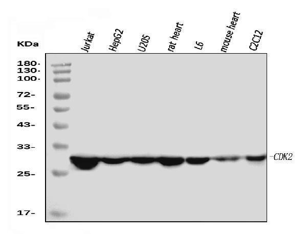
Click image to see more details
Western blot analysis of Cdk2 using anti-Cdk2 antibody (M00166-4).
Electrophoresis was performed on a 5-20% SDS-PAGE gel at 70V (Stacking gel) / 90V (Resolving gel) for 2-3 hours. The sample well of each lane was loaded with 30 ug of sample under reducing conditions.
Lane 1: human Jurkat whole cell lysates,
Lane 2: human HepG2 whole cell lysates,
Lane 3: human U20S whole cell lysates,
Lane 4: rat heart tissue lysates,
Lane 5: rat L6 whole cell lysates,
Lane 6: mouse heart tissue lysates,
Lane 7: mouse C2C12 whole cell lysates.
After electrophoresis, proteins were transferred to a nitrocellulose membrane at 150 mA for 50-90 minutes. Blocked the membrane with 5% non-fat milk/TBS for 1.5 hour at RT. The membrane was incubated with mouse anti-Cdk2 antigen affinity purified monoclonal antibody (Catalog # M00166-4) at 0.5 μg/mL overnight at 4°C, then washed with TBS-0.1%Tween 3 times with 5 minutes each and probed with a goat anti-mouse IgG-HRP secondary antibody at a dilution of 1:10000 for 1.5 hour at RT. The signal is developed using an Enhanced Chemiluminescent detection (ECL) kit (Catalog # EK1001) with Tanon 5200 system. A specific band was detected for Cdk2 at approximately 30 kDa. The expected band size for Cdk2 is at 30 kDa.

Click image to see more details
IHC analysis of Cdk2 using anti-Cdk2 antibody (M00166-4).
Cdk2 was detected in a paraffin-embedded section of human laryngeal squamous cell carcinomas tissue. Heat mediated antigen retrieval was performed in EDTA buffer (pH 8.0, epitope retrieval solution). The tissue section was blocked with 10% goat serum. The tissue section was then incubated with 2 μg/ml mouse anti-Cdk2 Antibody (M00166-4) overnight at 4°C. Peroxidase Conjugated Goat Anti-mouse IgG was used as secondary antibody and incubated for 30 minutes at 37°C. The tissue section was developed using HRP Conjugated Mouse IgG Super Vision Assay Kit (Catalog # SV0001) with DAB as the chromogen.

Click image to see more details
IHC analysis of Cdk2 using anti-Cdk2 antibody (M00166-4).
Cdk2 was detected in a paraffin-embedded section of human liver cancer tissue. Heat mediated antigen retrieval was performed in EDTA buffer (pH 8.0, epitope retrieval solution). The tissue section was blocked with 10% goat serum. The tissue section was then incubated with 2 μg/ml mouse anti-Cdk2 Antibody (M00166-4) overnight at 4°C. Peroxidase Conjugated Goat Anti-mouse IgG was used as secondary antibody and incubated for 30 minutes at 37°C. The tissue section was developed using HRP Conjugated Mouse IgG Super Vision Assay Kit (Catalog # SV0001) with DAB as the chromogen.

Click image to see more details
IHC analysis of Cdk2 using anti-Cdk2 antibody (M00166-4).
Cdk2 was detected in a paraffin-embedded section of human renal clear cell carcinoma tissue. Heat mediated antigen retrieval was performed in EDTA buffer (pH 8.0, epitope retrieval solution). The tissue section was blocked with 10% goat serum. The tissue section was then incubated with 2 μg/ml mouse anti-Cdk2 Antibody (M00166-4) overnight at 4°C. Peroxidase Conjugated Goat Anti-mouse IgG was used as secondary antibody and incubated for 30 minutes at 37°C. The tissue section was developed using HRP Conjugated Mouse IgG Super Vision Assay Kit (Catalog # SV0001) with DAB as the chromogen.

Click image to see more details
IHC analysis of Cdk2 using anti-Cdk2 antibody (M00166-4).
Cdk2 was detected in a paraffin-embedded section of human serous adenocarcinoma of ovary tissue. Heat mediated antigen retrieval was performed in EDTA buffer (pH 8.0, epitope retrieval solution). The tissue section was blocked with 10% goat serum. The tissue section was then incubated with 2 μg/ml mouse anti-Cdk2 Antibody (M00166-4) overnight at 4°C. Peroxidase Conjugated Goat Anti-mouse IgG was used as secondary antibody and incubated for 30 minutes at 37°C. The tissue section was developed using HRP Conjugated Mouse IgG Super Vision Assay Kit (Catalog # SV0001) with DAB as the chromogen.

Click image to see more details
IHC analysis of Cdk2 using anti-Cdk2 antibody (M00166-4).
Cdk2 was detected in a paraffin-embedded section of mouse colon tissue. Heat mediated antigen retrieval was performed in EDTA buffer (pH 8.0, epitope retrieval solution). The tissue section was blocked with 10% goat serum. The tissue section was then incubated with 2 μg/ml mouse anti-Cdk2 Antibody (M00166-4) overnight at 4°C. Peroxidase Conjugated Goat Anti-mouse IgG was used as secondary antibody and incubated for 30 minutes at 37°C. The tissue section was developed using HRP Conjugated Mouse IgG Super Vision Assay Kit (Catalog # SV0001) with DAB as the chromogen.

Click image to see more details
IHC analysis of Cdk2 using anti-Cdk2 antibody (M00166-4).
Cdk2 was detected in a paraffin-embedded section of rat colon tissue. Heat mediated antigen retrieval was performed in EDTA buffer (pH 8.0, epitope retrieval solution). The tissue section was blocked with 10% goat serum. The tissue section was then incubated with 2 μg/ml mouse anti-Cdk2 Antibody (M00166-4) overnight at 4°C. Peroxidase Conjugated Goat Anti-mouse IgG was used as secondary antibody and incubated for 30 minutes at 37°C. The tissue section was developed using HRP Conjugated Mouse IgG Super Vision Assay Kit (Catalog # SV0001) with DAB as the chromogen.
Specific Publications For Anti-Cdk2 Antibody Picoband® (monoclonal, 6D5B5) (M00166-4)
Loading publications
Recommended Resources
Here are featured tools and databases that you might find useful.
- Boster's Pathways Library
- Protein Databases
- Bioscience Research Protocol Resources
- Data Processing & Analysis Software
- Photo Editing Software
- Scientific Literature Resources
- Research Paper Management Tools
- Molecular Biology Software
- Primer Design Tools
- Bioinformatics Tools
- Phylogenetic Tree Analysis
Customer Reviews
Have you used Anti-Cdk2 Antibody Picoband® (monoclonal, 6D5B5)?
Share your experimental results or join a short interview to earn up to $1,000 in product credits or other rewards.
0 Reviews For Anti-Cdk2 Antibody Picoband® (monoclonal, 6D5B5)
Customer Q&As
Have a question?
Find answers in Q&As, reviews.
Can't find your answer?
Submit your question
