Click image to see more details
-

-
-
-
-
+5
Product Info Summary
| SKU: | A06266-1 |
|---|---|
| Size: | 100 μg/vial |
| Reactive Species: | Human, Rat |
| Host: | Rabbit |
| Application: | ELISA, Flow Cytometry, IP, IF, IHC, ICC, WB |
Customers Who Bought This Also Bought

Product info
Product Name
Anti-CHCHD2 Antibody Picoband®
SKU/Catalog Number
A06266-1
Size
100 μg/vial
Form
Lyophilized
Description
Boster Bio Anti-CHCHD2 Antibody Picoband® catalog # A06266-1. Tested in WB, IHC, ICC/IF, IP, Flow Cytometry, ELISA applications. This antibody reacts with Human, Rat. The brand Picoband indicates this is a premium antibody that guarantees superior quality, high affinity, and strong signals with minimal background in Western blot applications. Only our best-performing antibodies are designated as Picoband, ensuring unmatched performance.
Storage & Handling
At -20°C for one year from date of receipt. After reconstitution, at 4°C for one month. It can also be aliquotted and stored frozen at -20°C for six months. Avoid repeated freezing and thawing.
Cite This Product
Anti-CHCHD2 Antibody Picoband® (Boster Biological Technology, Pleasanton CA, USA, Catalog # A06266-1)
Host
Rabbit
Contents
Each vial contains 4 mg Trehalose, 0.9 mg NaCl, 0.2 mg Na2HPO4.
Clonality
Polyclonal
Immunogen
E.coli-derived human CHCHD2 recombinant protein (Position: H74-A151).
Reactive Species
A06266-1 is reactive to CHCHD2 in Human, Rat
Observed Molecular Weight
18 kDa
Calculated molecular weight
15.5 kDa
Background of CHCHD2
The protein encoded by this gene belongs to a class of eukaryotic CX(9)C proteins characterized by four cysteine residues spaced ten amino acids apart from one another. These residues form disulfide linkages that define a CHCH fold. In response to stress, the protein translocates from the mitochondrial intermembrane space to the nucleus where it binds to a highly conserved 13 nucleotide oxygen responsive element in the promoter of cytochrome oxidase 4I2, a subunit of the terminal enzyme of the electron transport chain. In concert with recombination signal sequence-binding protein J, binding of this protein activates the oxygen responsive element at four percent oxygen. In addition, it has been shown that this protein is a negative regulator of mitochondria-mediated apoptosis. In response to apoptotic stimuli, mitochondrial levels of this protein decrease, allowing BCL2-associated X protein to oligomerize and activate the caspase cascade. Pseudogenes of this gene are found on multiple chromosomes. Alternative splicing results in multiple transcript variants.
Antibody Validation
Boster validates all antibodies on WB, IHC, ICC, Immunofluorescence, and ELISA with known positive control and negative samples to ensure specificity and high affinity, including thorough antibody incubations.
Application & Images
Applications
A06266-1 is guaranteed for ELISA, Flow Cytometry, IP, IF, IHC, ICC, WB Boster Guarantee
Assay Dilutions Recommendation
The recommendations below provide a starting point for assay optimization. The actual working concentration varies and should be decided by the user.
Western blot, 0.25-0.5 μg/ml, Human
Immunohistochemistry(Paraffin-embedded Section), 2-5 μg/ml, Human, Rat
Immunocytochemistry/Immunofluorescence, 5 μg/ml, Human
Immunoprecipitation, 0.5-2 μg/ml, Human
Flow Cytometry (Fixed), 1-3 μg/1x106 cells, Human
ELISA, 0.1-0.5 μg/ml
Positive Control
WB: human 293T whole cell, human HT1080 whole cell, human HepG2 whole cell, human A431 whole cell
IHC: human lung cancer tissue
Validation Images & Assay Conditions
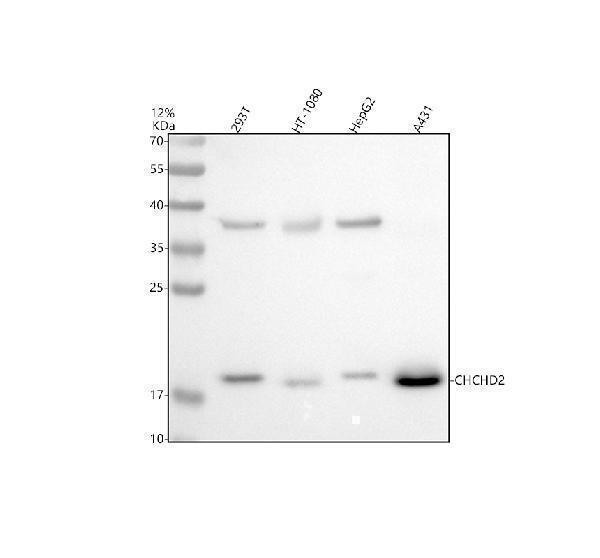
Click image to see more details
Western blot analysis of CHCHD2 using anti-CHCHD2 antibody (A06266-1).
Electrophoresis was performed on a 12% SDS-PAGE gel at 80V (Stacking gel) / 120V (Resolving gel) for 2 hours. The sample well of each lane was loaded with 30 ug of sample under reducing conditions.
Lane 1: human 293T whole cell lysates,
Lane 2: human HT1080 whole cell lysates,
Lane 3: human HepG2 whole cell lysates,
Lane 4: human A431 whole cell lysates.
After electrophoresis, proteins were transferred to a nitrocellulose membrane at 150 mA for 50-90 minutes. Blocked the membrane with 5% non-fat milk/TBS for 1.5 hour at RT. The membrane was incubated with rabbit anti-CHCHD2 antigen affinity purified polyclonal antibody (A06266-1) at 1:1000 overnight at 4°C, then washed with TBS-0.1%Tween 3 times with 5 minutes each and probed with a goat anti-rabbit IgG-HRP secondary antibody at a dilution of 1:5000 for 1.5 hour at RT. The signal is developed using an ECL Plus Western Blotting Substrate (Catalog # AR1196-200) with Tanon 5200 system. A specific band was detected for CHCHD2 at approximately 18 kDa. The expected band size for CHCHD2 is at 16 kDa.

Click image to see more details
IHC analysis of CHCHD2 using anti-CHCHD2 antibody (A06266-1).
CHCHD2 was detected in a paraffin-embedded section of human lung cancer tissue. Heat mediated antigen retrieval was performed in EDTA buffer (pH 8.0, epitope retrieval solution). The tissue section was blocked with 10% goat serum. The tissue section was then incubated with 1:200 rabbit anti-CHCHD2 Antibody (A06266-1) overnight at 4°C. Peroxidase Conjugated Goat Anti-rabbit IgG was used as secondary antibody and incubated for 30 minutes at 37°C. The tissue section was developed using HRP Conjugated Rabbit IgG Super Vision Assay Kit (Catalog # SV0002) with DAB as the chromogen.

Click image to see more details
Western blot analysis of CHCHD2 using anti-CHCHD2 antibody (A06266-1).
Electrophoresis was performed on a 12% SDS-PAGE gel at 80V (Stacking gel) / 120V (Resolving gel) for 2 hours. The sample well of each lane was loaded with 30 ug of sample under reducing conditions.
Lane 1: human A431 whole cell lysates,
Lane 2: human K562 whole cell lysates,
Lane 3: human THP-1 whole cell lysates,
Lane 4: human Hela whole cell lysates.
After electrophoresis, proteins were transferred to a nitrocellulose membrane at 150 mA for 50-90 minutes. Blocked the membrane with 5% non-fat milk/TBS for 1.5 hour at RT. The membrane was incubated with rabbit anti-CHCHD2 antigen affinity purified polyclonal antibody (A06266-1) at 0.5 μg/mL overnight at 4°C, then washed with TBS-0.1%Tween 3 times with 5 minutes each and probed with a goat anti-rabbit IgG-HRP secondary antibody at a dilution of 1:5000 for 1.5 hour at RT. The signal is developed using an ECL Plus Western Blotting Substrate (Catalog # AR1196-200) with Tanon 5200 system. A specific band was detected for CHCHD2 at approximately 18 kDa. The expected band size for CHCHD2 is at 16 kDa.

Click image to see more details
IHC analysis of CHCHD2 using anti-CHCHD2 antibody (A06266-1).
CHCHD2 was detected in a paraffin-embedded section of human pancreas cancer tissue. Heat mediated antigen retrieval was performed in EDTA buffer (pH 8.0, epitope retrieval solution). The tissue section was blocked with 10% goat serum. The tissue section was then incubated with 2 μg/ml rabbit anti-CHCHD2 Antibody (A06266-1) overnight at 4°C. Peroxidase Conjugated Goat Anti-rabbit IgG was used as secondary antibody and incubated for 30 minutes at 37°C. The tissue section was developed using HRP Conjugated Rabbit IgG Super Vision Assay Kit (Catalog # SV0002) with DAB as the chromogen.

Click image to see more details
IHC analysis of CHCHD2 using anti-CHCHD2 antibody (A06266-1).
CHCHD2 was detected in a paraffin-embedded section of rat brain tissue. Heat mediated antigen retrieval was performed in EDTA buffer (pH 8.0, epitope retrieval solution). The tissue section was blocked with 10% goat serum. The tissue section was then incubated with 2 μg/ml rabbit anti-CHCHD2 Antibody (A06266-1) overnight at 4°C. Peroxidase Conjugated Goat Anti-rabbit IgG was used as secondary antibody and incubated for 30 minutes at 37°C. The tissue section was developed using HRP Conjugated Rabbit IgG Super Vision Assay Kit (Catalog # SV0002) with DAB as the chromogen.

Click image to see more details
IHC analysis of CHCHD2 using anti-CHCHD2 antibody (A06266-1).
CHCHD2 was detected in a paraffin-embedded section of human lung cancer tissue. Heat mediated antigen retrieval was performed in EDTA buffer (pH 8.0, epitope retrieval solution). The tissue section was blocked with 10% goat serum. The tissue section was then incubated with 2 μg/ml rabbit anti-CHCHD2 Antibody (A06266-1) overnight at 4°C. Peroxidase Conjugated Goat Anti-rabbit IgG was used as secondary antibody and incubated for 30 minutes at 37°C. The tissue section was developed using HRP Conjugated Rabbit IgG Super Vision Assay Kit (Catalog # SV0002) with DAB as the chromogen.

Click image to see more details
IF analysis of CHCHD2 using anti-CHCHD2 antibody (A06266-1).
CHCHD2 was detected in an immunocytochemical section of A431 cells. Enzyme antigen retrieval was performed using IHC enzyme antigen retrieval reagent (AR0022) for 15 mins. The cells were blocked with 10% goat serum. And then incubated with 5 μg/mL rabbit anti-CHCHD2 Antibody (A06266-1) overnight at 4°C. Cy3 Conjugated Goat Anti-Rabbit IgG (BA1032) was used as secondary antibody at 1:500 dilution and incubated for 30 minutes at 37°C. The section was counterstained with DAPI. Visualize using a fluorescence microscope and filter sets appropriate for the label used.

Click image to see more details
Immunoprecipitating CHCHD2 in K562 whole cell lysate.
Western blot analysis of CHCHD2 using anti-CHCHD2 antibody (A06266-1).
Lane 1: K562 whole cell lysates (30ug),
Lane 2: Rabbit control IgG instead of anti-CHCHD2 antibody in K562 whole cell lysate,
Lane 3: anti-CHCHD2 antibody (2μg) + K562 whole cell lysate (500μg).
After electrophoresis, proteins were transferred to a membrane. Then the membrane was incubated with rabbit anti-CHCHD2 antigen affinity purified polyclonal antibody (A06266-1) at a dilution of 0.5 μg/mL and probed with a goat anti-rabbit IgG-HRP secondary antibody (Catalog # BA1054). The signal is developed using ECL Plus Western Blotting Substrate (Catalog # AR1197). A specific band was detected for CHCHD2 at approximately 18 kDa. The expected band size for CHCHD2 is at 16 kDa.

Click image to see more details
Flow Cytometry analysis of CACO-2 cells using anti-CHCHD2 antibody (A06266-1).
Overlay histogram showing CACO-2 cells stained with A06266-1 (Blue line). To facilitate intracellular staining, cells were fixed with 4% paraformaldehyde and permeabilized with permeabilization buffer. The cells were blocked with 10% normal goat serum. And then incubated with rabbit anti-CHCHD2 Antibody (A06266-1, 1 μg/1x106 cells) for 30 min at 20°C. Fluoro488 conjugated goat anti-rabbit IgG (BA1127, 5-10 μg/1x106 cells) was used as secondary antibody for 30 minutes at 20°C. Isotype control antibody (Green line) was rabbit IgG (1 μg/1x106) used under the same conditions. Unlabelled sample without incubation with primary antibody and secondary antibody (Red line) was used as a blank control.
Specific Publications For Anti-CHCHD2 Antibody Picoband® (A06266-1)
Loading publications
Recommended Resources
Here are featured tools and databases that you might find useful.
- Boster's Pathways Library
- Protein Databases
- Bioscience Research Protocol Resources
- Data Processing & Analysis Software
- Photo Editing Software
- Scientific Literature Resources
- Research Paper Management Tools
- Molecular Biology Software
- Primer Design Tools
- Bioinformatics Tools
- Phylogenetic Tree Analysis
Customer Reviews
Have you used Anti-CHCHD2 Antibody Picoband®?
Share your experimental results or join a short interview to earn up to $1,000 in product credits or other rewards.
0 Reviews For Anti-CHCHD2 Antibody Picoband®
Customer Q&As
Have a question?
Find answers in Q&As, reviews.
Can't find your answer?
Submit your question
