Click image to see more details
-

-
-
-
-
+1
Product Info Summary
| SKU: | A05300-1 |
|---|---|
| Size: | 100 μg/vial |
| Reactive Species: | Human, Rat |
| Host: | Rabbit |
| Application: | ELISA, Flow Cytometry, IF, IHC, ICC, WB |
Customers Who Bought This Also Bought

Product info
Product Name
Anti-CLCN3 Antibody Picoband®
SKU/Catalog Number
A05300-1
Size
100 μg/vial
Form
Lyophilized
Description
Boster Bio Anti-CLCN3 Antibody Picoband® catalog # A05300-1. Tested in WB, IF, IHC, ICC, Flow Cytometry, ELISA applications. This antibody reacts with Human, Rat. The brand Picoband indicates this is a premium antibody that guarantees superior quality, high affinity, and strong signals with minimal background in Western blot applications. Only our best-performing antibodies are designated as Picoband, ensuring unmatched performance.
Storage & Handling
At -20°C for one year from date of receipt. After reconstitution, at 4°C for one month. It can also be aliquotted and stored frozen at -20°C for six months. Avoid repeated freezing and thawing.
Cite This Product
Anti-CLCN3 Antibody Picoband® (Boster Biological Technology, Pleasanton CA, USA, Catalog # A05300-1)
Host
Rabbit
Contents
Each vial contains 4 mg Trehalose, 0.9 mg NaCl, 0.2 mg Na2HPO4.
Clonality
Polyclonal
Immunogen
E.coli-derived human CLCN3 recombinant protein (Position: G293-R752). Human CLCN3 shares 99.6% amino acid (aa) sequence identity with mouse CLCN3.
Reactive Species
A05300-1 is reactive to CLCN3 in Human, Rat
Observed Molecular Weight
170 kDa
Calculated molecular weight
91.0 kDa
Background of CLCN3
H+/Cl? exchange transporter 3 is a protein that in humans is encoded by the CLCN3 gene. This gene encodes a member of the voltage-gated chloride channel (ClC) family. The encoded protein is present in all cell types and localized in plasma membranes and in intracellular vesicles. It is a multi-pass membrane protein which contains a ClC domain and two additional C-terminal CBS (cystathionine beta-synthase) domains. The ClC domain catalyzes the selective flow of Cl- ions across cell membranes, and the CBS domain may have a regulatory function. This protein plays a role in both acidification and transmitter loading of GABAergic synaptic vesicles, and in smooth muscle cell activation and neointima formation. This protein is required for lysophosphatidic acid (LPA)-activated Cl- current activity and fibroblast-to-myofibroblast differentiation. The protein activity is regulated by Ca(2+)/calmodulin-dependent protein kinase II (CaMKII) in glioma cells. Multiple alternatively spliced transcript variants encoding different isoforms have been identified.
Antibody Validation
Boster validates all antibodies on WB, IHC, ICC, Immunofluorescence, and ELISA with known positive control and negative samples to ensure specificity and high affinity, including thorough antibody incubations.
Application & Images
Applications
A05300-1 is guaranteed for ELISA, Flow Cytometry, IF, IHC, ICC, WB Boster Guarantee
Assay Dilutions Recommendation
The recommendations below provide a starting point for assay optimization. The actual working concentration varies and should be decided by the user.
Western blot, 0.25-0.5 μg/ml, Human
Immunohistochemistry(Paraffin-embedded Section), 2-5 μg/ml, Human, Rat
Immunocytochemistry/Immunofluorescence, 5 μg/ml, Human
Flow Cytometry (Fixed), 1-3 μg/1x106 cells, Human
ELISA, 0.1-0.5 μg/ml, -
Positive Control
WB: human Jurkat whole cell, human U-87MG whole cell, human SH-SY6Y whole cell, human MCF-7 whole cell, human A549 whole cell, human Hela whole cell, human K562 whole cell
IHC: human prostatic cancer tissue, rat kidney tissue
ICC/IF: SiHa cell
FCM: JK cell
Validation Images & Assay Conditions
Click image to see more details
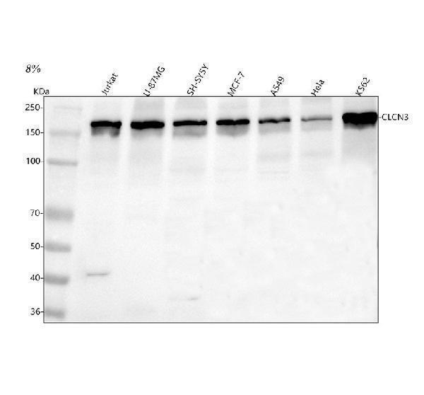
Western blot analysis of CLCN3 using anti-CLCN3 antibody (A05300-1).
Electrophoresis was performed on a 5-20% SDS-PAGE gel at 70V (Stacking gel) / 90V (Resolving gel) for 2-3 hours. The sample well of each lane was loaded with 30 ug of sample under reducing conditions.
Lane 1: human Jurkat whole cell lysates,
Lane 2: human U-87MG whole cell lysates,
Lane 3: human SH-SY6Y whole cell lysates,
Lane 4: human MCF-7 whole cell lysates,
Lane 5: human A549 whole cell lysates,
Lane 6: human Hela whole cell lysates,
Lane 7: human K562 whole cell lysates.
After electrophoresis, proteins were transferred to a nitrocellulose membrane at 150 mA for 50-90 minutes. Blocked the membrane with 5% non-fat milk/TBS for 1.5 hour at RT. The membrane was incubated with rabbit anti-CLCN3 antigen affinity purified polyclonal antibody (Catalog # A05300-1) at 0.5 μg/mL overnight at 4°C, then washed with TBS-0.1%Tween 3 times with 5 minutes each and probed with a goat anti-rabbit IgG-HRP secondary antibody at a dilution of 1:5000 for 1.5 hour at RT. The signal is developed using an Enhanced Chemiluminescent detection (ECL) kit (Catalog # EK1002) with Tanon 5200 system. A specific band was detected for CLCN3 at approximately 170 kDa. The expected band size for CLCN3 is at 91 kDa.

Click image to see more details
IHC analysis of CLCN3 using anti-CLCN3 antibody (A05300-1).
CLCN3 was detected in a paraffin-embedded section of human prostatic cancer tissue. Heat mediated antigen retrieval was performed in EDTA buffer (pH 8.0, epitope retrieval solution). The tissue section was blocked with 10% goat serum. The tissue section was then incubated with 2 μg/ml rabbit anti-CLCN3 Antibody (A05300-1) overnight at 4°C. Peroxidase Conjugated Goat Anti-rabbit IgG was used as secondary antibody and incubated for 30 minutes at 37°C. The tissue section was developed using HRP Conjugated Rabbit IgG Super Vision Assay Kit (Catalog # SV0002) with DAB as the chromogen.

Click image to see more details
IHC analysis of CLCN3 using anti-CLCN3 antibody (A05300-1).
CLCN3 was detected in a paraffin-embedded section of rat kidney tissue. Heat mediated antigen retrieval was performed in EDTA buffer (pH 8.0, epitope retrieval solution). The tissue section was blocked with 10% goat serum. The tissue section was then incubated with 2 μg/ml rabbit anti-CLCN3 Antibody (A05300-1) overnight at 4°C. Peroxidase Conjugated Goat Anti-rabbit IgG was used as secondary antibody and incubated for 30 minutes at 37°C. The tissue section was developed using HRP Conjugated Rabbit IgG Super Vision Assay Kit (Catalog # SV0002) with DAB as the chromogen.

Click image to see more details
IF analysis of CLCN3 using anti-CLCN3 antibody (A05300-1).
CLCN3 was detected in an immunocytochemical section of SiHa cells. Enzyme antigen retrieval was performed using IHC enzyme antigen retrieval reagent (AR0022) for 15 mins. The cells were blocked with 10% goat serum. And then incubated with 5 μg/mL rabbit anti-CLCN3 Antibody (A05300-1) overnight at 4°C. DyLight®488 Conjugated Goat Anti-Rabbit IgG (BA1127) was used as secondary antibody at 1:500 dilution and incubated for 30 minutes at 37°C. The section was counterstained with DAPI. Visualize using a fluorescence microscope and filter sets appropriate for the label used.

Click image to see more details
Flow Cytometry analysis of JK cells using anti-AXIN1 antibody (A05300-1).
Overlay histogram showing JK cells stained with A05300-1 (Blue line). To facilitate intracellular staining, cells were fixed with 4% paraformaldehyde and permeabilized with permeabilization buffer. The cells were blocked with 10% normal goat serum. And then incubated with rabbit anti-AXIN1 Antibody (A05300-1, 1 μg/1x106 cells) for 30 min at 20°C. DyLight®488 conjugated goat anti-rabbit IgG (BA1127, 5-10 μg/1x106 cells) was used as secondary antibody for 30 minutes at 20°C. Isotype control antibody (Green line) was rabbit IgG (1 μg/1x106) used under the same conditions. Unlabelled sample without incubation with primary antibody and secondary antibody (Red line) was used as a blank control.
Specific Publications For Anti-CLCN3 Antibody Picoband® (A05300-1)
Loading publications
Recommended Resources
Here are featured tools and databases that you might find useful.
- Boster's Pathways Library
- Protein Databases
- Bioscience Research Protocol Resources
- Data Processing & Analysis Software
- Photo Editing Software
- Scientific Literature Resources
- Research Paper Management Tools
- Molecular Biology Software
- Primer Design Tools
- Bioinformatics Tools
- Phylogenetic Tree Analysis
Customer Reviews
Have you used Anti-CLCN3 Antibody Picoband®?
Share your experimental results or join a short interview to earn up to $1,000 in product credits or other rewards.
0 Reviews For Anti-CLCN3 Antibody Picoband®
Customer Q&As
Have a question?
Find answers in Q&As, reviews.
Can't find your answer?
Submit your question
