Click image to see more details
-

-
-
-
-
+8
Product Info Summary
| SKU: | A00031-1 |
|---|---|
| Size: | 0.1 mg |
| Reactive Species: | Human, Mouse, Rat |
| Host: | Rabbit |
| Application: | ELISA, Flow Cytometry, IF, IHC-P, ICC, WB |
Customers Who Bought This Also Bought

Product info
Product Name
Anti-CXCR4 Antibody
SKU/Catalog Number
A00031-1
Size
0.1 mg
Form
Liquid
Description
Boster Bio Anti-CXCR4 Antibody (Catalog # A00031-1). Tested in ELISA, WB, ICC, IF, Flow Cytometry, IHC-P applications. This antibody reacts with Human, Mouse, Rat.
Storage & Handling
CXCR4 antibody can be stored at 4°C for three months and -20°C, stable for up to one year. Avoid repeated freeze-thaw cycles. Antibodies should not be exposed to prolonged high temperatures.
Cite This Product
Anti-CXCR4 Antibody (Boster Biological Technology, Pleasanton CA, USA, Catalog # A00031-1)
Host
Rabbit
Contents
CXCR4 Antibody is supplied in PBS containing 0.02% sodium azide.
Clonality
Polyclonal
Isotype
IgG
Immunogen
Anti-CXCR4 antibody was raised against a peptide corresponding to 14 amino acids near the amino terminus of human CXCR4 isoform b. The immunogen is located within the first 50 amino acids of CXCR4.
Cross-reactivity
CXCR4 Antibody is predicted to not cross-react with other CXCR familiy members.
Reactive Species
A00031-1 is reactive to CXCR4 in Human, Mouse, Rat
Observed Molecular Weight
68 kDa
Calculated molecular weight
39.7 kDa
Background of CXCR4
CXCR4, a G-protein coupled receptor (GPCR) with seven transmembrane domains, is a CXC chemokine receptor specific for stromal-derived-factor-1 (SDF-1 or CXCL12). CXCR4 was initially discovered as one of the co-receptors for HIV entry into CD4+ T cells (1). Blocking CXCR4 could be potentially used as novel therapeutics for HIV treatment.
CXCR4 signaling plays an important role in the migration, proliferation and quiescence of hematopoietic stem cell and their retention within the bone marrow, where it has high levels of SDF-1/CXCL12(2). It has been demonstrated that CXCR4 signaling mediates CD20 up-regulation on B cells (3).
CXCR4 is highly expressed in more than 23 types of cancer, including breast cancer, ovarian cancer, melanoma, and prostate cancer, while there is very less or no expression of CXCR4 in healthy tissues. CXCR4 expression in cancer cells has been reported to be associated with tumor survival, growth and metastasis in tissues with high levels of SDF-1/CXCL12, such as lungs, liver and bone marrow (4,5).
CXCR4 has been shown to regulate neuronal migration, cell positioning and axon wiring (6,7). CXCR4 mutant mice displayed aberrant neuronal distribution, which implicates the role in neuronal disorders such as epilepsy. CXCR4 is also involved in WHIM syndrome (8). WHIM mutations in CXCR4 were recently found in patients with Waldenstrom's macroglobulinemia, and these mutations are correlated to clinical resistance to ibrutinib (9,10).
Antibody Validation
Boster validates all antibodies on WB, IHC, ICC, Immunofluorescence, and ELISA with known positive control and negative samples to ensure specificity and high affinity, including thorough antibody incubations.
Application & Images
Applications
A00031-1 is guaranteed for ELISA, Flow Cytometry, IF, IHC-P, ICC, WB Boster Guarantee
Assay Dilutions Recommendation
The recommendations below provide a starting point for assay optimization. The actual working concentration varies and should be decided by the user.
WB: 1 - 2 μg/mL; IP/ ICC: 2 μg/mL; IHC-P: 5 μg/mL; IF: 20 μg/mL; Flow Cyt: 0.1 μg/mL.
Antibody validated: Western Blot in human, mouse, and rat samples; Immunohistochemistry, Immunocytochemistry and Immunofluorescence in human samples; Flow Cytometry in human and mouse samples. All other applications and species not yet tested. Optimal dilutions for each application should be determined by the researcher.
Validation Images & Assay Conditions
Click image to see more details
Western Blot Validation of CXCR4 in HeLa Cells
Loading: 15 μg of lysates per lane. Antibodies: A00031-1 (1 μg/mL), 1 h incubation at RT in 5% NFDM/TBST. Secondary: Goat anti-rabbit IgG HRP conjugate at 1:10000 dilution.

Click image to see more details
Independent Antibody Validation (IAV) via Protein Expression Profile in Cell Lines
Loading: 15 μg of lysates per lane. Antibodies: A00031-1 (1 μg/mL), 1012 (1 μg/mL), and beta-actin (1 μg/mL), 1 h incubation at RT in 5% NFDM/TBST. Secondary: Goat anti-rabbit IgG HRP conjugate at 1:10000 dilution.

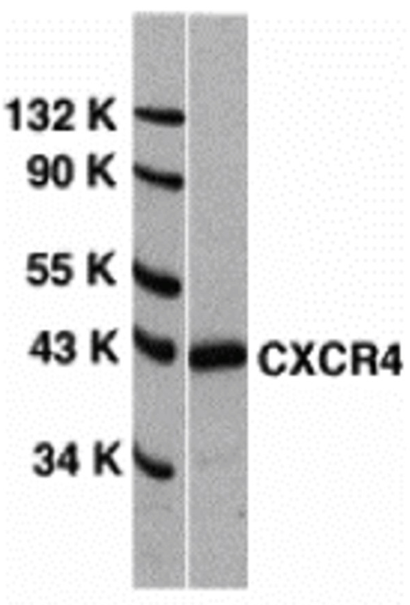
Click image to see more details
Validation with CXCR4 siRNA Knockdown in HeLa Cells
HeLa cells were transfected with control siRNAs (lane 1) or CXCR4 siRNAs (lane 2) Loading: 10 μg of HeLa whole cell lysates per lane. Antibodies: A00031-1 (2 μg/mL), 1 h incubation at RT in 5% NFDM/TBST. Secondary: Goat anti-rabbit IgG HRP conjugate at 1:10000 dilution.

Click image to see more details
Animal Species Reactivity
Loading: Lysates/proteins at 20 μg per lane. Antibodies: A00031-1 (2 μg/mL) or 1012 (2 μg/mL). 1 h incubation at RT in 5% NFDM/TBST. Secondary: Goat anti-rabbit IgG HRP conjugate at 1:10000 dilution.

Click image to see more details
Recombinant Protein Test
Loading: CXCR4 partial recombinant protein (Novus Biologicals, Cat# H00007852-Q01). Lane 1: Anti-CXCR4 antibody (0.1 μg/mL) 1 h incubation at RT in 5% NFDM/TBST. Lane 2: Coomassie blue staining. Secondary: Goat anti-rabbit IgG HRP conjugate at 1:10000 dilution.

Click image to see more details
Immunofluorescence Validation of CXCR4 in HeLa Cells
Immunofluorescent analysis of 4% paraformaldehyde-fixed HeLa cells labeling CXCR4 with A00031-1 at 20 μg/mL, followed by goat anti-rabbit IgG secondary antibody at 1/500 dilution (red). Image showing both membrane and cytoplasmic staining on HeLa cells.

Click image to see more details
Flow Cytometry Validation of CXCR4 in HeLa Cells
Overlay histogram showing HeLa cells stained with A00031-1 (red line, 1μg/1x106 cells). 1 h incubation at 4˚C in 2% FBS/PBS. Followed by secondary antibody 488 goat anti-rabbit IgG (H+L) at 1/500 dilution for 1 h 4˚C.
Isotype control antibody (Green line) was mouse IgG1 (1μg/1x106 cells) used under the same conditions.

Click image to see more details
Overexpression Validation of CXCR4 (Kozak et al., 2002)
U87MG and U87MG-CXCR4 extracts were included as negative and positive controls, respectively, for CXCR4 detection with anti-CXCR4 antibodies.

Click image to see more details
WB Validation of CXCR4 in Human Metastatic Melanoma (Scala et al., 2006)
CXCR4 protein was detected in the human metastatic melanoma cell lines and human melanoma cell line (colo38), but not in the human primary melanocytes (MPR1) with anti-CXCR4 antibodies.

Click image to see more details
Immunohistochemistry Validation of CXCR4 in Human Spleen
Immunohistochemical analysis of paraffin-embedded human spleen tissue using anti-CXCR4 antibody (A00031-1) at 5 μg/ml. Tissue was fixed with formaldehyde and blocked with 10% serum for 1 h at RT; antigen retrieval was by heat mediation with a citrate buffer (pH6). Samples were incubated with primary antibody overnight at 4˚C. A Goat anti-rabbit IgG H&L (HRP) at 1/250 was used as secondary. Counter stained with Hematoxylin.

Click image to see more details
Immunocytochemistry Validation of CXCR4 in HeLa Cells
Immunocytochemical analysis of HeLa cells using anti-CXCR4 antibody (A00031-1) at 2 μg/ml. Cells was fixed with formaldehyde and blocked with 10% serum for 1 h at RT; antigen retrieval was by heat mediation with a citrate buffer (pH6). Samples were incubated with primary antibody overnight at 4˚C. A goat anti-rabbit IgG H&L (HRP) at 1/250 was used as secondary. Counter stained with Hematoxylin.

Click image to see more details
KO Validation of CXCR4 by Flow Cytometry (?demis, et al., 2010)
Astrocytes from wild-type or CXCR4 knockout mice were stained with primary antibodies against CXCR4 and FITC-labeled secondary antibodies, and subsequently subjected to flow cytometry. CXCR4?/? astrocytes (red) showed loss of CXCR4 cell-surface expression compared with wild-type cells (black).
Specific Publications For Anti-CXCR4 Antibody (A00031-1)
Loading publications
Recommended Resources
Here are featured tools and databases that you might find useful.
- Boster's Pathways Library
- Protein Databases
- Bioscience Research Protocol Resources
- Data Processing & Analysis Software
- Photo Editing Software
- Scientific Literature Resources
- Research Paper Management Tools
- Molecular Biology Software
- Primer Design Tools
- Bioinformatics Tools
- Phylogenetic Tree Analysis
Customer Reviews
Have you used Anti-CXCR4 Antibody?
Share your experimental results or join a short interview to earn up to $1,000 in product credits or other rewards.
0 Reviews For Anti-CXCR4 Antibody
Customer Q&As
Have a question?
Find answers in Q&As, reviews.
Can't find your answer?
Submit your question
