Click image to see more details
-

-
-
-
-
+1
Product Info Summary
| SKU: | A15137 |
|---|---|
| Size: | 100 μg/vial |
| Reactive Species: | Human, Mouse, Rat |
| Host: | Rabbit |
| Application: | ELISA, Flow Cytometry, IHC, WB |
Customers Who Bought This Also Bought

Product info
Product Name
Anti-DNAJB13 Antibody Picoband®
SKU/Catalog Number
A15137
Size
100 μg/vial
Form
Lyophilized
Description
Boster Bio Anti-DNAJB13 Antibody Picoband® catalog # A15137. Tested in WB, IHC, Flow Cytometry, ELISA applications. This antibody reacts with Human, Mouse, Rat. The brand Picoband indicates this is a premium antibody that guarantees superior quality, high affinity, and strong signals with minimal background in Western blot applications. Only our best-performing antibodies are designated as Picoband, ensuring unmatched performance.
Storage & Handling
At -20°C for one year from date of receipt. After reconstitution, at 4°C for one month. It can also be aliquotted and stored frozen at -20°C for six months. Avoid repeated freezing and thawing.
Cite This Product
Anti-DNAJB13 Antibody Picoband® (Boster Biological Technology, Pleasanton CA, USA, Catalog # A15137)
Host
Rabbit
Contents
Each vial contains 4 mg Trehalose, 0.9 mg NaCl, 0.2 mg Na2HPO4.
Clonality
Polyclonal
Immunogen
E.coli-derived human DNAJB13 recombinant protein (Position: M1-T316).
Reactive Species
A15137 is reactive to DNAJB13 in Human, Mouse, Rat
Observed Molecular Weight
36 kDa
Calculated molecular weight
36.1 kDa
Background of DNAJB13
This gene encodes a member of the heat shock protein 40 co-chaperone family which is produced in large amounts in the testis and is located on the radial spokes of the axoneme in human sperm flagella and other flagellar structures. The encoded protein associates with the sperm annulus, as part of the septin complex, through direct interaction with septin 4, during sperm terminal differentiation. Naturally occurring mutations in this gene are associated with primary ciliary dyskinesia and male infertility.
Antibody Validation
Boster validates all antibodies on WB, IHC, ICC, Immunofluorescence, and ELISA with known positive control and negative samples to ensure specificity and high affinity, including thorough antibody incubations.
Application & Images
Applications
A15137 is guaranteed for ELISA, Flow Cytometry, IHC, WB Boster Guarantee
Assay Dilutions Recommendation
The recommendations below provide a starting point for assay optimization. The actual working concentration varies and should be decided by the user.
Western blot, 0.25-0.5 μg/ml, Mouse, Rat
Immunohistochemistry(Paraffin-embedded Section), 2-5 μg/ml, Human, Mouse, Rat
Flow Cytometry (Fixed), 1-3 μg/1x106 cells, Human
ELISA, 0.1-0.5 μg/ml
Validation Images & Assay Conditions
Click image to see more details
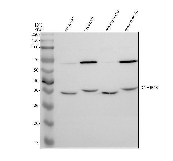
Western blot analysis of DNAJB13 using anti-DNAJB13 antibody (A15137).
Electrophoresis was performed on a 10% SDS-PAGE gel at 80V (Stacking gel) / 120V (Resolving gel) for 2 hours. The sample well of each lane was loaded with 30 ug of sample under reducing conditions.
Lane 1: rat testis tissue lysates,
Lane 2: rat brain tissue lysates,
Lane 3: mouse testis tissue lysates,
Lane 4: mouse brain tissue lysates.
After electrophoresis, proteins were transferred to a nitrocellulose membrane at 150 mA for 50-90 minutes. Blocked the membrane with 5% non-fat milk/TBS for 1.5 hour at RT. The membrane was incubated with rabbit anti-DNAJB13 antigen affinity purified polyclonal antibody (A15137) at 0.5 μg/mL overnight at 4°C, then washed with TBS-0.1%Tween 3 times with 5 minutes each and probed with a goat anti-rabbit IgG-HRP secondary antibody at a dilution of 1:5000 for 1.5 hour at RT. The signal is developed using an ECL Plus Western Blotting Substrate (Catalog # AR1196-200) with Tanon 5200 system. A specific band was detected for DNAJB13 at approximately 36 kDa. The expected band size for DNAJB13 is at 36 kDa.

Click image to see more details
IHC analysis of DNAJB13 using anti-DNAJB13 antibody (A15137).
DNAJB13 was detected in a paraffin-embedded section of human fallopian tube tissue. Heat mediated antigen retrieval was performed in EDTA buffer (pH 8.0, epitope retrieval solution). The tissue section was blocked with 10% goat serum. The tissue section was then incubated with 2 μg/ml rabbit anti-DNAJB13 Antibody (A15137) overnight at 4°C. Peroxidase Conjugated Goat Anti-rabbit IgG was used as secondary antibody and incubated for 30 minutes at 37°C. The tissue section was developed using HRP Conjugated Rabbit IgG Super Vision Assay Kit (Catalog # SV0002) with DAB as the chromogen.

Click image to see more details
Flow Cytometry analysis of Jurkat cells using anti-DNAJB13 antibody (A15137).
Overlay histogram showing Jurkat cells stained with A15137 (Blue line). The cells were fixed with 4% paraformaldehyde and blocked with 10% normal goat serum. And then incubated with rabbit anti-DNAJB13 Antibody (A15137, 1 μg/1x106 cells) for 30 min at 20°C. Fluoro488 conjugated goat anti-rabbit IgG (BA1127, 5-10 μg/1x106 cells) was used as secondary antibody for 30 minutes at 20°C. Isotype control antibody (Green line) was rabbit IgG (1 μg/1x106) used under the same conditions. Unlabelled sample without incubation with primary antibody and secondary antibody (Red line) was used as a blank control.
Specific Publications For Anti-DNAJB13 Antibody Picoband® (A15137)
Loading publications
Recommended Resources
Here are featured tools and databases that you might find useful.
- Boster's Pathways Library
- Protein Databases
- Bioscience Research Protocol Resources
- Data Processing & Analysis Software
- Photo Editing Software
- Scientific Literature Resources
- Research Paper Management Tools
- Molecular Biology Software
- Primer Design Tools
- Bioinformatics Tools
- Phylogenetic Tree Analysis
Customer Reviews
Have you used Anti-DNAJB13 Antibody Picoband®?
Share your experimental results or join a short interview to earn up to $1,000 in product credits or other rewards.
0 Reviews For Anti-DNAJB13 Antibody Picoband®
Customer Q&As
Have a question?
Find answers in Q&As, reviews.
Can't find your answer?
Submit your question
