Click image to see more details
-

-
-
-
-
+7
Product Info Summary
| SKU: | A05556 |
|---|---|
| Size: | 100 μg/vial |
| Reactive Species: | Human, Mouse, Rat |
| Host: | Rabbit |
| Application: | ELISA, IF, IHC, WB |
Customers Who Bought This Also Bought

Product info
Product Name
Anti-DYNLT1 Antibody Picoband®
SKU/Catalog Number
A05556
Size
100 μg/vial
Form
Lyophilized
Description
Boster Bio Anti-DYNLT1 Antibody Picoband® catalog # A05556. Tested in ELISA, IF, IHC, WB applications. This antibody reacts with Human, Mouse, Rat. The brand Picoband indicates this is a premium antibody that guarantees superior quality, high affinity, and strong signals with minimal background in Western blot applications. Only our best-performing antibodies are designated as Picoband, ensuring unmatched performance.
Storage & Handling
Store at -20˚C for one year from date of receipt. After reconstitution, at 4˚C for one month. It can also be aliquotted and stored frozen at -20˚C for six months. Avoid repeated freeze-thaw cycles.
Cite This Product
Anti-DYNLT1 Antibody Picoband® (Boster Biological Technology, Pleasanton CA, USA, Catalog # A05556)
Host
Rabbit
Contents
Each vial contains 4 mg Trehalose, 0.9 mg NaCl and 0.2 mg Na2HPO4.
Clonality
Polyclonal
Isotype
Rabbit IgG
Immunogen
E. coli-derived human DYNLT1 recombinant protein (Position: M1-A80).
Cross-reactivity
No cross-reactivity with other proteins.
Reactive Species
A05556 is reactive to DYNLT1 in Human, Mouse, Rat
Observed Molecular Weight
16 kDa
Calculated molecular weight
12.5 kDa
Background of DYNLT1
Dynein light chain Tctex-type 1 is a protein that in humans is encoded by the DYNLT1 gene. This gene encodes a component of the motor complex, cytoplasmic dynein, which transports cellular cargo along microtubules in the cell. The encoded protein regulates the length of primary cilia which are sensory organelles found on the surface of cells. The protein encoded by this gene interacts with viral proteins, like the minor capsid protein L2 of human papillomavirus, and is required for dynein-mediated delivery of the viral nucleic acid to the host nucleus. This protein interacts with oncogenic nucleoporins to disrupt gene regulation and cause leukemic transformation. Pseudogenes of this gene are present on chromosomes 4 and 17. Alternative splicing results in multiple transcript variants encoding different isoforms.
Antibody Validation
Boster validates all antibodies on WB, IHC, ICC, Immunofluorescence, and ELISA with known positive control and negative samples to ensure specificity and high affinity, including thorough antibody incubations.
Application & Images
Applications
A05556 is guaranteed for ELISA, IF, IHC, WB Boster Guarantee
Recommend Dilution
| Application | Dilution | Species |
|---|---|---|
| Western blot | 0.1-0.5μg/ml | Human |
| Immunohistochemistry (Paraffin-embedded Section) | 2-5μg/ml | Human, Mouse, Rat |
| Immunofluorescence | 5μg/ml | Human, Rat |
| ELISA | 0.1-0.5μg/ml | - |
Tested application
Suggested blocking solution with 5% non-fat milk or BSA; (*)Recommended protein loading: 20-40 µg per lane
Use TE buffer pH 9.0 for antigen retrieval; (*) citrate buffer pH 6.0 is an alternative.
Validation Images & Assay Conditions
Click image to see more details
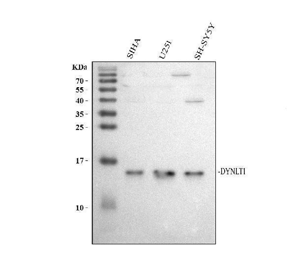
Western blot analysis of DYNLT1 using anti-DYNLT1 antibody (A05556).
Electrophoresis was performed on a 13% SDS-PAGE gel at 80V (Stacking gel) / 120V (Resolving gel) for 2 hours. The sample well of each lane was loaded with 30 ug of sample under reducing conditions.
Lane 1: human SiHa whole cell lysates,
Lane 2: human U251 whole cell lysates,
Lane 3: human SH-SY5Y whole cell lysates.
After electrophoresis, proteins were transferred to a nitrocellulose membrane at 150 mA for 50-90 minutes. Blocked the membrane with 5% non-fat milk/TBS for 1.5 hour at RT. The membrane was incubated with rabbit anti-DYNLT1 antigen affinity purified polyclonal antibody (Catalog # A05556) at 0.5 μg/mL overnight at 4°C, then washed with TBS-0.1%Tween 3 times with 5 minutes each and probed with a goat anti-rabbit IgG-HRP secondary antibody at a dilution of 1:5000 for 1.5 hour at RT. The signal is developed using an ECL Plus Western Blotting Substrate (Catalog # AR1196-200) with Tanon 5200 system. A specific band was detected for DYNLT1 at approximately 16 kDa. The expected band size for DYNLT1 is at 13 kDa.

Click image to see more details
IHC analysis of DYNLT1 using anti-DYNLT1 antibody (A05556).
DYNLT1 was detected in a paraffin-embedded section of human colon adenocarcinoma tissue. Heat mediated antigen retrieval was performed in EDTA buffer (pH 8.0, epitope retrieval solution). The tissue section was blocked with 10% goat serum. The tissue section was then incubated with 2 μg/ml rabbit anti-DYNLT1 Antibody (A05556) overnight at 4°C. Peroxidase Conjugated Goat Anti-rabbit IgG was used as secondary antibody and incubated for 30 minutes at 37°C. The tissue section was developed using HRP Conjugated Rabbit IgG Super Vision Assay Kit (Catalog # SV0002) with DAB as the chromogen.

Click image to see more details
IHC analysis of DYNLT1 using anti-DYNLT1 antibody (A05556).
DYNLT1 was detected in a paraffin-embedded section of human esophageal keratinizing squamous cell carcinoma tissue. Heat mediated antigen retrieval was performed in EDTA buffer (pH 8.0, epitope retrieval solution). The tissue section was blocked with 10% goat serum. The tissue section was then incubated with 2 μg/ml rabbit anti-DYNLT1 Antibody (A05556) overnight at 4°C. Peroxidase Conjugated Goat Anti-rabbit IgG was used as secondary antibody and incubated for 30 minutes at 37°C. The tissue section was developed using HRP Conjugated Rabbit IgG Super Vision Assay Kit (Catalog # SV0002) with DAB as the chromogen.

Click image to see more details
IHC analysis of DYNLT1 using anti-DYNLT1 antibody (A05556).
DYNLT1 was detected in a paraffin-embedded section of human testis cancer tissue. Heat mediated antigen retrieval was performed in EDTA buffer (pH 8.0, epitope retrieval solution). The tissue section was blocked with 10% goat serum. The tissue section was then incubated with 2 μg/ml rabbit anti-DYNLT1 Antibody (A05556) overnight at 4°C. Peroxidase Conjugated Goat Anti-rabbit IgG was used as secondary antibody and incubated for 30 minutes at 37°C. The tissue section was developed using HRP Conjugated Rabbit IgG Super Vision Assay Kit (Catalog # SV0002) with DAB as the chromogen.

Click image to see more details
IHC analysis of DYNLT1 using anti-DYNLT1 antibody (A05556).
DYNLT1 was detected in a paraffin-embedded section of human lung cancer tissue. Heat mediated antigen retrieval was performed in EDTA buffer (pH 8.0, epitope retrieval solution). The tissue section was blocked with 10% goat serum. The tissue section was then incubated with 2 μg/ml rabbit anti-DYNLT1 Antibody (A05556) overnight at 4°C. Peroxidase Conjugated Goat Anti-rabbit IgG was used as secondary antibody and incubated for 30 minutes at 37°C. The tissue section was developed using HRP Conjugated Rabbit IgG Super Vision Assay Kit (Catalog # SV0002) with DAB as the chromogen.

Click image to see more details
IHC analysis of DYNLT1 using anti-DYNLT1 antibody (A05556).
DYNLT1 was detected in a paraffin-embedded section of mouse epididymis tissue. Heat mediated antigen retrieval was performed in EDTA buffer (pH 8.0, epitope retrieval solution). The tissue section was blocked with 10% goat serum. The tissue section was then incubated with 2 μg/ml rabbit anti-DYNLT1 Antibody (A05556) overnight at 4°C. Peroxidase Conjugated Goat Anti-rabbit IgG was used as secondary antibody and incubated for 30 minutes at 37°C. The tissue section was developed using HRP Conjugated Rabbit IgG Super Vision Assay Kit (Catalog # SV0002) with DAB as the chromogen.

Click image to see more details
IHC analysis of DYNLT1 using anti-DYNLT1 antibody (A05556).
DYNLT1 was detected in a paraffin-embedded section of mouse testis tissue. Heat mediated antigen retrieval was performed in EDTA buffer (pH 8.0, epitope retrieval solution). The tissue section was blocked with 10% goat serum. The tissue section was then incubated with 2 μg/ml rabbit anti-DYNLT1 Antibody (A05556) overnight at 4°C. Peroxidase Conjugated Goat Anti-rabbit IgG was used as secondary antibody and incubated for 30 minutes at 37°C. The tissue section was developed using HRP Conjugated Rabbit IgG Super Vision Assay Kit (Catalog # SV0002) with DAB as the chromogen.

Click image to see more details
IHC analysis of DYNLT1 using anti-DYNLT1 antibody (A05556).
DYNLT1 was detected in a paraffin-embedded section of rat testis tissue. Heat mediated antigen retrieval was performed in EDTA buffer (pH 8.0, epitope retrieval solution). The tissue section was blocked with 10% goat serum. The tissue section was then incubated with 2 μg/ml rabbit anti-DYNLT1 Antibody (A05556) overnight at 4°C. Peroxidase Conjugated Goat Anti-rabbit IgG was used as secondary antibody and incubated for 30 minutes at 37°C. The tissue section was developed using HRP Conjugated Rabbit IgG Super Vision Assay Kit (Catalog # SV0002) with DAB as the chromogen.

Click image to see more details
IF analysis of DYNLT1 using anti-DYNLT1 antibody (A05556).
DYNLT1 was detected in a paraffin-embedded section of human placenta tissue. Heat mediated antigen retrieval was performed in EDTA buffer (pH 8.0, epitope retrieval solution). The tissue section was blocked with 10% goat serum. The tissue section was then incubated with 5 μg/mL rabbit anti-DYNLT1 Antibody (A05556) overnight at 4°C. Cy2 Conjugated Goat Anti-Rabbit IgG (BA1032) was used as secondary antibody at 1:500 dilution and incubated for 30 minutes at 37°C. The section was counterstained with DAPI. Visualize using a fluorescence microscope and filter sets appropriate for the label used.

Click image to see more details
IF analysis of DYNLT1 using anti-DYNLT1 antibody (A05556).
DYNLT1 was detected in a paraffin-embedded section of human testicular cancer tissue. Heat mediated antigen retrieval was performed in EDTA buffer (pH 8.0, epitope retrieval solution). The tissue section was blocked with 10% goat serum. The tissue section was then incubated with 5 μg/mL rabbit anti-DYNLT1 Antibody (A05556) overnight at 4°C. Cy2 Conjugated Goat Anti-Rabbit IgG (BA1032) was used as secondary antibody at 1:500 dilution and incubated for 30 minutes at 37°C. The section was counterstained with DAPI. Visualize using a fluorescence microscope and filter sets appropriate for the label used.

Click image to see more details
IF analysis of DYNLT1 using anti-DYNLT1 antibody (A05556).
DYNLT1 was detected in a paraffin-embedded section of rat testis tissue. Heat mediated antigen retrieval was performed in EDTA buffer (pH 8.0, epitope retrieval solution). The tissue section was blocked with 10% goat serum. The tissue section was then incubated with 5 μg/mL rabbit anti-DYNLT1 Antibody (A05556) overnight at 4°C. Cy2 Conjugated Goat Anti-Rabbit IgG (BA1032) was used as secondary antibody at 1:500 dilution and incubated for 30 minutes at 37°C. The section was counterstained with DAPI. Visualize using a fluorescence microscope and filter sets appropriate for the label used.
Specific Publications For Anti-DYNLT1 Antibody Picoband® (A05556)
Loading publications
Recommended Resources
Here are featured tools and databases that you might find useful.
- Boster's Pathways Library
- Protein Databases
- Bioscience Research Protocol Resources
- Data Processing & Analysis Software
- Photo Editing Software
- Scientific Literature Resources
- Research Paper Management Tools
- Molecular Biology Software
- Primer Design Tools
- Bioinformatics Tools
- Phylogenetic Tree Analysis
Customer Reviews
Have you used Anti-DYNLT1 Antibody Picoband®?
Share your experimental results or join a short interview to earn up to $1,000 in product credits or other rewards.
0 Reviews For Anti-DYNLT1 Antibody Picoband®
Customer Q&As
Have a question?
Find answers in Q&As, reviews.
Can't find your answer?
Submit your question
3 Customer Q&As for Anti-DYNLT1 Antibody Picoband®
Question
We are currently using anti-DYNLT1 antibody A05556 for human tissue, and we are satisfied with the ELISA results. The species of reactivity given in the datasheet says human, mouse, rat. Is it likely that the antibody can work on zebrafish tissues as well?
K. Collins
Verified customer
Asked: 2017-01-18
Answer
The anti-DYNLT1 antibody (A05556) has not been validated for cross reactivity specifically with zebrafish tissues, but there is a good chance of cross reactivity. We have an innovator award program that if you test this antibody and show it works in zebrafish you can get your next antibody for free. Please contact me if I can help you with anything.
Boster Scientific Support
Answered: 2017-01-18
Question
I see that the anti-DYNLT1 antibody A05556 works with ELISA, what is the protocol used to produce the result images on the product page?
V. Jones
Verified customer
Asked: 2016-06-15
Answer
You can find protocols for ELISA on the "support/technical resources" section of our navigation menu. If you have any further questions, please send an email to support@bosterbio.com
Boster Scientific Support
Answered: 2016-06-15
Question
Is there a BSA free version of anti-DYNLT1 antibody A05556 available?
B. Jones
Verified customer
Asked: 2015-07-08
Answer
Thank you for your recent telephone inquiry. I can confirm that some lots of this anti-DYNLT1 antibody A05556 are BSA free. For now, these lots are available and we can make a BSA free formula for you free of charge. It will take 3 extra days to prepare. If you require this antibody BSA free again in future, please do not hesitate to contact me and I will be pleased to check which lots we have in stock that are BSA free.
Boster Scientific Support
Answered: 2015-07-08
