Click image to see more details
Product Info Summary
| SKU: | A00023-2 |
|---|---|
| Size: | 100 μg/vial |
| Reactive Species: | Mouse, Rat |
| Host: | Rabbit |
| Application: | ELISA, IHC, WB |
Customers Who Bought This Also Bought

Product info
Product Name
Anti-EGFR Picoband® Antibody
SKU/Catalog Number
A00023-2
Size
100 μg/vial
Form
Lyophilized
Description
Boster Bio Anti-EGFR Picoband® Antibody catalog # A00023-2. Tested in ELISA, IHC, WB applications. This antibody reacts with Mouse, Rat. The brand Picoband indicates this is a premium antibody that guarantees superior quality, high affinity, and strong signals with minimal background in Western blot applications. Only our best-performing antibodies are designated as Picoband, ensuring unmatched performance.
Storage & Handling
Store at -20˚C for one year from date of receipt. After reconstitution, at 4˚C for one month. It can also be aliquotted and stored frozen at -20˚C for six months. Avoid repeated freeze-thaw cycles.
Cite This Product
Anti-EGFR Picoband® Antibody (Boster Biological Technology, Pleasanton CA, USA, Catalog # A00023-2)
Host
Rabbit
Contents
Each vial contains 4 mg Trehalose, 0.9 mg NaCl, 0.2 mg Na2HPO4, 0.01 mg NaN3.
Clonality
Polyclonal
Isotype
Rabbit IgG
Immunogen
E.coli-derived mouse EGFR recombinant protein (Position: L25-P596).
Cross-reactivity
No cross-reactivity with other proteins.
Reactive Species
A00023-2 is reactive to Egfr in Mouse, Rat
Observed Molecular Weight
180 kDa
Calculated molecular weight
134.9 kDa
Background of Egfr
The epidermal growth factor receptor (EGFR; ErbB-1; HER1 in humans) is a transmembrane protein that is a receptor for members of the epidermal growth factor family (EGF family) of extracellular protein ligands. It is mapped to 11 A2; 11 9.41 cM. The protein encoded by this gene is a transmembrane glycoprotein that is a member of the protein kinase superfamily. This protein is a receptor for members of the epidermal growth factor family. EGFR is a cell surface protein that binds to epidermal growth factor. Binding of the protein to a ligand induces receptor dimerization and tyrosine autophosphorylation and leads to cell proliferation. Mutations in this gene are associated with lung cancer.
Antibody Validation
Boster validates all antibodies on WB, IHC, ICC, Immunofluorescence, and ELISA with known positive control and negative samples to ensure specificity and high affinity, including thorough antibody incubations.
Application & Images
Applications
A00023-2 is guaranteed for ELISA, IHC, WB Boster Guarantee
Recommend Dilution
| Application | Dilution | Species |
|---|---|---|
| Western blot | 0.25-0.5μg/ml | Human, Mouse, Rat |
| Immunohistochemistry (Paraffin-embedded Section) | 0.5-1μg/ml | Human, Mouse, Rat |
| ELISA | 0.1-0.5μg/ml | - |
Tested application
Suggested blocking solution with 5% non-fat milk or BSA; (*)Recommended protein loading: 20-40 µg per lane
Use TE buffer pH 9.0 for antigen retrieval; (*) citrate buffer pH 6.0 is an alternative.
Validation Images & Assay Conditions
Click image to see more details
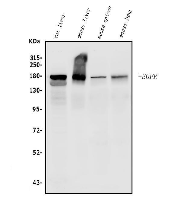
Western blot analysis of EGFR using anti-EGFR antibody (A00023-2).
Electrophoresis was performed on a 5-20% SDS-PAGE gel at 70V (Stacking gel) / 90V (Resolving gel) for 2-3 hours. The sample well of each lane was loaded with 50ug of sample under reducing conditions.
Lane 1: rat liver tissue lysates,
Lane 2: mouse liver tissue lysates,
Lane 3: mouse spleen tissue lysates,
Lane 4: mouse lung tissue lysates.
After Electrophoresis, proteins were transferred to a Nitrocellulose membrane at 150mA for 50-90 minutes. Blocked the membrane with 5% Non-fat Milk/ TBS for 1.5 hour at RT. The membrane was incubated with rabbit anti-EGFR antigen affinity purified polyclonal antibody (Catalog # A00023-2) at 0.5 μg/mL overnight at 4°C, then washed with TBS-0.1%Tween 3 times with 5 minutes each and probed with a goat anti-rabbit IgG-HRP secondary antibody at a dilution of 1:5000 for 1.5 hour at RT. The signal is developed using an Enhanced Chemiluminescent detection (ECL) kit (Catalog # EK1002) with Tanon 5200 system. A specific band was detected for EGFR at approximately 180KD. The expected band size for EGFR is at 180KD.

Click image to see more details
RHBDF2 related functions were mediated by EGFR signaling pathway. a , b Phosphorylation of EGFR and PD-L1 protein level in 786-O and 769-P cells were detected by western blot. Significance testing of gray statistics was analyzed by two-way ANOVA. c Growth rate of the transplanted tumor in the control group and RHBDF2 knockdown group (data were presented as the mean ± SEM and subjected to two-way ANOVA for significance test). d Representative images of xenografts in nude mice. e Immunofluorescent staining of phosphorylation of EGFR in the graft sections (The horizontal line at the bottom right represents 50 microns). f The mean fluorescence intensity of the phosphorylation staining of EGFR in the graft sections (t-test). g The migratory ability testing of 786-O cells and 769-P cells after Gefitinib treatment. h Statistics of the cell migratory ability after Gefitinib treatment (two-way ANOVA). i The detection of pEGFR and PD-L1 level in 786-O and 769-P cells after Gefitinib treatment. j Gray statistics of pEGFR and PD-L1 level in 786-O and 769-P cells (two-way ANOVA, * p < 0.05, ** p < 0.01, *** p < 0.005, **** p < 0.001)
Index in PubMed under a CC BY license. PMID: 34736454

Click image to see more details
IHC analysis of EGFR using anti-EGFR antibody (A00023-2).
EGFR was detected in paraffin-embedded section of mouse liver tissue. Heat mediated antigen retrieval was performed in EDTA buffer (pH8.0, epitope retrieval solution). The tissue section was blocked with 10% goat serum. The tissue section was then incubated with 1μg/ml rabbit anti-EGFR Antibody (A00023-2) overnight at 4°C. Biotinylated goat anti-rabbit IgG was used as secondary antibody and incubated for 30 minutes at 37°C. The tissue section was developed using Strepavidin-Biotin-Complex (SABC)(Catalog # SA1022) with DAB as the chromogen.

Click image to see more details
IHC analysis of EGFR using anti-EGFR antibody (A00023-2).
EGFR was detected in paraffin-embedded section of rat liver tissue. Heat mediated antigen retrieval was performed in EDTA buffer (pH8.0, epitope retrieval solution). The tissue section was blocked with 10% goat serum. The tissue section was then incubated with 1μg/ml rabbit anti-EGFR Antibody (A00023-2) overnight at 4°C. Biotinylated goat anti-rabbit IgG was used as secondary antibody and incubated for 30 minutes at 37°C. The tissue section was developed using Strepavidin-Biotin-Complex (SABC)(Catalog # SA1022) with DAB as the chromogen.
Specific Publications For Anti-EGFR Picoband® Antibody (A00023-2)
Loading publications
Recommended Resources
Here are featured tools and databases that you might find useful.
- Boster's Pathways Library
- Protein Databases
- Bioscience Research Protocol Resources
- Data Processing & Analysis Software
- Photo Editing Software
- Scientific Literature Resources
- Research Paper Management Tools
- Molecular Biology Software
- Primer Design Tools
- Bioinformatics Tools
- Phylogenetic Tree Analysis
Customer Reviews
Have you used Anti-EGFR Picoband® Antibody?
Share your experimental results or join a short interview to earn up to $1,000 in product credits or other rewards.
0 Reviews For Anti-EGFR Picoband® Antibody
Customer Q&As
Have a question?
Find answers in Q&As, reviews.
Can't find your answer?
Submit your question
