Click image to see more details
Product Info Summary
| SKU: | A00851-2 |
|---|---|
| Size: | 100 μg/vial |
| Reactive Species: | Human, Mouse, Rat |
| Host: | Rabbit |
| Application: | ELISA, Flow Cytometry, WB |
Customers Who Bought This Also Bought

Product info
Product Name
Anti-GH1 Antibody Picoband®
SKU/Catalog Number
A00851-2
Size
100 μg/vial
Form
Lyophilized
Description
Boster Bio Anti-GH1 Antibody Picoband® catalog # A00851-2. Tested in WB, FCM, ELISA applications. This antibody reacts with Human, Mouse, Rat. The brand Picoband indicates this is a premium antibody that guarantees superior quality, high affinity, and strong signals with minimal background in Western blot applications. Only our best-performing antibodies are designated as Picoband, ensuring unmatched performance.
Storage & Handling
At -20°C for one year from date of receipt. After reconstitution, at 4°C for one month. It can also be aliquotted and stored frozen at -20°C for six months. Avoid repeated freezing and thawing.
Cite This Product
Anti-GH1 Antibody Picoband® (Boster Biological Technology, Pleasanton CA, USA, Catalog # A00851-2)
Host
Rabbit
Contents
Each vial contains 4 mg Trehalose, 0.9 mg NaCl, 0.2 mg Na2HPO4.
Clonality
Polyclonal
Immunogen
E.coli-derived human GH1 recombinant protein (Position: Q22-F217).
Reactive Species
A00851-2 is reactive to GH1 in Human, Mouse, Rat
Observed Molecular Weight
22 kDa
Calculated molecular weight
24.8 kDa
Background of GH1
Growth Hormone(GH) is mapped to 17q22-q24. Human growth hormone has a molecular mass of 22,005 and contains 191 amino acid residues with 2 disulfide bridges. Rat GH shares 98% amino acid sequence homology with mouse. It binds two receptor molecules and thereby induces signal transduction through receptor dimerization. At high concentrations, GH acts as an antagonist because of a large difference in affinities at the respective binding sites.
Antibody Validation
Boster validates all antibodies on WB, IHC, ICC, Immunofluorescence, and ELISA with known positive control and negative samples to ensure specificity and high affinity, including thorough antibody incubations.
Application & Images
Applications
A00851-2 is guaranteed for ELISA, Flow Cytometry, WB Boster Guarantee
Recommend Dilution
| Application | Dilution | Species |
|---|---|---|
| Western blot | 0.25-0.5 μg/ml | Human, Mouse, Rat |
| Flow Cytometry (Fixed) | 1-3 μg/1x106 cells | Human |
| ELISA | 0.1-0.5 μg/ml | - |
Tested application
Suggested blocking solution with 5% non-fat milk or BSA; (*)Recommended protein loading: 20-40 µg per lane
Validation Images & Assay Conditions
Click image to see more details
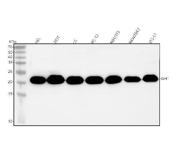
Western blot analysis of GH1 using anti-GH1 antibody (A00851-2).
Electrophoresis was performed on a 5-20% SDS-PAGE gel at 70V (Stacking gel) / 90V (Resolving gel) for 2-3 hours. The sample well of each lane was loaded with 30 ug of sample under reducing conditions.
Lane 1: human HEL whole cell lysates,
Lane 2: human 293T whole cell lysates,
Lane 3: rat C6 whole cell lysates,
Lane 4: rat PC-12 whole cell lysates,
Lane 5: mouse NIH/3T3 whole cell lysates,
Lane 6: mouse RAW264.7 whole cell lysates,
Lane 7: mouse 3T3-L1 whole cell lysates.
After electrophoresis, proteins were transferred to a nitrocellulose membrane at 150 mA for 50-90 minutes. Blocked the membrane with 5% non-fat milk/TBS for 1.5 hour at RT. The membrane was incubated with rabbit anti-GH1 antigen affinity purified polyclonal antibody (Catalog # A00851-2) at 0.5 μg/mL overnight at 4°C, then washed with TBS-0.1%Tween 3 times with 5 minutes each and probed with a goat anti-rabbit IgG-HRP secondary antibody at a dilution of 1:5000 for 1.5 hour at RT. The signal is developed using an Enhanced Chemiluminescent detection (ECL) kit (Catalog # EK1002) with Tanon 5200 system. A specific band was detected for GH1 at approximately 22 kDa. The expected band size for GH1 is at 25 kDa.

Click image to see more details
Western blot analysis of GH1 using anti-GH1 antibody (A00851-2).
Electrophoresis was performed on a 5-20% SDS-PAGE gel at 70V (Stacking gel) / 90V (Resolving gel) for 2-3 hours. The sample well of each lane was loaded with 30 ug of sample under reducing conditions.
Lane 1-2: mouse hippocampal tissue lysates.
After electrophoresis, proteins were transferred to a nitrocellulose membrane at 150 mA for 50-90 minutes. Blocked the membrane with 5% non-fat milk/TBS for 1.5 hour at RT. The membrane was incubated with rabbit anti-GH1 antigen affinity purified polyclonal antibody (Catalog # A00851-2) at 1:4000 overnight at 4°C, then washed with TBS-0.1%Tween 3 times with 5 minutes each and probed with a goat anti-rabbit IgG-HRP secondary antibody at a dilution of 1:10000 for 1 hour at RT. The signal is developed using an Enhanced Chemiluminescent detection (ECL) kit (Catalog # EK1002) with ChemiDoc MP system. The expected band size for GH1 is at 25 kDa.

Click image to see more details
Flow Cytometry analysis of HEL cells using anti-GH1 antibody (A00851-2).
Overlay histogram showing HEL cells stained with A00851-2 (Blue line). The cells were fixed with 4% paraformaldehyde and blocked with 10% normal goat serum. And then incubated with rabbit anti-GH1 Antibody (A00851-2, 1 μg/1x106 cells) for 30 min at 20°C. DyLight®488 conjugated goat anti-rabbit IgG (BA1127, 5-10 μg/1x106 cells) was used as secondary antibody for 30 minutes at 20°C. Isotype control antibody (Green line) was rabbit IgG (1 μg/1x106) used under the same conditions. Unlabelled sample (Red line) was also used as a control.
Specific Publications For Anti-GH1 Antibody Picoband® (A00851-2)
Loading publications
Recommended Resources
Here are featured tools and databases that you might find useful.
- Boster's Pathways Library
- Protein Databases
- Bioscience Research Protocol Resources
- Data Processing & Analysis Software
- Photo Editing Software
- Scientific Literature Resources
- Research Paper Management Tools
- Molecular Biology Software
- Primer Design Tools
- Bioinformatics Tools
- Phylogenetic Tree Analysis
Customer Reviews
Have you used Anti-GH1 Antibody Picoband®?
Share your experimental results or join a short interview to earn up to $1,000 in product credits or other rewards.
1 Reviews For Anti-GH1 Antibody Picoband®
WB analysis using Anti-GH antibody (A00851-2) in mouse hippocampal tissue revealed a specific band at the expected molecular weight with negligible non-specific signals.
Excellent

| SKU | A00851-2 |
|---|---|
| Application | Western Blot |
| Sample | mouse hippocampal tissue |
| Sample Processing Description | Total protein was extracted from the left hippocampus of normal mouse brain. |
| Other Reagents | RIPA lysis buffer, Protease inhibitor, Electrophoresis buffer, Transfer buffer, Blocking buffer |
| Primary Antibody | GH1 Antibody Picoband® |
| Primary Incubation | 1:4000, overnight at 4 ℃ |
| Secondary Antibody | HRP-conjugated goat anti-rabbit IgG |
| Secondary Incubation | 1:10000, 1h in RT |
| Detection | Substrate: ECL substrate; Image system: ChemiDoc MP |
| Results Summary | Growth hormone (GH) serves as a central integrative signal that coordinates growth, metabolism, and tissue repair in response to changes in nutritional status. It is not only the primary driver of linear growth during puberty but also plays a critical role throughout life in maintaining muscle mass, bone strength, and metabolic flexibility. In this study, hippocampal tissues from two normal mouse brains were used to evaluate the performance of the GH antibody. The results showed a band at the expected position with good specificity, indicating that the antibody performs well in WB applications. |
Yajun Qiao, Qinghai University
Verified customer
Submitted 2026-05-06
Customer Q&As
Have a question?
Find answers in Q&As, reviews.
Can't find your answer?
Submit your question
