Click image to see more details
-

-
-
-
-
+18
Product Info Summary
| SKU: | M33947 |
|---|---|
| Size: | 100ul |
| Reactive Species: | Human, Mouse, Rat |
| Host: | Mouse |
| Application: | ELISA, IF, WB |
Customers Who Bought This Also Bought

Product info
Product Name
Anti-Histone H3 Monoclonal Antibody
SKU/Catalog Number
M33947
Size
100ul
Form
Liquid
Description
Boster Bio Anti-Histone H3 Monoclonal Antibody catalog # M33947. Tested in IP, IF, WB applications. This antibody reacts with Human, Mouse, Rat, Yeast.
Storage & Handling
Store at -20°C for one year. For short term storage and frequent use, store at 4°C for up to one month. Avoid repeated freeze-thaw cycles.
Cite This Product
Anti-Histone H3 Monoclonal Antibody (Boster Biological Technology, Pleasanton CA, USA, Catalog # M33947)
Host
Mouse
Contents
Liquid in PBS containing 50% glycerol, 0.5% stabilizing protein and 0.02% sodium azide.
This antibody is supplied in a stabilized formulation.
Compatibility with conjugation reactions depends on the chemistry of the conjugation method used.
For conjugation methods that are not compatible with the stabilizing components present in this formulation, a carrier-free antibody format is required.
Clonality
Monoclonal
Clone Number
1G1
Isotype
IgG
Immunogen
Recombinant Protein of Histone H3.AA range: 1-100
Reactive Species
M33947 is reactive to in Human, Mouse, Rat
Antibody Validation
Boster validates all antibodies on WB, IHC, ICC, Immunofluorescence, and ELISA with known positive control and negative samples to ensure specificity and high affinity, including thorough antibody incubations.
Application & Images
Applications
M33947 is guaranteed for ELISA, IF, WB Boster Guarantee
Assay Dilutions Recommendation
The recommendations below provide a starting point for assay optimization. The actual working concentration varies and should be decided by the user.
WB 1:500-2000
IF 1:100-500
ELISA 1:1000-5000
Validation Images & Assay Conditions
Click image to see more details
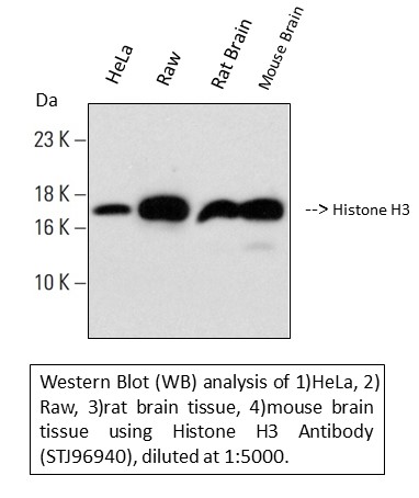
Western blotting validation for Anti-Histone H3 Monoclonal Antibody M33947
Western blot (WB) analysis of Histone H3 monoclonal antibody.Electrophoresis was performed on a SDS-PAGE gel. To determine SDS-PAGE gel concentration

Click image to see more details
Immunohistochemistry validation of Histone H3 using Anti-Histone H3 Monoclonal Antibody (M33947).
Immunohistochemical analysis of human-appendix tissue. Anti-Histone H3 at 1:200 (4°C
Click image to see more details
Immunohistochemistry validation of Histone H3 using Anti-Histone H3 Monoclonal Antibody (M33947).
Immunohistochemical analysis of human colon tissue. Anti-Histone H3 at 1:200 (4°C
Click image to see more details
Immunohistochemistry validation of Histone H3 using Anti-Histone H3 Monoclonal Antibody (M33947).
Immunohistochemical analysis of human liver cancer tissue. Anti-Histone H3 at 1:200 (4°C
Click image to see more details
Immunohistochemistry validation of Histone H3 using Anti-Histone H3 Monoclonal Antibody (M33947).
Immunohistochemical analysis of human lung cancer tissue. Anti-Histone H3 at 1:200 (4°C
Click image to see more details
Immunohistochemistry validation of Histone H3 using Anti-Histone H3 Monoclonal Antibody (M33947).
Immunohistochemical analysis of human lung tissue. Anti-Histone H3 at 1:200 (4°C
Click image to see more details
Immunohistochemistry validation of Histone H3 using Anti-Histone H3 Monoclonal Antibody (M33947).
Immunohistochemical analysis of human stomach cancer tissue. Anti-Histone H3 at 1:200 (4°C
Click image to see more details
Immunohistochemistry validation of Histone H3 using Anti-Histone H3 Monoclonal Antibody (M33947).
Immunohistochemical analysis of human stomach tissue. M33947 was diluted at 1:200 (4°C
Click image to see more details
Immunohistochemistry validation of Histone H3 using Anti-Histone H3 Monoclonal Antibody (M33947).
Immunohistochemical analysis of human tonsil tissue. M33947 was diluted at 1:200 (4°C
Click image to see more details
Immunohistochemistry validation of Histone H3 using Anti-Histone H3 Monoclonal Antibody (M33947).
Immunohistochemical analysis of human uterus cancer tissue. M33947 was diluted at 1:200 (4°C
Click image to see more details
Immunohistochemistry validation of Histone H3 using Anti-Histone H3 Monoclonal Antibody (M33947).
Immunohistochemical analysis of human uterus tissue. M33947 was diluted at 1:200 (4°C
Click image to see more details
Immunohistochemistry validation of Histone H3 using Anti-Histone H3 Monoclonal Antibody (M33947).
Immunohistochemical analysis of mouse brain tissue. M33947 was diluted at 1:200 (4°C
Click image to see more details
Immunohistochemistry validation of Histone H3 using Anti-Histone H3 Monoclonal Antibody (M33947).
Immunohistochemical analysis of mouse colon tissue. M33947 was diluted at 1:200 (4°C
Click image to see more details
Immunohistochemistry validation of Histone H3 using Anti-Histone H3 Monoclonal Antibody (M33947).
Immunohistochemical analysis of mouse kidney tissue. M33947 was diluted at 1:200 (4°C
Click image to see more details
Immunohistochemistry validation of Histone H3 using Anti-Histone H3 Monoclonal Antibody (M33947).
Immunohistochemical analysis of mouse liver tissue. M33947 was diluted at 1:200 (4°C
Click image to see more details
Immunohistochemistry validation of Histone H3 using Anti-Histone H3 Monoclonal Antibody (M33947).
Immunohistochemical analysis of mouse testis tissue. M33947 was diluted at 1:200 (4°C
Click image to see more details
Immunohistochemistry validation of Histone H3 using Anti-Histone H3 Monoclonal Antibody (M33947).
Immunohistochemical analysis of rat brain tissue. Anti-Cleaved-Caspase-1 (D210) antibody was diluted at 1:200 (4°C
Click image to see more details
Immunohistochemistry validation of Histone H3 using Anti-Histone H3 Monoclonal Antibody (M33947).
Immunohistochemical analysis of rat kidney tissue. Anti-Cleaved-Caspase-1 (D210) antibody was diluted at 1:200 (4°C
Click image to see more details
Immunohistochemistry validation of Histone H3 using Anti-Histone H3 Monoclonal Antibody (M33947).
Immunohistochemical analysis of rat spinal cord tissue. Anti-Cleaved-Caspase-1 (D210) antibody was diluted at 1:200 (4°C
Click image to see more details
Immunohistochemistry validation of Histone H3 using Anti-Histone H3 Monoclonal Antibody (M33947).
Immunohistochemical analysis of rat spleen tissue. Anti-Cleaved-Caspase-1 (D210) antibody was diluted at 1:200 (4°C
Click image to see more details
Immunohistochemistry validation of Histone H3 using Anti-Histone H3 Monoclonal Antibody (M33947).
Immunohistochemical analysis of rat testis tissue. Anti-Cleaved-Caspase-1 (D210) antibody was diluted at 1:200 (4°C
Click image to see more details
Immunofluorescent staining data of Histone H3 using Anti-Histone H3 Monoclonal Antibody (M33947).
Immunofluorescence (IF) analysis of HeLaSpecific Publications For Anti-Histone H3 Monoclonal Antibody (M33947)
Loading publications
Recommended Resources
Here are featured tools and databases that you might find useful.
- Boster's Pathways Library
- Protein Databases
- Bioscience Research Protocol Resources
- Data Processing & Analysis Software
- Photo Editing Software
- Scientific Literature Resources
- Research Paper Management Tools
- Molecular Biology Software
- Primer Design Tools
- Bioinformatics Tools
- Phylogenetic Tree Analysis
Customer Reviews
Have you used Anti-Histone H3 Monoclonal Antibody?
Share your experimental results or join a short interview to earn up to $1,000 in product credits or other rewards.
0 Reviews For Anti-Histone H3 Monoclonal Antibody
Customer Q&As
Have a question?
Find answers in Q&As, reviews.
Can't find your answer?
Submit your question
