Click image to see more details
-

-
-
-
-
+25
Product Info Summary
| SKU: | A12477-2 |
|---|---|
| Size: | 100 μg/vial |
| Reactive Species: | Human, Mouse, Rat |
| Host: | Rabbit |
| Application: | ELISA, Flow Cytometry, IF, IHC, ICC, WB |
Customers Who Bought This Also Bought

Product info
Product Name
Anti-Histone H3 HIST1H3A/B/C/D/E/F/G/H/I/J Antibody Picoband®
View all HIST1H3A/B/C/D/E/F/G/H/I/J Antibodies
SKU/Catalog Number
A12477-2
Size
100 μg/vial
Form
Lyophilized
Description
Boster Bio Anti-Histone H3 HIST1H3A/B/C/D/E/F/G/H/I/J Antibody Picoband® catalog # A12477-2. Tested in ELISA, Flow Cytometry, IF, IHC, ICC, WB applications. This antibody reacts with Human, Mouse, Rat. The brand Picoband indicates this is a premium antibody that guarantees superior quality, high affinity, and strong signals with minimal background in Western blot applications. Only our best-performing antibodies are designated as Picoband, ensuring unmatched performance.
Storage & Handling
Store at -20˚C for one year from date of receipt. After reconstitution, at 4˚C for one month. It can also be aliquotted and stored frozen at -20˚C for six months. Avoid repeated freeze-thaw cycles.
Cite This Product
Anti-Histone H3 HIST1H3A/B/C/D/E/F/G/H/I/J Antibody Picoband® (Boster Biological Technology, Pleasanton CA, USA, Catalog # A12477-2)
Host
Rabbit
Contents
Each vial contains 4mg Trehalose, 0.9mg NaCl, 0.2mg Na2HPO4.
Clonality
Polyclonal
Isotype
Rabbit IgG
Immunogen
E.coli-derived human Histone H3 recombinant protein (Position: Q56—R117).
Cross-reactivity
No cross-reactivity with other proteins.
Reactive Species
A12477-2 is reactive to HIST1H3A/B/C/D/E/F/G/H/I/J in Human, Mouse, Rat
Observed Molecular Weight
17 kDa
Calculated molecular weight
15.4 kDa
Background of HIST1H3A/B/C/D/E/F/G/H/I/J
Histone H3.1 is a protein that in humans is encoded by the HIST1H3A gene. Histones are basic nuclear proteins that are responsible for the nucleosome structure of the chromosomal fiber in eukaryotes. This structure consists of approximately 146 bp of DNA wrapped around a nucleosome, an octamer composed of pairs of each of the four core histones (H2A, H2B, H3, and H4). The chromatin fiber is further compacted through the interaction of a linker histone, H1, with the DNA between the nucleosomes to form higher order chromatin structures. This gene is intronless and encodes a replication-dependent histone that is a member of the histone H3 family. Transcripts from this gene lack polyA tails; instead, they contain a palindromic termination element. This gene is found in the large histone gene cluster on chromosome 6p22-p21.3.
Antibody Validation
Boster validates all antibodies on WB, IHC, ICC, Immunofluorescence, and ELISA with known positive control and negative samples to ensure specificity and high affinity, including thorough antibody incubations.
Application & Images
Applications
A12477-2 is guaranteed for ELISA, Flow Cytometry, IF, IHC, ICC, WB Boster Guarantee
Recommend Dilution
| Application | Dilution | Species |
|---|---|---|
| Western blot | 0.1-0.25μg/ml | Human, Mouse, Rat |
| Immunohistochemistry (Paraffin-embedded Section) | 2-5μg/ml | Human, Mouse, Rat |
| Immunocytochemistry/Immunofluorescence | 5μg/ml | Human |
| Flow Cytometry (Fixed) | 1-3μg/1x106 cells | Mouse, Rat |
| ELISA | 0.1-0.5μg/ml | - |
Tested application
Suggested blocking solution with 5% non-fat milk or BSA; (*)Recommended protein loading: 20-40 µg per lane
Use TE buffer pH 9.0 for antigen retrieval; (*) citrate buffer pH 6.0 is an alternative.
Validation Images & Assay Conditions
Click image to see more details
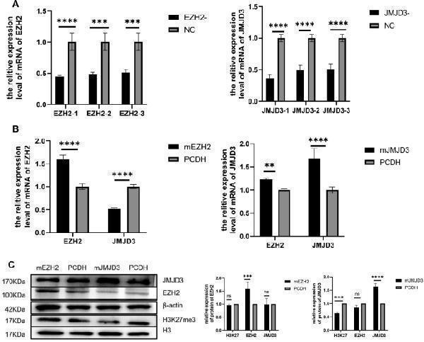
Expression levels of EZH2, JMJD3, H3K27me3 in spermatogonia. ( A ) qRT-PCR was used to detect the expression of EZH2 siRNA and JMJD3 siRNA in spermatogonia after interference. ( B ) measurements of EZH2 and JMJD3 mRNA levels in spermatogonia after overexpression. ( C ) The protein levels of EZH2, JMJD3 and H3K27me3 in spermatogonia were detected and statistically analyzed by Western blot. The membrane is lysed prior to hybridization with the antibody and the image has been cropped for a more aesthetically pleasing display. The full- length blots can be obtained from Additional file 2: Fig
Index in PubMed under a CC BY license. PMID: 38424516

Click image to see more details
TET1 coordinates with H3K27me3 to target Pramel3 to promote its activation and expression. ( A ) qRT-PCR was used to detect the expression of genes enriched by Chip-seq after JMJD3 overexpression. ( B ) Peak plots of 2610002M06Rik and Pramel3 target genes obtained from TET1 overexpressing cells deposited with H3K27me3 antibody
Index in PubMed under a CC BY license. PMID: 38424516

Click image to see more details
TET1-H3K27me3 regulated through PI3K-AKT pathway. ( A ) KEGG enrichment analysis. ( B ) Western blot was used to detect the protein expressions of AKT and P-AKT in the PI3K-AKT pathway after JMJD3 overexpression. The membrane is lysed prior to hybridization with the antibody and the image has been cropped for a more aesthetically pleasing display. The full- length blots can be obtained from Additional file 2: Fig.
Index in PubMed under a CC BY license. PMID: 38424516

Click image to see more details
In vivo functional validation of JMJD3. ( A ) Spermatogenesis disorder model mice transplantation of control PCDH and JMJD3 Positive Cells. ( B ) Chart of comparison of control PCDH with testes transplanted with JMJD3 positive cells. ( C ) HE staining plot of JMJD3 overexpression versus control PCDH. ( D ) Expression of JMJD3 in testicular spermatogonia after overexpression of JMJD3. ( E ) Expression of H3K27me3 and PCNA in testicular spermatogonia after JMJD3 overexpression. ( F ) Statistical analysis of JMJD3 + cells/H3K27m3 + cells and PCNA + cells in immunohistochemistry. Scale bar = 50 μm. n = 3
Index in PubMed under a CC BY license. PMID: 38424516

Click image to see more details
Western blot analysis of Histone H3 using anti-Histone H3 antibody (A12477-2).
Electrophoresis was performed on a 5-20% SDS-PAGE gel at 70V (Stacking gel) / 90V (Resolving gel) for 2-3 hours. The sample well of each lane was loaded with 30 ug of sample under reducing conditions.
Lane 1: human 293T whole cell lysates,
Lane 2: human 22RV1 whole cell lysates,
Lane 3: human CACO-2 whole cell lysates,
Lane 4: human CCRF-CEM whole cell lysates,
Lane 5: human Hela whole cell lysates,
Lane 6: human HepG2 whole cell lysates,
Lane 7: human THP-1 whole cell lysates,
Lane 8: human U2OS whole cell lysates.
After Electrophoresis, proteins were transferred to a Nitrocellulose membrane at 150mA for 50-90 minutes. Blocked the membrane with 5% Non-fat Milk/ TBS for 1.5 hour at RT. The membrane was incubated with rabbit anti-Histone H3 antigen affinity purified polyclonal antibody (Catalog # A12477-2) at 0.25 μg/mL overnight at 4°C, then washed with TBS-0.1%Tween 3 times with 5 minutes each and probed with a goat anti-rabbit IgG-HRP secondary antibody at a dilution of 1:5000 for 1.5 hour at RT. The signal is developed using an Enhanced Chemiluminescent detection (ECL) kit (Catalog # EK1002) with Tanon 5200 system. A specific band was detected for Histone H3 at approximately 17 kDa. The expected band size for Histone H3 is at 15 kDa.

Click image to see more details
Western blot analysis of Histone H3 using anti-Histone H3 antibody (A12477-2).
Electrophoresis was performed on a 5-20% SDS-PAGE gel at 70V (Stacking gel) / 90V (Resolving gel) for 2-3 hours. The sample well of each lane was loaded with 30 ug of sample under reducing conditions.
Lane 1: rat brain tissue lysates,
Lane 2: rat liver tissue lysates,
Lane 3: rat PC-12 whole cell lysates,
Lane 4: rat C6 whole cell lysates,
Lane 5: mouse brain tissue lysates,
Lane 6: mouse liver tissue lysates,
Lane 7: mouse HEPA1-6 whole cell lysates,
Lane 8: mouse RAW264.7 whole cell lysates.
After Electrophoresis, proteins were transferred to a Nitrocellulose membrane at 150mA for 50-90 minutes. Blocked the membrane with 5% Non-fat Milk/ TBS for 1.5 hour at RT. The membrane was incubated with rabbit anti-Histone H3 antigen affinity purified polyclonal antibody (Catalog # A12477-2) at 0.25 μg/mL overnight at 4°C, then washed with TBS-0.1%Tween 3 times with 5 minutes each and probed with a goat anti-rabbit IgG-HRP secondary antibody at a dilution of 1:5000 for 1.5 hour at RT. The signal is developed using an Enhanced Chemiluminescent detection (ECL) kit (Catalog # EK1002) with Tanon 5200 system. A specific band was detected for Histone H3 at approximately 17 kDa. The expected band size for Histone H3 is at 15 kDa.

Click image to see more details
IHC analysis of Histone H3 using anti-Histone H3 antibody (A12477-2).
Histone H3 was detected in a paraffin-embedded section of human colon adenocarcinoma tissue. Heat mediated antigen retrieval was performed in EDTA buffer (pH 8.0, epitope retrieval solution). The tissue section was blocked with 10% goat serum. The tissue section was then incubated with 2 μg/ml rabbit anti-Histone H3 Antibody (A12477-2) overnight at 4°C. Peroxidase Conjugated Goat Anti-rabbit IgG was used as secondary antibody and incubated for 30 minutes at 37°C. The tissue section was developed using HRP Conjugated Rabbit IgG Super Vision Assay Kit (Catalog # SV0002) with DAB as the chromogen.

Click image to see more details
IHC analysis of Histone H3 using anti-Histone H3 antibody (A12477-2).
Histone H3 was detected in a paraffin-embedded section of human esophageal squamous carcinoma tissue. Heat mediated antigen retrieval was performed in EDTA buffer (pH 8.0, epitope retrieval solution). The tissue section was blocked with 10% goat serum. The tissue section was then incubated with 2 μg/ml rabbit anti-Histone H3 Antibody (A12477-2) overnight at 4°C. Peroxidase Conjugated Goat Anti-rabbit IgG was used as secondary antibody and incubated for 30 minutes at 37°C. The tissue section was developed using HRP Conjugated Rabbit IgG Super Vision Assay Kit (Catalog # SV0002) with DAB as the chromogen.

Click image to see more details
IHC analysis of Histone H3 using anti-Histone H3 antibody (A12477-2).
Histone H3 was detected in a paraffin-embedded section of human lung cancer tissue. Heat mediated antigen retrieval was performed in EDTA buffer (pH 8.0, epitope retrieval solution). The tissue section was blocked with 10% goat serum. The tissue section was then incubated with 2 μg/ml rabbit anti-Histone H3 Antibody (A12477-2) overnight at 4°C. Peroxidase Conjugated Goat Anti-rabbit IgG was used as secondary antibody and incubated for 30 minutes at 37°C. The tissue section was developed using HRP Conjugated Rabbit IgG Super Vision Assay Kit (Catalog # SV0002) with DAB as the chromogen.

Click image to see more details
IHC analysis of Histone H3 using anti-Histone H3 antibody (A12477-2).
Histone H3 was detected in a paraffin-embedded section of human placenta tissue. Heat mediated antigen retrieval was performed in EDTA buffer (pH 8.0, epitope retrieval solution). The tissue section was blocked with 10% goat serum. The tissue section was then incubated with 2 μg/ml rabbit anti-Histone H3 Antibody (A12477-2) overnight at 4°C. Peroxidase Conjugated Goat Anti-rabbit IgG was used as secondary antibody and incubated for 30 minutes at 37°C. The tissue section was developed using HRP Conjugated Rabbit IgG Super Vision Assay Kit (Catalog # SV0002) with DAB as the chromogen.

Click image to see more details
IHC analysis of Histone H3 using anti-Histone H3 antibody (A12477-2).
Histone H3 was detected in a paraffin-embedded section of human spleen tissue. Heat mediated antigen retrieval was performed in EDTA buffer (pH 8.0, epitope retrieval solution). The tissue section was blocked with 10% goat serum. The tissue section was then incubated with 2 μg/ml rabbit anti-Histone H3 Antibody (A12477-2) overnight at 4°C. Peroxidase Conjugated Goat Anti-rabbit IgG was used as secondary antibody and incubated for 30 minutes at 37°C. The tissue section was developed using HRP Conjugated Rabbit IgG Super Vision Assay Kit (Catalog # SV0002) with DAB as the chromogen.

Click image to see more details
IHC analysis of Histone H3 using anti-Histone H3 antibody (A12477-2).
Histone H3 was detected in a paraffin-embedded section of human testicular seminoma tissue. Heat mediated antigen retrieval was performed in EDTA buffer (pH 8.0, epitope retrieval solution). The tissue section was blocked with 10% goat serum. The tissue section was then incubated with 2 μg/ml rabbit anti-Histone H3 Antibody (A12477-2) overnight at 4°C. Peroxidase Conjugated Goat Anti-rabbit IgG was used as secondary antibody and incubated for 30 minutes at 37°C. The tissue section was developed using HRP Conjugated Rabbit IgG Super Vision Assay Kit (Catalog # SV0002) with DAB as the chromogen.

Click image to see more details
IHC analysis of Histone H3 using anti-Histone H3 antibody (A12477-2).
Histone H3 was detected in a paraffin-embedded section of mouse brain tissue. Heat mediated antigen retrieval was performed in EDTA buffer (pH 8.0, epitope retrieval solution). The tissue section was blocked with 10% goat serum. The tissue section was then incubated with 2 μg/ml rabbit anti-Histone H3 Antibody (A12477-2) overnight at 4°C. Peroxidase Conjugated Goat Anti-rabbit IgG was used as secondary antibody and incubated for 30 minutes at 37°C. The tissue section was developed using HRP Conjugated Rabbit IgG Super Vision Assay Kit (Catalog # SV0002) with DAB as the chromogen.

Click image to see more details
IHC analysis of Histone H3 using anti-Histone H3 antibody (A12477-2).
Histone H3 was detected in a paraffin-embedded section of rat brain tissue. Heat mediated antigen retrieval was performed in EDTA buffer (pH 8.0, epitope retrieval solution). The tissue section was blocked with 10% goat serum. The tissue section was then incubated with 2 μg/ml rabbit anti-Histone H3 Antibody (A12477-2) overnight at 4°C. Peroxidase Conjugated Goat Anti-rabbit IgG was used as secondary antibody and incubated for 30 minutes at 37°C. The tissue section was developed using HRP Conjugated Rabbit IgG Super Vision Assay Kit (Catalog # SV0002) with DAB as the chromogen.

Click image to see more details
IF analysis of Histone H3 using anti-Histone H3 antibody (A12477-2) and anti-Beta Tubulin antibody (M01857-3).
Histone H3 was detected in immunocytochemical section of U2OS cell. Enzyme antigen retrieval was performed using IHC enzyme antigen retrieval reagent (AR0022) for 15 mins. The cells were blocked with 10% goat serum. And then incubated with 5 μg/mL rabbit anti-Histone H3 Antibody (A12477-2) and mouse anti-Beta Tubulin antibody (M01857-3) overnight at 4°C. DyLight®488 Conjugated Goat Anti-Rabbit IgG (BA1127) and Cy3 Conjugated Goat Anti-Mouse IgG (BA1031) were used as secondary antibody at 1:500 dilution and incubated for 30 minutes at 37°C. Visualize using a fluorescence microscope and filter sets appropriate for the label used.

Click image to see more details
Flow Cytometry analysis of RAW264.7 cells using anti-Histone H3 antibody (A12477-2).
Overlay histogram showing RAW264.7 cells stained with A12477-2 (Blue line). To facilitate intracellular staining, cells were fixed with 4% paraformaldehyde and permeabilized with permeabilization buffer. The cells were blocked with 10% normal goat serum. And then incubated with rabbit anti-Histone H3 Antibody (A12477-2, 1 μg/1x106 cells) for 30 min at 20°C. DyLight®488 conjugated goat anti-rabbit IgG (BA1127, 5-10 μg/1x106 cells) was used as secondary antibody for 30 minutes at 20°C. Isotype control antibody (Green line) was rabbit IgG (1 μg/1x106) used under the same conditions. Unlabelled sample without incubation with primary antibody and secondary antibody (Red line) was used as a blank control.

Click image to see more details
Flow Cytometry analysis of C6 cells using anti-Histone H3 antibody (A12477-2).
Overlay histogram showing C6 cells stained with A12477-2 (Blue line). To facilitate intracellular staining, cells were fixed with 4% paraformaldehyde and permeabilized with permeabilization buffer. The cells were blocked with 10% normal goat serum. And then incubated with rabbit anti-Histone H3 Antibody (A12477-2, 1 μg/1x106 cells) for 30 min at 20°C. DyLight®488 conjugated goat anti-rabbit IgG (BA1127, 5-10 μg/1x106 cells) was used as secondary antibody for 30 minutes at 20°C. Isotype control antibody (Green line) was rabbit IgG (1 μg/1x106) used under the same conditions. Unlabelled sample without incubation with primary antibody and secondary antibody (Red line) was used as a blank control.
Specific Publications For Anti-Histone H3 HIST1H3A/B/C/D/E/F/G/H/I/J Antibody Picoband® (A12477-2)
Loading publications
Recommended Resources
Here are featured tools and databases that you might find useful.
- Boster's Pathways Library
- Protein Databases
- Bioscience Research Protocol Resources
- Data Processing & Analysis Software
- Photo Editing Software
- Scientific Literature Resources
- Research Paper Management Tools
- Molecular Biology Software
- Primer Design Tools
- Bioinformatics Tools
- Phylogenetic Tree Analysis
Customer Reviews
Have you used Anti-Histone H3 HIST1H3A/B/C/D/E/F/G/H/I/J Antibody Picoband®?
Share your experimental results or join a short interview to earn up to $1,000 in product credits or other rewards.
0 Reviews For Anti-Histone H3 HIST1H3A/B/C/D/E/F/G/H/I/J Antibody Picoband®
Customer Q&As
Have a question?
Find answers in Q&As, reviews.
Can't find your answer?
Submit your question
1 Customer Q&As for Anti-Histone H3 HIST1H3A/B/C/D/E/F/G/H/I/J Antibody Picoband®
Question
Our lab want to know about using your anti-Histone H3 antibody for formation of the beta-catenin:tcf transactivating complex studies. Has this antibody been tested with western blotting on hela whole cell lysates? We would like to see some validation images before ordering.
A. Williams
Verified customer
Asked: 2015-09-11
Answer
I appreciate your inquiry. This A12477-2 anti-Histone H3 antibody is tested on human hela, 22rv1 whole cell lysates, hela whole cell lysates, hepg2 whole cell lysates, u2os whole cell lysates, rat brain tissue, mouse liver tissue. It is guaranteed to work for ELISA, WB in human, mouse, rat. Our Boster guarantee will cover your intended experiment even if the sample type has not been be directly tested.
Boster Scientific Support
Answered: 2015-09-11
