Click image to see more details
-

-
-
-
-
+3
Product Info Summary
| SKU: | PB9325 |
|---|---|
| Size: | 100 μg/vial |
| Reactive Species: | Human, Mouse, Rat |
| Host: | Rabbit |
| Application: | Flow Cytometry, IF, IHC, ICC, WB |
Customers Who Bought This Also Bought

Product info
Product Name
Anti-Hsp47/SERPINH1 Antibody Picoband®
SKU/Catalog Number
PB9325
Size
100 μg/vial
Form
Lyophilized
Description
Boster Bio Anti-Hsp47/SERPINH1 Antibody Picoband® catalog # PB9325. Tested in Flow Cytometry, ICC/IF, IHC, WB applications. This antibody reacts with Human, Mouse, Rat. The brand Picoband indicates this is a premium antibody that guarantees superior quality, high affinity, and strong signals with minimal background in Western blot applications. Only our best-performing antibodies are designated as Picoband, ensuring unmatched performance.
Storage & Handling
Store at -20˚C for one year from date of receipt. After reconstitution, at 4˚C for one month. It can also be aliquotted and stored frozen at -20˚C for six months. Avoid repeated freeze-thaw cycles.
Cite This Product
Anti-Hsp47/SERPINH1 Antibody Picoband® (Boster Biological Technology, Pleasanton CA, USA, Catalog # PB9325)
Host
Rabbit
Contents
Each vial contains 4 mg Trehalose, 0.9 mg NaCl and 0.2 mg Na2HPO4.
Clonality
Polyclonal
Isotype
Rabbit IgG
Immunogen
E.coli-derived human Hsp47 recombinant protein (Position: D247-L418). Human Hsp47 shares 97% amino acid (aa) sequence identity with both mouse and rat Hsp47.
Cross-reactivity
No cross-reactivity with other proteins
Reactive Species
PB9325 is reactive to SERPINH1 in Human, Mouse, Rat
Observed Molecular Weight
46 kDa
Calculated molecular weight
46.4 kDa
Background of SERPINH1
Heat shock protein 47, also known as SERPINH1 or HSP47, is a serpin which serves as a human chaperone protein for collagen. This protein is a member of the serpin superfamily of serine proteinase inhibitors. Its expression is induced by heat shock. The protein localizes to the endoplasmic reticulum lumen and binds collagen; thus it is thought to be a molecular chaperone involved in the maturation of collagen molecules. Autoantibodies to this protein have been found in patients with rheumatoid arthritis. It has been found that HSP47 monitors the integrity of the triple helix of type I procollagen at the ER/cis-Golgi boundary and, when absent, the rate of transit from the ER to the Golgi is increased and the helical structure is compromised.
Antibody Validation
Boster validates all antibodies on WB, IHC, ICC, Immunofluorescence, and ELISA with known positive control and negative samples to ensure specificity and high affinity, including thorough antibody incubations.
Application & Images
Applications
PB9325 is guaranteed for Flow Cytometry, IF, IHC, ICC, WB Boster Guarantee
Recommend Dilution
| Application | Dilution | Species |
|---|---|---|
| Western blot | 0.1-0.5μg/ml | Human, Mouse, Rat |
| Immunohistochemistry (Paraffin-embedded Section) | 2-5μg/ml | Human |
| Immunocytochemistry/Immunofluorescence | 5 μg/ml | Human |
| Flow Cytometry(Fixed) | 1-3 μg/1x106 cells | Human |
Tested application
Suggested blocking solution with 5% non-fat milk or BSA; (*)Recommended protein loading: 20-40 µg per lane
Use TE buffer pH 9.0 for antigen retrieval; (*) citrate buffer pH 6.0 is an alternative.
Validation Images & Assay Conditions
Click image to see more details
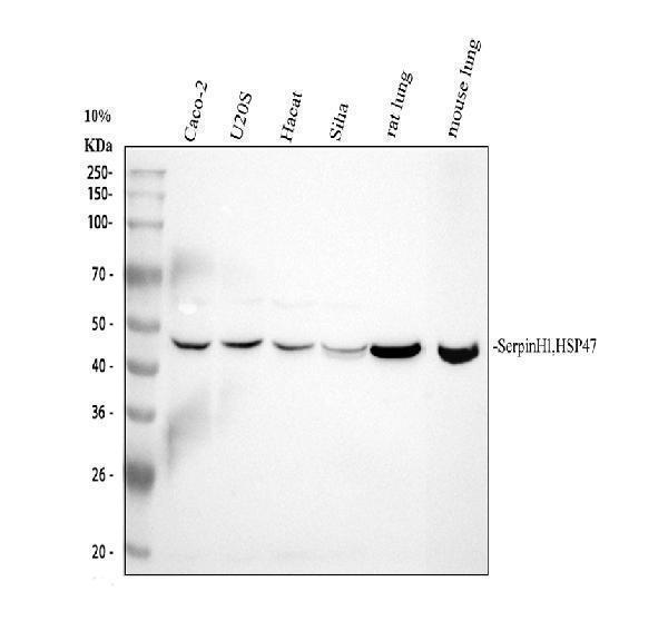
Western blot analysis of Hsp47 using anti-Hsp47 antibody (PB9325).
Electrophoresis was performed on a 10% SDS-PAGE gel at 80V (Stacking gel) / 120V (Resolving gel) for 2 hours. The sample well of each lane was loaded with 30 ug of sample under reducing conditions.
Lane 1: human CACO-2 whole cell lysates,
Lane 2: human U2OS whole cell lysates,
Lane 3: human Hacat whole cell lysates,
Lane 4: human SiHa whole cell lysates,
Lane 5: rat lung tissue lysates,
Lane 6: mouse lung tissue lysates.
After electrophoresis, proteins were transferred to a nitrocellulose membrane at 150 mA for 50-90 minutes. Blocked the membrane with 5% non-fat milk/TBS for 1.5 hour at RT. The membrane was incubated with rabbit anti-Hsp47 antigen affinity purified polyclonal antibody (PB9325) at 0.5 μg/mL overnight at 4°C, then washed with TBS-0.1%Tween 3 times with 5 minutes each and probed with a goat anti-rabbit IgG-HRP secondary antibody (Catalog # BA1054) at a dilution of 1:5000 for 1.5 hour at RT. The signal is developed using an ECL Plus Western Blotting Substrate (Catalog # AR1196-200) with Tanon 5200 system. A specific band was detected for Hsp47 at approximately 46 kDa. The expected band size for Hsp47 is at 46 kDa.

Click image to see more details
IHC analysis of Hsp47 using anti-Hsp47 antibody (PB9325).
Hsp47 was detected in a paraffin-embedded section of human esophageal squamous cancer tissue. Heat mediated antigen retrieval was performed in EDTA buffer (pH 8.0, epitope retrieval solution). The tissue section was blocked with 10% goat serum. The tissue section was then incubated with 2 μg/ml rabbit anti-Hsp47 Antibody (PB9325) overnight at 4°C. Peroxidase Conjugated Goat Anti-rabbit IgG was used as secondary antibody and incubated for 30 minutes at 37°C. The tissue section was developed using HRP Conjugated Rabbit IgG Super Vision Assay Kit (Catalog # SV0002) with DAB as the chromogen.

Click image to see more details
IHC analysis of Hsp47 using anti-Hsp47 antibody (PB9325).
Hsp47 was detected in a paraffin-embedded section of human pendiceal adenocarcinama tissue. Heat mediated antigen retrieval was performed in EDTA buffer (pH 8.0, epitope retrieval solution). The tissue section was blocked with 10% goat serum. The tissue section was then incubated with 2 μg/ml rabbit anti-Hsp47 Antibody (PB9325) overnight at 4°C. Peroxidase Conjugated Goat Anti-rabbit IgG was used as secondary antibody and incubated for 30 minutes at 37°C. The tissue section was developed using HRP Conjugated Rabbit IgG Super Vision Assay Kit (Catalog # SV0002) with DAB as the chromogen.

Click image to see more details
IHC analysis of Hsp47 using anti-Hsp47 antibody (PB9325).
Hsp47 was detected in a paraffin-embedded section of human breast cancer tissue. Heat mediated antigen retrieval was performed in EDTA buffer (pH 8.0, epitope retrieval solution). The tissue section was blocked with 10% goat serum. The tissue section was then incubated with 2 μg/ml rabbit anti-Hsp47 Antibody (PB9325) overnight at 4°C. Peroxidase Conjugated Goat Anti-rabbit IgG was used as secondary antibody and incubated for 30 minutes at 37°C. The tissue section was developed using HRP Conjugated Rabbit IgG Super Vision Assay Kit (Catalog # SV0002) with DAB as the chromogen.

Click image to see more details
IHC analysis of Hsp47 using anti-Hsp47 antibody (PB9325).
Hsp47 was detected in a paraffin-embedded section of human colon cancer tissue. Heat mediated antigen retrieval was performed in EDTA buffer (pH 8.0, epitope retrieval solution). The tissue section was blocked with 10% goat serum. The tissue section was then incubated with 2 μg/ml rabbit anti-Hsp47 Antibody (PB9325) overnight at 4°C. Peroxidase Conjugated Goat Anti-rabbit IgG was used as secondary antibody and incubated for 30 minutes at 37°C. The tissue section was developed using HRP Conjugated Rabbit IgG Super Vision Assay Kit (Catalog # SV0002) with DAB as the chromogen.

Click image to see more details
IF analysis of Hsp47 using anti-Hsp47 antibody (PB9325).
Hsp47 was detected in an immunocytochemical section of A549 cells. Enzyme antigen retrieval was performed using IHC enzyme antigen retrieval reagent (AR0022) for 15 mins. The cells were blocked with 10% goat serum. And then incubated with 5 μg/mL rabbit anti-Hsp47 Antibody (PB9325) overnight at 4°C. Fluoro488 Conjugated Goat Anti-Rabbit IgG (BA1127) was used as secondary antibody at 1:500 dilution and incubated for 30 minutes at 37°C. The section was counterstained with DAPI. Visualize using a fluorescence microscope and filter sets appropriate for the label used.

Click image to see more details
Flow Cytometry analysis of A431 cells using anti-Hsp47 antibody (PB9325).
Overlay histogram showing A431 cells stained with PB9325 (Blue line). To facilitate intracellular staining, cells were fixed with 4% paraformaldehyde and permeabilized with permeabilization buffer. The cells were blocked with 10% normal goat serum. And then incubated with rabbit anti-Hsp47 Antibody (PB9325, 1 μg/1x106 cells) for 30 min at 20°C. Fluoro488 conjugated goat anti-rabbit IgG (BA1127, 5-10 μg/1x106 cells) was used as secondary antibody for 30 minutes at 20°C. Isotype control antibody (Green line) was rabbit IgG (1 μg/1x106) used under the same conditions. Unlabelled sample without incubation with primary antibody and secondary antibody (Red line) was used as a blank control.
Specific Publications For Anti-Hsp47/SERPINH1 Antibody Picoband® (PB9325)
Loading publications
Recommended Resources
Here are featured tools and databases that you might find useful.
- Boster's Pathways Library
- Protein Databases
- Bioscience Research Protocol Resources
- Data Processing & Analysis Software
- Photo Editing Software
- Scientific Literature Resources
- Research Paper Management Tools
- Molecular Biology Software
- Primer Design Tools
- Bioinformatics Tools
- Phylogenetic Tree Analysis
Customer Reviews
Have you used Anti-Hsp47/SERPINH1 Antibody Picoband®?
Share your experimental results or join a short interview to earn up to $1,000 in product credits or other rewards.
0 Reviews For Anti-Hsp47/SERPINH1 Antibody Picoband®
Customer Q&As
Have a question?
Find answers in Q&As, reviews.
Can't find your answer?
Submit your question
6 Customer Q&As for Anti-Hsp47/SERPINH1 Antibody Picoband®
Question
Does anti-Hsp47/SERPINH1 antibody PB9325 work for WB with brain?
Verified Customer
Verified customer
Asked: 2020-04-06
Answer
According to the expression profile of brain, SERPINH1 is highly expressed in brain. So, it is likely that anti-Hsp47/SERPINH1 antibody PB9325 will work for WB with brain.
Boster Scientific Support
Answered: 2020-04-06
Question
We are currently using anti-Hsp47/SERPINH1 antibody PB9325 for human tissue, and we are content with the WB results. The species of reactivity given in the datasheet says human, rat. Is it likely that the antibody can work on monkey tissues as well?
Verified Customer
Verified customer
Asked: 2020-02-20
Answer
The anti-Hsp47/SERPINH1 antibody (PB9325) has not been tested for cross reactivity specifically with monkey tissues, though there is a good chance of cross reactivity. We have an innovator award program that if you test this antibody and show it works in monkey you can get your next antibody for free. Please contact me if I can help you with anything.
Boster Scientific Support
Answered: 2020-02-20
Question
Will anti-Hsp47/SERPINH1 antibody PB9325 work on zebrafish IHC with liver?
Verified Customer
Verified customer
Asked: 2020-01-10
Answer
Our lab technicians have not validated anti-Hsp47/SERPINH1 antibody PB9325 on zebrafish. You can run a BLAST between zebrafish and the immunogen sequence of anti-Hsp47/SERPINH1 antibody PB9325 to see if they may cross-react. If the sequence homology is close, then you can perform a pilot test. Keep in mind that since we have not validated zebrafish samples, this use of the antibody is not covered by our guarantee. However we have an innovator award program that if you test this antibody and show it works in zebrafish liver in IHC, you can get your next antibody for free.
Boster Scientific Support
Answered: 2020-01-10
Question
I am interested in to test anti-Hsp47/SERPINH1 antibody PB9325 on human brain for research purposes, then I may be interested in using anti-Hsp47/SERPINH1 antibody PB9325 for diagnostic purposes as well. Is the antibody suitable for diagnostic purposes?
Verified Customer
Verified customer
Asked: 2019-08-30
Answer
The products we sell, including anti-Hsp47/SERPINH1 antibody PB9325, are only intended for research use. They would not be suitable for use in diagnostic work. If you have the means to develop a product into diagnostic use, and are interested in collaborating with us and develop our product into an IVD product, please contact us for more discussions.
Boster Scientific Support
Answered: 2019-08-30
Question
Is there a BSA free version of anti-Hsp47/SERPINH1 antibody PB9325 available?
Verified Customer
Verified customer
Asked: 2018-01-11
Answer
We appreciate your recent telephone inquiry. I can confirm that some lots of this anti-Hsp47/SERPINH1 antibody PB9325 are BSA free. For now, these lots are available and we can make a BSA free formula for you free of charge. It will take 3 extra days to prepare. If you require this antibody BSA free again in future, please do not hesitate to contact me and I will be pleased to check which lots we have in stock that are BSA free.
Boster Scientific Support
Answered: 2018-01-11
Question
I was wanting to use your anti-Hsp47/SERPINH1 antibody for WB for human brain on frozen tissues, but I want to know if it has been validated for this particular application. Has this antibody been validated and is this antibody a good choice for human brain identification?
S. Li
Verified customer
Asked: 2016-03-28
Answer
You can see on the product datasheet, PB9325 anti-Hsp47/SERPINH1 antibody has been validated for IHC, WB on human, rat tissues. We have an innovator award program that if you test this antibody and show it works in human brain in IHC-frozen, you can get your next antibody for free.
Boster Scientific Support
Answered: 2016-03-28
