Click image to see more details
-

-
-
-
-
+2
Product Info Summary
| SKU: | PB9603 |
|---|---|
| Size: | 100 μg/vial |
| Reactive Species: | Human |
| Host: | Rabbit |
| Application: | Flow Cytometry, IF, IHC, ICC, WB |
Customers Who Bought This Also Bought

Product info
Product Name
Anti-Indoleamine 2, 3-dioxygenase/IDO1 Antibody Picoband®
SKU/Catalog Number
PB9603
Size
100 μg/vial
Form
Lyophilized
Description
Boster Bio Anti-Indoleamine 2, 3-dioxygenase/IDO1 Antibody Picoband® catalog # PB9603. Tested in Flow Cytometry, IF, IHC, ICC, WB applications. This antibody reacts with Human. The brand Picoband indicates this is a premium antibody that guarantees superior quality, high affinity, and strong signals with minimal background in Western blot applications. Only our best-performing antibodies are designated as Picoband, ensuring unmatched performance.
Storage & Handling
Store at -20˚C for one year from date of receipt. After reconstitution, at 4˚C for one month. It can also be aliquotted and stored frozen at -20˚C for six months. Avoid repeated freeze-thaw cycles.
Cite This Product
Anti-Indoleamine 2, 3-dioxygenase/IDO1 Antibody Picoband® (Boster Biological Technology, Pleasanton CA, USA, Catalog # PB9603)
Host
Rabbit
Contents
Each vial contains antibody formulated with stabilizing components, 0.9 mg NaCl, 0.2 mg Na2HPO4, and 0.05 mg NaN3.
*This antibody is supplied in a stabilized formulation.
Compatibility with conjugation reactions depends on the chemistry of the conjugation method used.
For conjugation methods that are not compatible with the stabilizing components present in this formulation, a carrier-free antibody format is required.
Clonality
Polyclonal
Isotype
Rabbit IgG
Immunogen
A synthetic peptide corresponding to a sequence at the N-terminus of human IDO1, different from the related mouse sequence by fourteen amino acids, and from the related rat sequence by seventeen amino acids.
Cross-reactivity
No cross-reactivity with other proteins
Reactive Species
PB9603 is reactive to IDO1 in Human
Observed Molecular Weight
45 kDa
Calculated molecular weight
45.3 kDa
Background of IDO1
IDO1 (INDOLEAMINE 2,3-DIOXYGENASE), INDO or IDO, is an immunomodulatory enzyme produced by some alternatively activated macrophages and other immunoregulatory cells. This enzyme catalyzes the degradation of the essential amino acid L-tryptophan to N-formyl-kynurenine. By fluorescence in situ hybridization, the assignment is narrowed to chromosome 8p12-p11. INDO Interferon-gamma has an antiproliferative effect on many tumor cells and inhibits intracellular pathogens such as Toxoplasma and chlamydia, at least partly because of the induction of indoleamine 2,3-dioxygenase. During inflammation, IDO is upregulated in dendritic cells and phagocytes by proinflammatory stimuli, most notably IFNG, and the enzyme then uses superoxide as a 'cofactor' for oxidative cleavage of the indole ring of tryptophan, yielding an intermediate that deformylates to L-kynurenine.
Antibody Validation
Boster validates all antibodies on WB, IHC, ICC, Immunofluorescence, and ELISA with known positive control and negative samples to ensure specificity and high affinity, including thorough antibody incubations.
Application & Images
Applications
PB9603 is guaranteed for Flow Cytometry, IF, IHC, ICC, WB Boster Guarantee
Assay Dilutions Recommendation
The recommendations below provide a starting point for assay optimization. The actual working concentration varies and should be decided by the user.
Western blot, 0.1-0.5μg/ml, Human
Immunohistochemistry (Paraffin-embedded Section), 0.5-1μg/ml, Human
Immunocytochemistry/Immunofluorescence, 2μg/ml, Human
Flow Cytometry (Fixed), 1-3μg/1x106 cells, Human
Positive Control
WB: human Hela whole cell, human K562 whole cell, human PC-3 whole cell
IHC: Human Lung Cancer tissue
ICC/IF: A431 cell
FCM: A431 cell
Validation Images & Assay Conditions
Click image to see more details
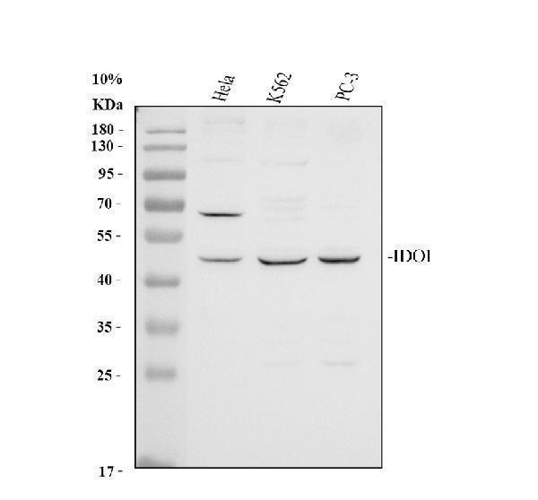
Western blot analysis of IDO1 using anti-IDO1 antibody (PB9603).
Electrophoresis was performed on a 10% SDS-PAGE gel at 80V (Stacking gel) / 120V (Resolving gel) for 2 hours. The sample well of each lane was loaded with 30 ug of sample under reducing conditions.
Lane 1: human Hela whole cell lysates,
Lane 2: human K562 whole cell lysates,
Lane 3: human PC-3 whole cell lysates.
After electrophoresis, proteins were transferred to a nitrocellulose membrane at 150 mA for 50-90 minutes. Blocked the membrane with 5% non-fat milk/TBS for 1.5 hour at RT. The membrane was incubated with rabbit anti-IDO1 antigen affinity purified polyclonal antibody (PB9603) at 0.5 μg/mL overnight at 4°C, then washed with TBS-0.1%Tween 3 times with 5 minutes each and probed with a goat anti-rabbit IgG-HRP secondary antibody at a dilution of 1:5000 for 1.5 hour at RT. The signal is developed using an ECL Plus Western Blotting Substrate (Catalog # AR1196-200) with Tanon 5200 system. A specific band was detected for IDO1 at approximately 45 kDa. The expected band size for IDO1 is at 45 kDa.

Click image to see more details
IHC analysis of IDO1 using anti-IDO1 antibody (PB9603).
IDO1 was detected in paraffin-embedded section of Human Lung Cancer Tissue. Heat mediated antigen retrieval was performed in citrate buffer (pH6, epitope retrieval solution) for 20 mins. The tissue section was blocked with 10% goat serum. The tissue section was then incubated with 1μg/ml rabbit anti-IDO1 Antibody (PB9603) overnight at 4°C. Biotinylated goat anti-rabbit IgG was used as secondary antibody and incubated for 30 minutes at 37°C. The tissue section was developed using Strepavidin-Biotin-Complex (SABC)(Catalog # SA1022) with DAB as the chromogen.

Click image to see more details
Flow Cytometry analysis of A431 cells using anti-IDO1 antibody (PB9603).
Overlay histogram showing A431 cells stained with PB9603 (Blue line). To facilitate intracellular staining, cells were fixed with 4% paraformaldehyde and permeabilized with permeabilization buffer. The cells were blocked with 10% normal goat serum. And then incubated with rabbit anti-IDO1 Antibody (PB9603,1μg/1x106 cells) for 30 min at 20°C. DyLight®488 conjugated goat anti-rabbit IgG (BA1127, 5-10μg/1x106 cells) was used as secondary antibody for 30 minutes at 20°C. Isotype control antibody (Green line) was rabbit IgG (1μg/1x106) used under the same conditions. Unlabelled sample without incubation with primary antibody and secondary antibody (Red line) was used as a blank control.

Click image to see more details
IF analysis of IDO1 using anti-IDO1 antibody (PB9603).
IDO1 was detected in immunocytochemical section of A431 cells. Enzyme antigen retrieval was performed using IHC enzyme antigen retrieval reagent (AR0022) for 15 mins. The cells were blocked with 10% goat serum. And then incubated with 2μg/mL rabbit anti-IDO1 Antibody (PB9603) overnight at 4°C. DyLight®488 Conjugated Goat Anti-Rabbit IgG (BA1127) was used as secondary antibody at 1:100 dilution and incubated for 30 minutes at 37°C. The section was counterstained with DAPI. Visualize using a fluorescence microscope and filter sets appropriate for the label used.

Click image to see more details
The heterogeneity within the myeloid cells. a 4 subclusters of myeloid cells were identified by UMAP analysis. b Marker gene expression of each cell type. c Comparison of cell count proportion of each cell type between imatinib resistant and sensitive patients. d mIHC staining of panCK (red), LYZ (green), IDO1 (magenta) and DAPI in GIST TME.
Index in PubMed under a CC BY license. PMID: 38443340

Click image to see more details
Transcription characteristics and heterogeneity among the six malignant cell types. a UMAP analysis showing the six cell type of malignant cells. b Colored cells by each sample. c Bar plot showing the cell count proportion of each maligant subcluster in GIST. d GO enrichment analysis of marker genes form BASP1_fib (left), DUSP1_fib (middle), IDO1_fib (right).
Index in PubMed under a CC BY license. PMID: 38443340
Specific Publications For Anti-Indoleamine 2, 3-dioxygenase/IDO1 Antibody Picoband® (PB9603)
Loading publications
Recommended Resources
Here are featured tools and databases that you might find useful.
- Boster's Pathways Library
- Protein Databases
- Bioscience Research Protocol Resources
- Data Processing & Analysis Software
- Photo Editing Software
- Scientific Literature Resources
- Research Paper Management Tools
- Molecular Biology Software
- Primer Design Tools
- Bioinformatics Tools
- Phylogenetic Tree Analysis
Customer Reviews
Have you used Anti-Indoleamine 2, 3-dioxygenase/IDO1 Antibody Picoband®?
Share your experimental results or join a short interview to earn up to $1,000 in product credits or other rewards.
0 Reviews For Anti-Indoleamine 2, 3-dioxygenase/IDO1 Antibody Picoband®
Customer Q&As
Have a question?
Find answers in Q&As, reviews.
Can't find your answer?
Submit your question
1 Customer Q&As for Anti-Indoleamine 2, 3-dioxygenase/IDO1 Antibody Picoband®
Question
We are currently using anti-Indoleamine 2, 3-dioxygenase/IDO1 antibody PB9603 for human tissue, and we are happy with the ICC results. The species of reactivity given in the datasheet says human. Is it likely that the antibody can work on zebrafish tissues as well?
Verified Customer
Verified customer
Asked: 2018-12-26
Answer
The anti-Indoleamine 2, 3-dioxygenase/IDO1 antibody (PB9603) has not been tested for cross reactivity specifically with zebrafish tissues, though there is a good chance of cross reactivity. We have an innovator award program that if you test this antibody and show it works in zebrafish you can get your next antibody for free. Please contact me if I can help you with anything.
Boster Scientific Support
Answered: 2018-12-26
