Click image to see more details
Product Info Summary
| SKU: | M00021-1 |
|---|---|
| Size: | 100μg/vial |
| Reactive Species: | Mouse |
| Host: | Rat |
| Application: | IHC-P, WB |
Customers Who Bought This Also Bought


Product info
Product Name
Anti- IL-10 Monoclonal Antibody
SKU/Catalog Number
M00021-1
Size
100μg/vial
Form
Lyophilized
Description
Boster Bio Anti- IL-10 Monoclonal Antibody catalog # M00021-1. Tested in IHC-P, WB applications. This antibody reacts with Mouse.
Storage & Handling
Store at -20°C for one year. For short term storage and frequent use, store at 4°C for up to one month. Avoid repeated freeze-thaw cycles.
Cite This Product
Anti- IL-10 Monoclonal Antibody (Boster Biological Technology, Pleasanton CA, USA, Catalog # M00021-1)
Host
Rat
Contents
PBS, pH 7.0. Contains no stabilizers or preservatives
Clonality
Monoclonal
Clone Number
AFIE-9
Isotype
Rat IgG1, κ
Immunogen
Recombinant mouse IL-10
Cross-reactivity
No cross reactivity with other proteins
Reactive Species
M00021-1 is reactive to Il10 in Mouse
Calculated molecular weight
20.6 kDa
Background of Il10
Interleukin-10 (IL-10 or IL10), also known as human cytokine synthesis inhibitory factor (CSIF), is an anti-inflammatory cytokine. In humans IL-10 is encoded by the IL10 gene. It is capable of inhibiting synthesis of pro-inflammatory cytokines like IFN-gamma, IL-2, IL-3, TNFalpha and GM-CSF made by cells such as macrophages and regulatory T-cells.IL-10 also displays potent abilities to suppress the antigen presentation capacity of antigen presenting cells. Kim et al. (1992) showed that the mouse IL 10 gene contains 5 exons and spans about 5.2 kb of genomic DNA. Eskdale et al. (1997) mapped the IL10 gene to the junction between 1q31 and 1q32.
Antibody Validation
Boster validates all antibodies on WB, IHC, ICC, Immunofluorescence, and ELISA with known positive control and negative samples to ensure specificity and high affinity, including thorough antibody incubations.
Application & Images
Applications
M00021-1 is guaranteed for IHC-P, WB Boster Guarantee
Recommend Dilution
| Application | Dilution | Species |
|---|---|---|
| Western blot | 0.25-0.5&mu:g/ml | Mouse |
| Immunohistochemistry (Paraffin-embedded Section) | 2-5μg/ml | Mouse |
Tested application
Use TE buffer pH 9.0 for antigen retrieval; (*) citrate buffer pH 6.0 is an alternative.
Validation Images & Assay Conditions
Click image to see more details
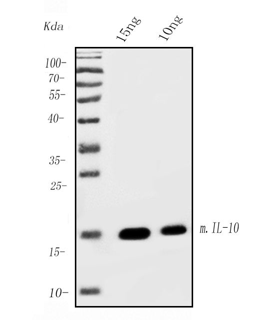
Western blot analysis of IL-10 using anti-IL-10 antibody (M00021-1).
Electrophoresis was performed on a 5-20% SDS-PAGE gel at 70V (Stacking gel) / 90V (Resolving gel) for 2-3 hours.
Lane 1: recombinant mouse IL-10 protein 15ng,
Lane 2: recombinant mouse IL-10 protein 10ng.
After Electrophoresis, proteins were transferred to a Nitrocellulose membrane at 150mA for 50-90 minutes. Blocked the membrane with 5% Non-fat Milk/ TBS for 1.5 hour at RT. The membrane was incubated with rat anti-IL-10 antigen affinity purified monoclonal antibody (Catalog # M00021-1) at 0.5 μg/mL overnight at 4°C, then washed with TBS-0.1%Tween 3 times with 5 minutes each and probed with a goat anti-rat IgG-HRP secondary antibody at a dilution of 1:10000 for 1.5 hour at RT. The signal is developed using an Enhanced Chemiluminescent detection (ECL) kit (Catalog # EK1001) with Tanon 5200 system.

Click image to see more details
IHC analysis of IL-10 using anti-IL-10 antibody (M00021-1).
IL-10 was detected in paraffin-embedded section of mouse spleen tissue. Heat mediated antigen retrieval was performed in EDTA buffer (pH8.0, epitope retrieval solution). The tissue section was blocked with 10% goat serum. The tissue section was then incubated with 5μg/ml rat anti-IL-10 Antibody (M00021-1) overnight at 4°C. Biotinylated goat anti-rat IgG was used as secondary antibody and incubated for 30 minutes at 37°C. The tissue section was developed using Strepavidin-Biotin-Complex (SABC) (Catalog # SA1021) with DAB as the chromogen.

Click image to see more details
IHC analysis of IL-10 using anti-IL-10 antibody (M00021-1).
IL-10 was detected in paraffin-embedded section of mouse lymphaden tissue. Heat mediated antigen retrieval was performed in EDTA buffer (pH8.0, epitope retrieval solution). The tissue section was blocked with 10% goat serum. The tissue section was then incubated with 5μg/ml rat anti-IL-10 Antibody (M00021-1) overnight at 4°C. Biotinylated goat anti-rat IgG was used as secondary antibody and incubated for 30 minutes at 37°C. The tissue section was developed using Strepavidin-Biotin-Complex (SABC) (Catalog # SA1021) with DAB as the chromogen.
Specific Publications For Anti- IL-10 Monoclonal Antibody (M00021-1)
Loading publications
Recommended Resources
Here are featured tools and databases that you might find useful.
- Boster's Pathways Library
- Protein Databases
- Bioscience Research Protocol Resources
- Data Processing & Analysis Software
- Photo Editing Software
- Scientific Literature Resources
- Research Paper Management Tools
- Molecular Biology Software
- Primer Design Tools
- Bioinformatics Tools
- Phylogenetic Tree Analysis
Customer Reviews
Have you used Anti- IL-10 Monoclonal Antibody?
Share your experimental results or join a short interview to earn up to $1,000 in product credits or other rewards.
0 Reviews For Anti- IL-10 Monoclonal Antibody
Customer Q&As
Have a question?
Find answers in Q&As, reviews.
Can't find your answer?
Submit your question
